A dislocation of the knee (tibiofemoral jointTibiofemoral jointKnee Joint: Anatomy) is a rare injury but is important to recognize because of limb-threatening trauma. Knee dislocations (KDs) are emergent cases that require immediate reduction and evaluation of the neurovascular system. Clinical presentation includes precedent trauma, obvious deformityDeformityExamination of the Upper Limbs, effusion, ecchymosisEcchymosisExtravasation of blood into the skin, resulting in a nonelevated, rounded or irregular, blue or purplish patch, larger than a petechia.Orbital Fractures, and possible signs of vascular injury to the popliteal arteryPopliteal ArteryThe continuation of the femoral artery coursing through the popliteal fossa; it divides into the anterior and posterior tibial arteries.Popliteal Fossa: Anatomy. Management includes reduction of the dislocation, serial examinations for arterial perfusion, imaging of the vascular system, and orthopedic and vascular consultations.
A dislocation of the knee (tibiofemoral jointTibiofemoral jointKnee Joint: Anatomy) is a rare injury that is important to recognize due to the frequent association with vascular injury and associated risk of limb amputationAmputationAn amputation is the separation of a portion of the limb or the entire limb from the body, along with the bone. Amputations are generally indicated for conditions that compromise the viability of the limb or promote the spread of a local process that could manifest systemically. Amputation. These are emergent cases that require immediate reduction and evaluation of the neurovascular system.
Epidemiology
0.02% of all musculoskeletal injuries (very uncommon)
IncidenceIncidenceThe number of new cases of a given disease during a given period in a specified population. It also is used for the rate at which new events occur in a defined population. It is differentiated from prevalence, which refers to all cases in the population at a given time.Measures of Disease Frequency may be higher, as the actual percentage of cases that dislocate and spontaneously reduce is unknown.
ObesityObesityObesity is a condition associated with excess body weight, specifically with the deposition of excessive adipose tissue. Obesity is considered a global epidemic. Major influences come from the western diet and sedentary lifestyles, but the exact mechanisms likely include a mixture of genetic and environmental factors. Obesity is an independent risk factor.
High-velocity dislocations are more commonly associated with vascular injury.
Vascular injuries reported in approximately 5%–50% of patientsPatientsIndividuals participating in the health care system for the purpose of receiving therapeutic, diagnostic, or preventive procedures.Clinician–Patient Relationship
Peroneal nervePeroneal nerveThe lateral of the two terminal branches of the sciatic nerve. The peroneal (or fibular) nerve provides motor and sensory innervation to parts of the leg and foot.Popliteal Fossa: Anatomy injuries reported in approximately 20% of patientsPatientsIndividuals participating in the health care system for the purpose of receiving therapeutic, diagnostic, or preventive procedures.Clinician–Patient Relationship
Potentially limb-threatening secondary to vascular injury
Low-velocity trauma: associated with morbid obesityObesityObesity is a condition associated with excess body weight, specifically with the deposition of excessive adipose tissue. Obesity is considered a global epidemic. Major influences come from the western diet and sedentary lifestyles, but the exact mechanisms likely include a mixture of genetic and environmental factors. Obesity
Classification
Knee dislocations (KDs) can be classified based on the position of the tibiaTibiaThe second longest bone of the skeleton. It is located on the medial side of the lower leg, articulating with the fibula laterally, the talus distally, and the femur proximally.Knee Joint: Anatomy in relation to the femur, etiology, or on the pattern of ligament tears (Schenck classification).
Classification based on the position of the tibiaTibiaThe second longest bone of the skeleton. It is located on the medial side of the lower leg, articulating with the fibula laterally, the talus distally, and the femur proximally.Knee Joint: Anatomy in relation to the femur:
Types:
Anterior dislocation
Posterior dislocation
Medial dislocation
Lateral dislocation
Rotatory dislocation
Posterior and anterior KDs: most common, highest risk of associated popliteal arteryPopliteal ArteryThe continuation of the femoral artery coursing through the popliteal fossa; it divides into the anterior and posterior tibial arteries.Popliteal Fossa: Anatomy injury
Posterior dislocations: primarily occur secondary to direct traumaDirect TraumaToddler’s Fractures to the anterior tibiaTibiaThe second longest bone of the skeleton. It is located on the medial side of the lower leg, articulating with the fibula laterally, the talus distally, and the femur proximally.Knee Joint: Anatomy of a flexed knee (dashboard injury)
Associated with morbid obesityObesityObesity is a condition associated with excess body weight, specifically with the deposition of excessive adipose tissue. Obesity is considered a global epidemic. Major influences come from the western diet and sedentary lifestyles, but the exact mechanisms likely include a mixture of genetic and environmental factors. Obesity
Schenck classification of KDs:based on pattern of ligament tears
Usually a high-energy mechanism of injury is required for multiple ligaments to fail in order for dislocation to occur.
KDKDAn acute, febrile, mucocutaneous condition accompanied by swelling of cervical lymph nodes in infants and young children. The principal symptoms are fever, congestion of the ocular conjunctivae, reddening of the lips and oral cavity, protuberance of tongue papillae, and edema or erythema of the extremities.Kawasaki Disease I: involvement of the anterior cruciate ligamentAnterior Cruciate LigamentA strong ligament of the knee that originates from the posteromedial portion of the lateral condyle of the femur, passes anteriorly and inferiorly between the condyles, and attaches to the depression in front of the intercondylar eminence of the tibia.Knee Joint: Anatomy (ACLACLA strong ligament of the knee that originates from the posteromedial portion of the lateral condyle of the femur, passes anteriorly and inferiorly between the condyles, and attaches to the depression in front of the intercondylar eminence of the tibia.Knee Joint: Anatomy) or posterior cruciate ligamentPosterior Cruciate LigamentA strong ligament of the knee that originates from the anterolateral surface of the medial condyle of the femur, passes posteriorly and inferiorly between the condyles, and attaches to the posterior intercondylar area of the tibia.Knee Joint: Anatomy (PCLPCLA strong ligament of the knee that originates from the anterolateral surface of the medial condyle of the femur, passes posteriorly and inferiorly between the condyles, and attaches to the posterior intercondylar area of the tibia.Knee Joint: Anatomy)
KDKDAn acute, febrile, mucocutaneous condition accompanied by swelling of cervical lymph nodes in infants and young children. The principal symptoms are fever, congestion of the ocular conjunctivae, reddening of the lips and oral cavity, protuberance of tongue papillae, and edema or erythema of the extremities.Kawasaki Disease II: injury to both ACLACLA strong ligament of the knee that originates from the posteromedial portion of the lateral condyle of the femur, passes anteriorly and inferiorly between the condyles, and attaches to the depression in front of the intercondylar eminence of the tibia.Knee Joint: Anatomy/PCLPCLA strong ligament of the knee that originates from the anterolateral surface of the medial condyle of the femur, passes posteriorly and inferiorly between the condyles, and attaches to the posterior intercondylar area of the tibia.Knee Joint: Anatomy with both collaterals intact (rare)
KDKDAn acute, febrile, mucocutaneous condition accompanied by swelling of cervical lymph nodes in infants and young children. The principal symptoms are fever, congestion of the ocular conjunctivae, reddening of the lips and oral cavity, protuberance of tongue papillae, and edema or erythema of the extremities.Kawasaki Disease III: injury to ACLACLA strong ligament of the knee that originates from the posteromedial portion of the lateral condyle of the femur, passes anteriorly and inferiorly between the condyles, and attaches to the depression in front of the intercondylar eminence of the tibia.Knee Joint: Anatomy and PCLPCLA strong ligament of the knee that originates from the anterolateral surface of the medial condyle of the femur, passes posteriorly and inferiorly between the condyles, and attaches to the posterior intercondylar area of the tibia.Knee Joint: Anatomy and either medial collateral ligamentMedial collateral ligamentKnee Joint: Anatomy (MCLMCLKnee Joint: Anatomy) or lateral collateral ligamentLateral collateral ligamentKnee Joint: Anatomy (LCL) (not both)
KDKDAn acute, febrile, mucocutaneous condition accompanied by swelling of cervical lymph nodes in infants and young children. The principal symptoms are fever, congestion of the ocular conjunctivae, reddening of the lips and oral cavity, protuberance of tongue papillae, and edema or erythema of the extremities.Kawasaki Disease IIIM: MCLMCLKnee Joint: Anatomy torn
KDKDAn acute, febrile, mucocutaneous condition accompanied by swelling of cervical lymph nodes in infants and young children. The principal symptoms are fever, congestion of the ocular conjunctivae, reddening of the lips and oral cavity, protuberance of tongue papillae, and edema or erythema of the extremities.Kawasaki Disease IIIL: LCL torn
KDKDAn acute, febrile, mucocutaneous condition accompanied by swelling of cervical lymph nodes in infants and young children. The principal symptoms are fever, congestion of the ocular conjunctivae, reddening of the lips and oral cavity, protuberance of tongue papillae, and edema or erythema of the extremities.Kawasaki Disease IV: all 4 ligaments torn (ACLACLA strong ligament of the knee that originates from the posteromedial portion of the lateral condyle of the femur, passes anteriorly and inferiorly between the condyles, and attaches to the depression in front of the intercondylar eminence of the tibia.Knee Joint: Anatomy, PCLPCLA strong ligament of the knee that originates from the anterolateral surface of the medial condyle of the femur, passes posteriorly and inferiorly between the condyles, and attaches to the posterior intercondylar area of the tibia.Knee Joint: Anatomy, MCLMCLKnee Joint: Anatomy, LCL), highest rate of vascular injury
KDKDAn acute, febrile, mucocutaneous condition accompanied by swelling of cervical lymph nodes in infants and young children. The principal symptoms are fever, congestion of the ocular conjunctivae, reddening of the lips and oral cavity, protuberance of tongue papillae, and edema or erythema of the extremities.Kawasaki Disease V: multiple ligamentous injuries with periarticular fractureFractureA fracture is a disruption of the cortex of any bone and periosteum and is commonly due to mechanical stress after an injury or accident. Open fractures due to trauma can be a medical emergency. Fractures are frequently associated with automobile accidents, workplace injuries, and trauma.Overview of Bone Fractures (knee fracture-dislocation)
Direct force to the anterior tibiaTibiaThe second longest bone of the skeleton. It is located on the medial side of the lower leg, articulating with the fibula laterally, the talus distally, and the femur proximally.Knee Joint: Anatomy, with the knee flexed at 90 degrees
Seen in motor vehicle accidentsMotor Vehicle AccidentsSpinal Cord Injuries: tibiaTibiaThe second longest bone of the skeleton. It is located on the medial side of the lower leg, articulating with the fibula laterally, the talus distally, and the femur proximally.Knee Joint: Anatomy striking the dashboard
Anterior:
Hyperextension injury
Disruption of the posterior structures of the knee
Lateral:
Varus/valgus stress
Commonly associated with a tibial plateauPlateauCardiac PhysiologyfractureFractureA fracture is a disruption of the cortex of any bone and periosteum and is commonly due to mechanical stress after an injury or accident. Open fractures due to trauma can be a medical emergency. Fractures are frequently associated with automobile accidents, workplace injuries, and trauma.Overview of Bone Fractures
Commonly associated with a tibial plateauPlateauCardiac PhysiologyfractureFractureA fracture is a disruption of the cortex of any bone and periosteum and is commonly due to mechanical stress after an injury or accident. Open fractures due to trauma can be a medical emergency. Fractures are frequently associated with automobile accidents, workplace injuries, and trauma.Overview of Bone Fractures
2 or more ligaments (cruciate or collateral) must be compromised to allow the knee to dislocate.
Most commonly, both cruciate ligaments and at least 1 collateral ligament are disrupted.
The popliteal arteryPopliteal ArteryThe continuation of the femoral artery coursing through the popliteal fossa; it divides into the anterior and posterior tibial arteries.Popliteal Fossa: Anatomy:
Traverses the posterior portion of the knee
Attaches proximally at the adductor hiatus
Attaches distally to the proximal arch of the soleusSoleusLeg: Anatomy muscle
This tethering makes it vulnerable to injury with a KDKDAn acute, febrile, mucocutaneous condition accompanied by swelling of cervical lymph nodes in infants and young children. The principal symptoms are fever, congestion of the ocular conjunctivae, reddening of the lips and oral cavity, protuberance of tongue papillae, and edema or erythema of the extremities.Kawasaki Disease.
The peroneal nervePeroneal nerveThe lateral of the two terminal branches of the sciatic nerve. The peroneal (or fibular) nerve provides motor and sensory innervation to parts of the leg and foot.Popliteal Fossa: Anatomy:
Winds laterally around the proximal fibulaFibulaThe bone of the lower leg lateral to and smaller than the tibia. In proportion to its length, it is the most slender of the long bones.Leg: Anatomy
Tethered above and below the fibular head by fascial/ligamentous structures
This tethering makes it vulnerable to injury with a KDKDAn acute, febrile, mucocutaneous condition accompanied by swelling of cervical lymph nodes in infants and young children. The principal symptoms are fever, congestion of the ocular conjunctivae, reddening of the lips and oral cavity, protuberance of tongue papillae, and edema or erythema of the extremities.Kawasaki Disease.
KDs can be open or closed.
Posterolateral dislocations are generally irreducible and require surgical management.
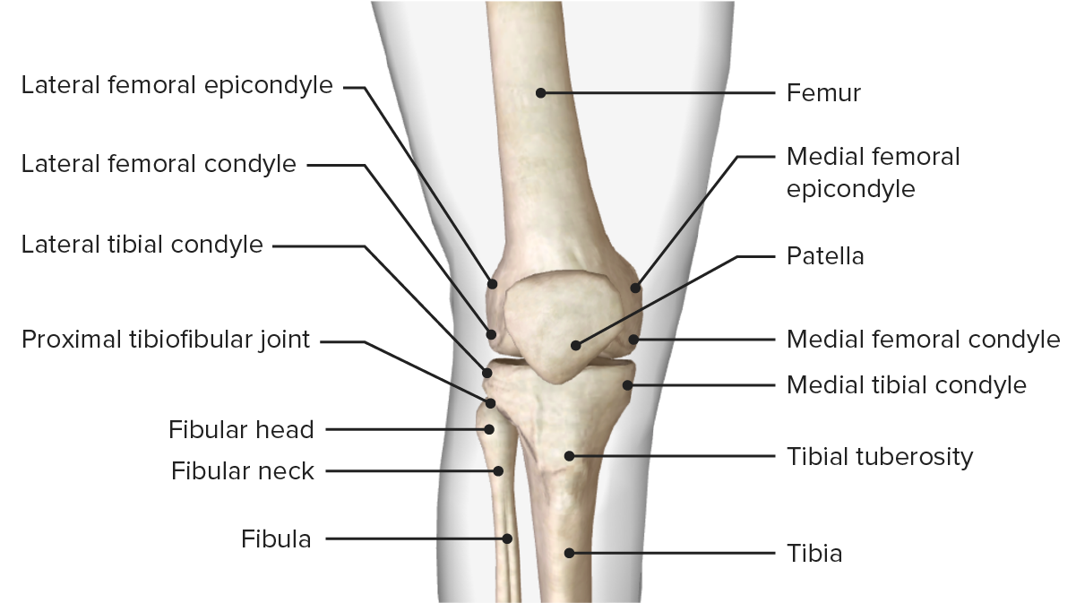
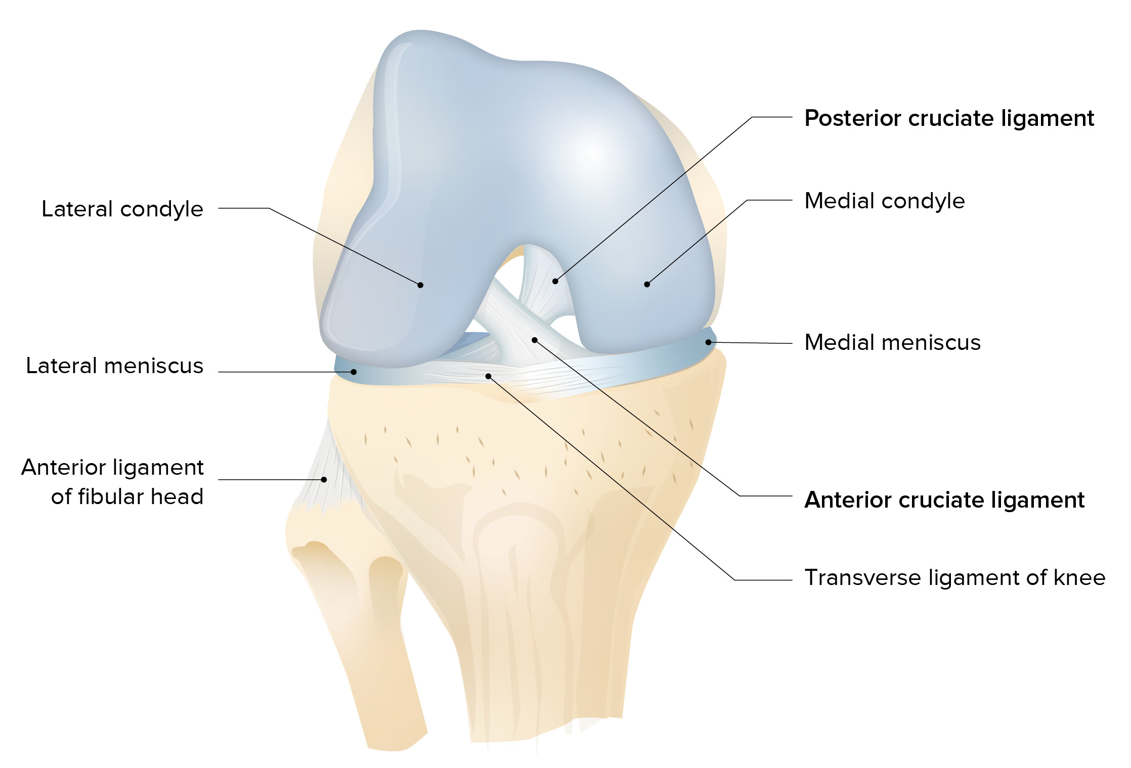
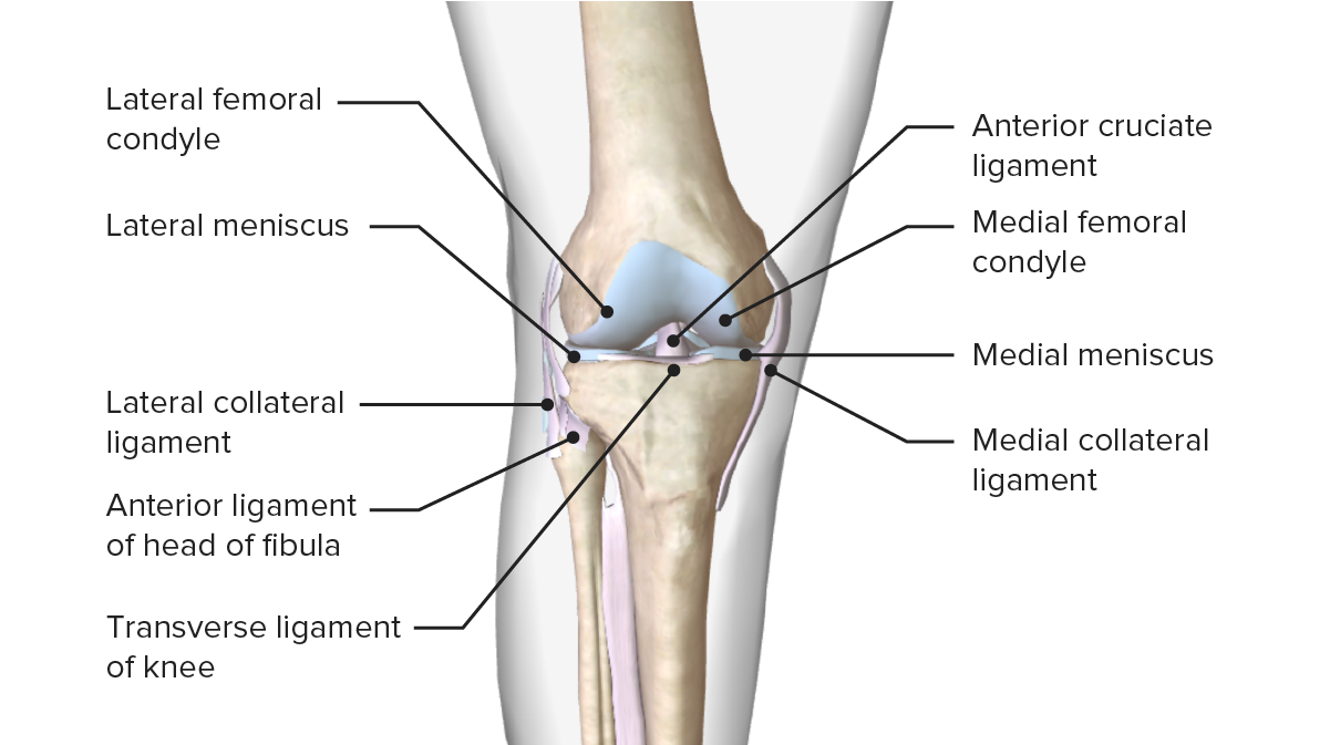
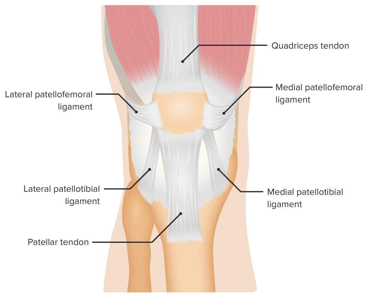
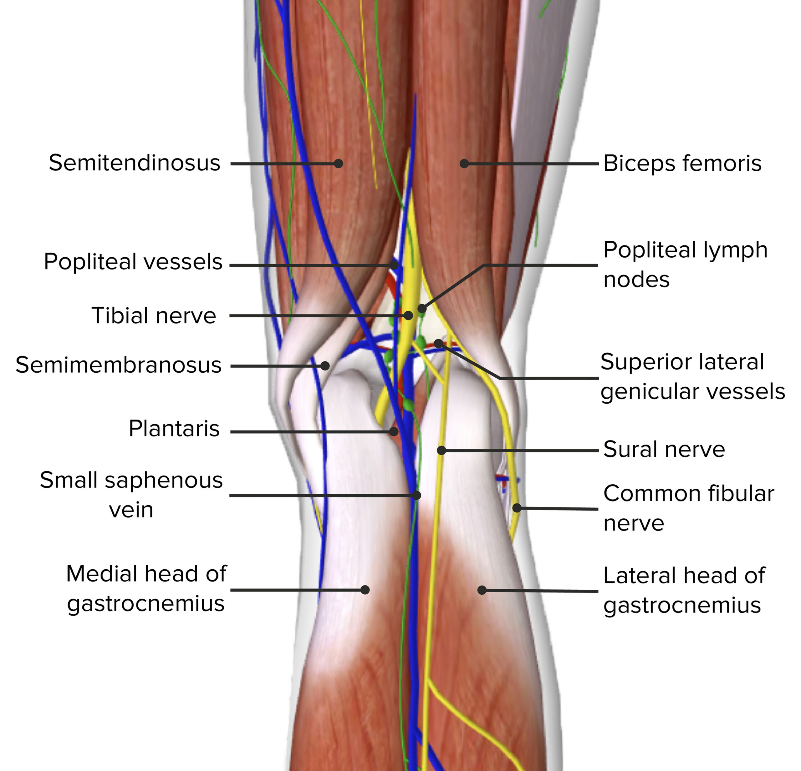
Image displaying the menisci, ligaments, and bony surfaces and their relation to one another
Knee dislocations are an uncommon injury but require emergent evaluation and management secondary to the possibility of limb ischemiaIschemiaA hypoperfusion of the blood through an organ or tissue caused by a pathologic constriction or obstruction of its blood vessels, or an absence of blood circulation.Ischemic Cell Damage, which may require surgical intervention to avoid amputationAmputationAn amputation is the separation of a portion of the limb or the entire limb from the body, along with the bone. Amputations are generally indicated for conditions that compromise the viability of the limb or promote the spread of a local process that could manifest systemically. Amputation.
Important considerations
If dislocation occurred in the context of high-energy trauma: PatientsPatientsIndividuals participating in the health care system for the purpose of receiving therapeutic, diagnostic, or preventive procedures.Clinician–Patient Relationship may require simultaneous evaluation and management following the advanced trauma life support (ATLS) method.
Consider the possibility of spontaneous reductions: In the absence of obvious deformityDeformityExamination of the Upper Limbs, the clinicianClinicianA physician, nurse practitioner, physician assistant, or another health professional who is directly involved in patient care and has a professional relationship with patients.Clinician–Patient Relationship may overlook the high risk of an associated vascular injury.
History
In isolated trauma, the patient may be able to describe the mechanism.
PatientsPatientsIndividuals participating in the health care system for the purpose of receiving therapeutic, diagnostic, or preventive procedures.Clinician–Patient Relationship or first responders will usually report a high-energy blunt trauma (e.g., motorMotorNeurons which send impulses peripherally to activate muscles or secretory cells.Nervous System: Histology vehicle accident, industrial accident, sports injury).
As these injuries are often high-energy trauma, evaluation for other life-threatening injuries is essential.
Limited range of motionRange of motionThe distance and direction to which a bone joint can be extended. Range of motion is a function of the condition of the joints, muscles, and connective tissues involved. Joint flexibility can be improved through appropriate muscle strength exercises.Examination of the Upper Limbs
Appearance less dramatic in morbidly obese patientsPatientsIndividuals participating in the health care system for the purpose of receiving therapeutic, diagnostic, or preventive procedures.Clinician–Patient Relationship
Up to 50% of KDs may have spontaneously reduced prior to presentation (exact percentage is unknown).
Meticulous vascular examination:
Popliteal arteryPopliteal ArteryThe continuation of the femoral artery coursing through the popliteal fossa; it divides into the anterior and posterior tibial arteries.Popliteal Fossa: Anatomy injury:
Decreased distal pulses (always check distal pulses and perfusion)
Ankle-brachial indexAnkle-brachial indexComparison of the blood pressure between the brachial artery and the posterior tibial artery. It is a predictor of peripheral arterial disease.Cardiovascular Examination < 0.9
Compare to contralateral limb.
Signs of severe vascular compromise:
Absent pulses
Pale or cool extremity
ParesthesiasParesthesiasSubjective cutaneous sensations (e.g., cold, warmth, tingling, pressure, etc.) that are experienced spontaneously in the absence of stimulation.Posterior Cord Syndrome
Paralysis
Palpable thrill or audible bruit
Visible expanding hematomaHematomaA collection of blood outside the blood vessels. Hematoma can be localized in an organ, space, or tissue.Intussusception
Neurological examinationNeurological examinationA neurological exam is a systematic assessment of cognitive, sensory, and motor responses to identify pathologies of the nervous system. A neurological exam allows for the localization of neurologic lesions to narrow the differential diagnosis and focus on subsequent laboratory and imaging examinations. The exam should include assessments of the subject’s mental status, speech, cranial nerves, motor system, deep tendon reflexes, sensation, balance, and coordination.Neurological Examination:
Peroneal nerve injuryNerve InjurySurgical Complications common with KDKDAn acute, febrile, mucocutaneous condition accompanied by swelling of cervical lymph nodes in infants and young children. The principal symptoms are fever, congestion of the ocular conjunctivae, reddening of the lips and oral cavity, protuberance of tongue papillae, and edema or erythema of the extremities.Kawasaki Disease
Peroneal nervePeroneal nerveThe lateral of the two terminal branches of the sciatic nerve. The peroneal (or fibular) nerve provides motor and sensory innervation to parts of the leg and foot.Popliteal Fossa: Anatomy exam:
Evaluate sensation: 1st web space
Evaluate motorMotorNeurons which send impulses peripherally to activate muscles or secretory cells.Nervous System: Histology involvement: eversionEversionChronic Apophyseal Injury of the footFootThe foot is the terminal portion of the lower limb, whose primary function is to bear weight and facilitate locomotion. The foot comprises 26 bones, including the tarsal bones, metatarsal bones, and phalanges. The bones of the foot form longitudinal and transverse arches and are supported by various muscles, ligaments, and tendons.Foot: Anatomy and footFootThe foot is the terminal portion of the lower limb, whose primary function is to bear weight and facilitate locomotion. The foot comprises 26 bones, including the tarsal bones, metatarsal bones, and phalanges. The bones of the foot form longitudinal and transverse arches and are supported by various muscles, ligaments, and tendons.Foot: Anatomy/toe dorsiflexion
No associated deep tendon reflex
Ligamentous examination:
Evaluate ligamentous integrity of the knee after neurovascular integrity.
Rule out associated fractureFractureA fracture is a disruption of the cortex of any bone and periosteum and is commonly due to mechanical stress after an injury or accident. Open fractures due to trauma can be a medical emergency. Fractures are frequently associated with automobile accidents, workplace injuries, and trauma.Overview of Bone Fractures in high-energy trauma prior to ligament evaluation.
Evidence of multiple ligamentous disruption in the appropriate clinical scenario may indicate a KDKDAn acute, febrile, mucocutaneous condition accompanied by swelling of cervical lymph nodes in infants and young children. The principal symptoms are fever, congestion of the ocular conjunctivae, reddening of the lips and oral cavity, protuberance of tongue papillae, and edema or erythema of the extremities.Kawasaki Disease with a spontaneous reduction.
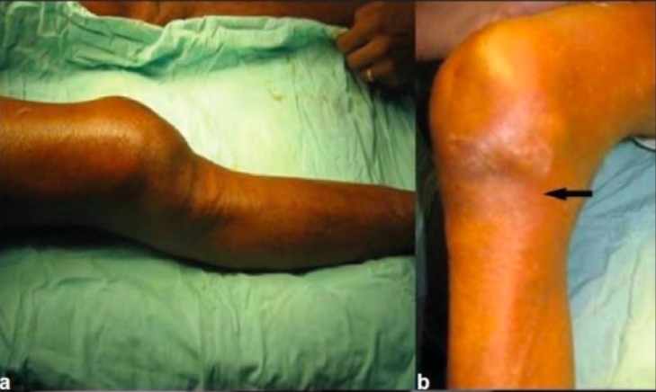
Posterior knee dislocation: Note the obvious deformity as the leg is displaced downward due to gravity.
Image: “Neglected irreducible posterolateral knee dislocation” by Department of Orthopaedics, Postgraduate Institute of Medical Education and Research, Sector 12, Chandigarh – 160 012, India. License: CC BY 2.0
Diagnosis
The majority of complete KDs are clinically obvious with a history of significant trauma and gross deformityDeformityExamination of the Upper Limbs of the knee. Diagnosis is made clinically, although imaging studies may be indicated to confirm suspected vascular injury or fractureFractureA fracture is a disruption of the cortex of any bone and periosteum and is commonly due to mechanical stress after an injury or accident. Open fractures due to trauma can be a medical emergency. Fractures are frequently associated with automobile accidents, workplace injuries, and trauma.Overview of Bone Fractures and/or for surgical planning.
Steps
Determine the direction of the KDKDAn acute, febrile, mucocutaneous condition accompanied by swelling of cervical lymph nodes in infants and young children. The principal symptoms are fever, congestion of the ocular conjunctivae, reddening of the lips and oral cavity, protuberance of tongue papillae, and edema or erythema of the extremities.Kawasaki Disease.
Consider potential dislocation with spontaneous reduction:
Gross instability of the knee in the setting of trauma
Evidence of significant hyperextension of the knee in the setting of trauma
Imaging studies are indicated for evaluation of associated fractureFractureA fracture is a disruption of the cortex of any bone and periosteum and is commonly due to mechanical stress after an injury or accident. Open fractures due to trauma can be a medical emergency. Fractures are frequently associated with automobile accidents, workplace injuries, and trauma.Overview of Bone Fractures, ligamentous injury, and/or arterial injuryArterial InjuryHemothorax.
X-rayX-rayPenetrating electromagnetic radiation emitted when the inner orbital electrons of an atom are excited and release radiant energy. X-ray wavelengths range from 1 pm to 10 nm. Hard x-rays are the higher energy, shorter wavelength x-rays. Soft x-rays or grenz rays are less energetic and longer in wavelength. The short wavelength end of the x-ray spectrum overlaps the gamma rays wavelength range. The distinction between gamma rays and x-rays is based on their radiation source.Pulmonary Function Tests
Plain X-raysX-raysX-rays are high-energy particles of electromagnetic radiation used in the medical field for the generation of anatomical images. X-rays are projected through the body of a patient and onto a film, and this technique is called conventional or projectional radiography. X-rays: indicated post-reduction or prior to any ligamentous evaluation
Anteroposterior (AP) and lateral projections of the knee: ideal to visualize the dislocation
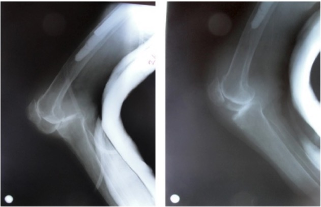
Posterior knee dislocation seen on radiography before and after reduction with underlying knee osteoarthritis
Image: “Posterior knee dislocation on X-ray” by Department of Orthopaedics and Traumatology, Ankara Numune Training and Research Hospital, 06100 Ankara, Türkiye. License: CC BY 3.0
Vascular evaluation
Ankle-brachial indices evaluation:
Required for patientsPatientsIndividuals participating in the health care system for the purpose of receiving therapeutic, diagnostic, or preventive procedures.Clinician–Patient Relationship without obvious severe vascular compromise (all reduced KDs without obvious signs of arterial injuryArterial InjuryHemothorax)
Ankle-brachial indexAnkle-brachial indexComparison of the blood pressure between the brachial artery and the posterior tibial artery. It is a predictor of peripheral arterial disease.Cardiovascular Examination < 0.9:
High incidenceIncidenceThe number of new cases of a given disease during a given period in a specified population. It also is used for the rate at which new events occur in a defined population. It is differentiated from prevalence, which refers to all cases in the population at a given time.Measures of Disease Frequency of vascular injury
Vascular imaging indicated
Duplex ultrasound:
Noninvasive alternative to direct angiographyAngiographyRadiography of blood vessels after injection of a contrast medium.Cardiac Surgery for vascular assessment
Can be performed at bedside; does not require transport to CT scanner or angiographyAngiographyRadiography of blood vessels after injection of a contrast medium.Cardiac Surgery suite
Lowest cost
CT angiogramCT angiogramA non-invasive method that uses a ct scanner for capturing images of blood vessels and tissues. A contrast material is injected, which helps produce detailed images that aid in diagnosing vascular diseases.Pulmonary Function Tests:
Indicated in cases of asymmetric pulses, decreased ankle-brachial indexAnkle-brachial indexComparison of the blood pressure between the brachial artery and the posterior tibial artery. It is a predictor of peripheral arterial disease.Cardiovascular Examination, or abnormal duplex ultrasound
Noninvasive alternative to direct arteriography
Requires transport to CT scanner; does not require an angiographyAngiographyRadiography of blood vessels after injection of a contrast medium.Cardiac Surgery suite
Intermediate cost
Direct arteriography:
Criterion standard method of assessing vascular integrity
Formerly indicated for all KDs after reduction:
There is debate over the appropriate application of imaging options.
Some experts utilize advanced imaging in all instances of KDKDAn acute, febrile, mucocutaneous condition accompanied by swelling of cervical lymph nodes in infants and young children. The principal symptoms are fever, congestion of the ocular conjunctivae, reddening of the lips and oral cavity, protuberance of tongue papillae, and edema or erythema of the extremities.Kawasaki Disease since vascular damage can go undetected.
Risk of injury related to procedure (requires arterial puncture)
Requires interventional radiologist or vascular surgeon
Highest cost
Management
Closed reductionClosed ReductionRadial Head Subluxation (Nursemaid’s Elbow) of a KDKDAn acute, febrile, mucocutaneous condition accompanied by swelling of cervical lymph nodes in infants and young children. The principal symptoms are fever, congestion of the ocular conjunctivae, reddening of the lips and oral cavity, protuberance of tongue papillae, and edema or erythema of the extremities.Kawasaki Disease should not be delayed, especially in a limb with obvious vascular impairment. As these injuries are often high-energy trauma, evaluation for other life-threatening injuries is essential.
Reduction should not be delayed if the patient has any evidence for vascular compromise.
Procedure:
Longitudinal or axialAxialComputed Tomography (CT) traction followed by translationTranslationTranslation is the process of synthesizing a protein from a messenger RNA (mRNA) transcript. This process is divided into three primary stages: initiation, elongation, and termination. Translation is catalyzed by structures known as ribosomes, which are large complexes of proteins and ribosomal RNA (rRNA). Stages and Regulation of Translation of the tibiaTibiaThe second longest bone of the skeleton. It is located on the medial side of the lower leg, articulating with the fibula laterally, the talus distally, and the femur proximally.Knee Joint: Anatomy
Anterior and posterior dislocations usually reduce easily.
Immediate orthopedic consultation for dislocations that are not easily reduced
Posterolateral dislocations are generally irreducible; this may be indicated by a skinSkinThe skin, also referred to as the integumentary system, is the largest organ of the body. The skin is primarily composed of the epidermis (outer layer) and dermis (deep layer). The epidermis is primarily composed of keratinocytes that undergo rapid turnover, while the dermis contains dense layers of connective tissue.Skin: Structure and Functions dimple in the anteromedial aspect of the knee.
Immediate post-reduction evaluation of pulses and perfusion
Bedside X-raysX-raysX-rays are high-energy particles of electromagnetic radiation used in the medical field for the generation of anatomical images. X-rays are projected through the body of a patient and onto a film, and this technique is called conventional or projectional radiography. X-rays post-reduction to confirm reduction and evaluate for fractureFractureA fracture is a disruption of the cortex of any bone and periosteum and is commonly due to mechanical stress after an injury or accident. Open fractures due to trauma can be a medical emergency. Fractures are frequently associated with automobile accidents, workplace injuries, and trauma.Overview of Bone Fractures
Advanced imaging indicated if suspected vascular compromise
Immediate post-reduction evaluation of pulses and perfusion
Serial perfusion evaluations:
Presence of normal distal pulses alone does not rule out popliteal arteryPopliteal ArteryThe continuation of the femoral artery coursing through the popliteal fossa; it divides into the anterior and posterior tibial arteries.Popliteal Fossa: Anatomy injury.
Thrombus related to arterial injuryArterial InjuryHemothorax may present with delayed findings of poor perfusion.
Arterial-brachial index assessments
Bedside duplex ultrasonographyDuplex ultrasonographyUltrasonography applying the doppler effect combined with real-time imaging. The real-time image is created by rapid movement of the ultrasound beam. A powerful advantage of this technique is the ability to estimate the velocity of flow from the doppler shift frequency.Hypercoagulable States
PatientsPatientsIndividuals participating in the health care system for the purpose of receiving therapeutic, diagnostic, or preventive procedures.Clinician–Patient Relationship with any abnormalities on vascular exam, ankle-brachial indexAnkle-brachial indexComparison of the blood pressure between the brachial artery and the posterior tibial artery. It is a predictor of peripheral arterial disease.Cardiovascular Examination, or duplex ultrasound: CT angiographyAngiographyRadiography of blood vessels after injection of a contrast medium.Cardiac Surgery or direct arteriography
Post-reduction imaging:
Bedside X-raysX-raysX-rays are high-energy particles of electromagnetic radiation used in the medical field for the generation of anatomical images. X-rays are projected through the body of a patient and onto a film, and this technique is called conventional or projectional radiography. X-rays post-reduction to confirm reduction and evaluate for fractureFractureA fracture is a disruption of the cortex of any bone and periosteum and is commonly due to mechanical stress after an injury or accident. Open fractures due to trauma can be a medical emergency. Fractures are frequently associated with automobile accidents, workplace injuries, and trauma.Overview of Bone Fractures
Advanced imaging indicated if suspected vascular compromise
Consultations:
Immediate vascular surgeryVascular surgeryVascular surgery is the specialized field of medicine that focuses on the surgical management of the pathologies of the peripheral circulation. The main goal of most vascular procedures is to restore circulatory function to the affected vessels by relieving occlusions or by redirecting blood flow (e.g., bypass).Vascular Surgery consultation for injuries with any signs or concerns of vascular compromise
Immediate orthopedic consultation:
Indicated for complicated reduction or irreducible reductions
Indicated for management of the fractures and/or ligamentous injury
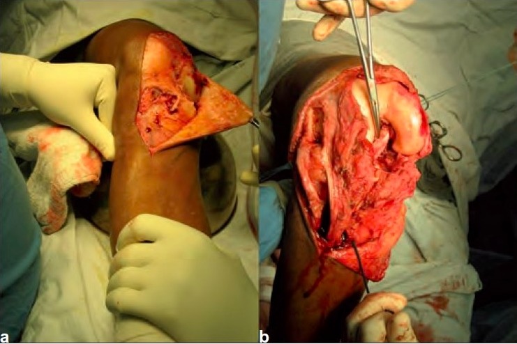
Operative repair of an irreducible posterolateral knee dislocation
Image: “Peroperative photograph” by Indian Journal of Orthopaedics. License: CC BY 4.0
Further management
Long legLegThe lower leg, or just “leg” in anatomical terms, is the part of the lower limb between the knee and the ankle joint. The bony structure is composed of the tibia and fibula bones, and the muscles of the leg are grouped into the anterior, lateral, and posterior compartments by extensions of fascia.Leg: Anatomy splint at 20–30 degrees of flexionFlexionExamination of the Upper Limbs
After initial management of acute injury, orthopedic management is related to the other specific structural injuries to the knee.
Complications
Injury to the popliteal arteryPopliteal ArteryThe continuation of the femoral artery coursing through the popliteal fossa; it divides into the anterior and posterior tibial arteries.Popliteal Fossa: Anatomy or vein
Arthrofibrosis (stiffness): most common complication
Compartment syndromeCompartment SyndromeCompartment syndrome is a surgical emergency usually occurring secondary to trauma. The condition is marked by increased pressure within a compartment that compromises the circulation and function of the tissues within that space.Compartment Syndrome
AmputationAmputationAn amputation is the separation of a portion of the limb or the entire limb from the body, along with the bone. Amputations are generally indicated for conditions that compromise the viability of the limb or promote the spread of a local process that could manifest systemically. Amputation
PrognosisPrognosisA prediction of the probable outcome of a disease based on a individual’s condition and the usual course of the disease as seen in similar situations.Non-Hodgkin Lymphomas
Depends on the velocity of the injury, neurovascular damage, and associated injuries
Athletes with a high-velocity injury are unlikely to return to pre-injury levels of sport.
Low-velocity dislocations have a relatively better prognosisPrognosisA prediction of the probable outcome of a disease based on a individual’s condition and the usual course of the disease as seen in similar situations.Non-Hodgkin Lymphomas.
PrognosisPrognosisA prediction of the probable outcome of a disease based on a individual’s condition and the usual course of the disease as seen in similar situations.Non-Hodgkin Lymphomas improves with timely repair of vascular injuries.
Clinical Relevance
Anterior cruciate ligamentAnterior Cruciate LigamentA strong ligament of the knee that originates from the posteromedial portion of the lateral condyle of the femur, passes anteriorly and inferiorly between the condyles, and attaches to the depression in front of the intercondylar eminence of the tibia.Knee Joint: Anatomy (ACLACLA strong ligament of the knee that originates from the posteromedial portion of the lateral condyle of the femur, passes anteriorly and inferiorly between the condyles, and attaches to the depression in front of the intercondylar eminence of the tibia.Knee Joint: Anatomy) injury: frequently injured, important stabilizing ligament of the knee. The ACLACLA strong ligament of the knee that originates from the posteromedial portion of the lateral condyle of the femur, passes anteriorly and inferiorly between the condyles, and attaches to the depression in front of the intercondylar eminence of the tibia.Knee Joint: Anatomy is most commonly injured in sporting endeavors and frequently torn when the knee is dislocated.
Meniscus tearMeniscus tearThe menisci are fibrocartilaginous wedge-shaped structures between the distal femur and proximal tibia that stabilize and dissipate weight-bearing forces at the knee joint. A meniscus tear is an injury to the meniscus caused by rotational or shearing forces across the tibiofemoral joint. Meniscus Tear: injury to the meniscus occurs due to rotational or shearing forcesShearing forcesVascular Resistance, Flow, and Mean Arterial Pressure placed across the knee jointKnee jointThe knee joint is made up of the articulations between the femur, tibia, and patella bones, and is one of the largest and most complex joints of the human body. The knee is classified as a synovial hinge joint, which primarily allows for flexion and extension with a more limited degree of translation and rotation. Knee Joint: Anatomy. Meniscus tears may be associated with an ACL tearACL tearKnee Pain and/or dislocation.
Patellar instability and dislocation: a spectrum of conditions affecting the patellaPatellaThe flat, triangular bone situated at the anterior part of the knee.Knee Joint: Anatomy secondary to trauma or activity. The conditions are characterized by peripatellar painPainAn unpleasant sensation induced by noxious stimuli which are detected by nerve endings of nociceptive neurons.Pain: Types and Pathways and knee instability. With dislocation of the patellaPatellaThe flat, triangular bone situated at the anterior part of the knee.Knee Joint: Anatomy, there may be obvious deformityDeformityExamination of the Upper Limbs and an inability to extend the knee.
AmputationAmputationAn amputation is the separation of a portion of the limb or the entire limb from the body, along with the bone. Amputations are generally indicated for conditions that compromise the viability of the limb or promote the spread of a local process that could manifest systemically. Amputation: amputationAmputationAn amputation is the separation of a portion of the limb or the entire limb from the body, along with the bone. Amputations are generally indicated for conditions that compromise the viability of the limb or promote the spread of a local process that could manifest systemically. Amputation may be required after prolonged ischemiaIschemiaA hypoperfusion of the blood through an organ or tissue caused by a pathologic constriction or obstruction of its blood vessels, or an absence of blood circulation.Ischemic Cell Damage associated with vascular compromise during dislocation.
Injury to the popliteal arteryPopliteal ArteryThe continuation of the femoral artery coursing through the popliteal fossa; it divides into the anterior and posterior tibial arteries.Popliteal Fossa: Anatomy or vein: the vessels of the popliteal fossaPopliteal fossaThe popliteal fossa or the “knee pit” is a diamond-shaped, fat-filled, shallow depression on the posterior aspect of the knee joint. The popliteal fossa is located at the dorsal aspect of the knee and contains an increased number of lymph nodes as well as structures of the neurovascular system that travel from the thigh to the lower leg.Popliteal Fossa: Anatomy may be injured with KDKDAn acute, febrile, mucocutaneous condition accompanied by swelling of cervical lymph nodes in infants and young children. The principal symptoms are fever, congestion of the ocular conjunctivae, reddening of the lips and oral cavity, protuberance of tongue papillae, and edema or erythema of the extremities.Kawasaki Disease. Delay in diagnosis or recognition, treatment with reduction, and/or surgical repair may lead to arterial injuryArterial InjuryHemothorax and amputationAmputationAn amputation is the separation of a portion of the limb or the entire limb from the body, along with the bone. Amputations are generally indicated for conditions that compromise the viability of the limb or promote the spread of a local process that could manifest systemically. Amputation.
Compartment syndromeCompartment SyndromeCompartment syndrome is a surgical emergency usually occurring secondary to trauma. The condition is marked by increased pressure within a compartment that compromises the circulation and function of the tissues within that space.Compartment Syndrome: a surgical emergencySurgical EmergencyAcute Abdomen usually occurring secondary to trauma. Compartment syndromeCompartment SyndromeCompartment syndrome is a surgical emergency usually occurring secondary to trauma. The condition is marked by increased pressure within a compartment that compromises the circulation and function of the tissues within that space.Compartment Syndrome is marked by increased pressure within a fascial compartment, which compromises the circulationCirculationThe movement of the blood as it is pumped through the cardiovascular system.ABCDE Assessment and function of the tissues within that space.
References
Mohseni, M, & Simon, LV. (2024). Knee dislocation. StatPearls. Treasure Island (FL): StatPearls Publishing. Retrieved June 28, 2026, from http://www.ncbi.nlm.nih.gov/books/NBK470595/
Raj, MA, Mabrouk, A, & Varacallo, M. (2023). Posterior cruciate ligament knee injuries. StatPearls. Treasure Island (FL): StatPearls Publishing. Retrieved June 28, 2026, from http://www.ncbi.nlm.nih.gov/books/NBK430726/
Duprey, K, & Lin, M. (2010). Posterior knee dislocation. The Western Journal of Emergency Medicine, 11(1), 103–104.
Create your free account or log in to continue reading!