A hordeolum is an acute infection affecting the meibomian, Zeis, or Moll glands of the eyelid. Stasis of the gland secretions predisposes the glands to bacterial infection. Staphylococcus aureus is the most common pathogen. The condition presents as a painful, localized, erythematous nodule in the anterior (external hordeolum) or posterior (internal hordeolum) lamella of the eyelid. A hordeolum usually resolves spontaneously and can be managed with warm compresses, massage, and lid hygiene. In certain cases of significant swelling, topical antibiotics with steroids may be needed. If a hordeolum does not resolve, the patient should be referred to ophthalmology for incision and drainage.
Last updated: Dec 15, 2025
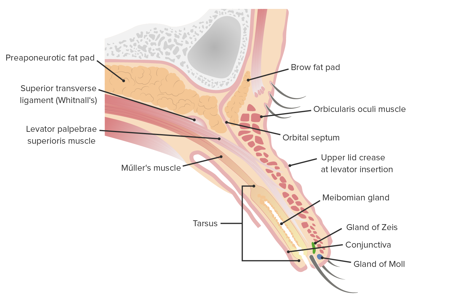
Sagittal cut of the upper eyelid
Image by Lecturio.| Name | Type | Opening location | Infection |
|---|---|---|---|
| Gland of Zeis | Sebaceous gland Sebaceous Gland Small, sacculated organs found within the dermis. Each gland has a single duct that emerges from a cluster of oval alveoli. Each alveolus consists of a transparent basement membrane enclosing epithelial cells. The ducts from most sebaceous glands open into a hair follicle, but some open on the general surface of the skin. Sebaceous glands secrete sebum. Hordeolum (Stye) | Directly into the eyelash follicle | External hordeolum External Hordeolum Hordeolum (Stye) |
| Gland of Moll Gland of Moll Blepharitis | Modified sweat glands Modified Sweat Glands Hordeolum (Stye) | Between adjacent lashes | External hordeolum External Hordeolum Hordeolum (Stye) |
| Meibomian gland Meibomian Gland Chalazion | Modified sebaceous gland Modified Sebaceous Gland Hordeolum (Stye) | Behind eyelashes | Internal hordeolum Internal Hordeolum Hordeolum (Stye) |
Clinical presentation[1,2]
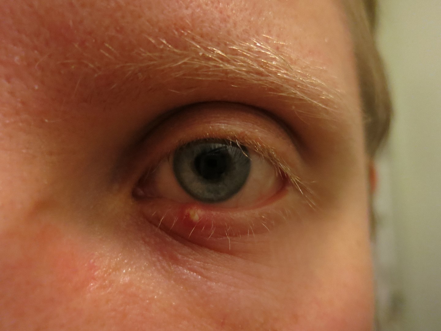
Man suffering from external hordeolum of the lower lid.
Image: “Stye” by Palosirkka. License: CC0 1.0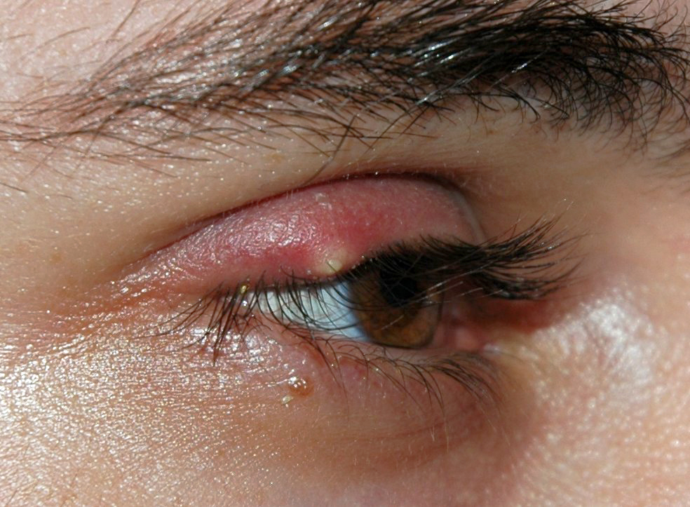
Upper eyelid hordeolum, illustrating a localized erythematous and swollen area.
Image: “Stye” by Andre Riemann. License: Public DomainDiagnosis[4-6,8]
Clinical trials and guidelines involving the management of hordeola are lacking; however, the following information is based on US and UK society recommendations. Additionally, management may vary depending on practice location. Please see your local practice guidelines.
Conservative management[4-6,8]
Most hordeola resolve spontaneously, lasting up to 1–2 weeks.
Topical antibiotic and/or steroid ointment[1,5,6,8]
Systemic antibiotics[2,5,6,8]
Incision and drainage Incision And Drainage Chalazion[5,6]
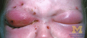
Photograph showing orbital cellulitis, a bacterial infection of the periocular tissues.
Image: “Orbital cellulitis” by Jonathan Trobe. License: CC BY 3.0Diagnosis Codes:
This code is used to diagnose a
hordeolum
Hordeolum
A hordeolum is an acute infection affecting the meibomian, Zeiss, or Moll glands of the eyelid. Stasis of the gland secretions predisposes to bacterial infection. Staphylococcus aureus is the most common pathogen.
Hordeolum (Stye), commonly known as a
stye
Stye
A hordeolum is an acute infection affecting the meibomian, Zeiss, or Moll glands of the eyelid. Stasis of the gland secretions predisposes to bacterial infection. Staphylococcus aureus is the most common pathogen.
Hordeolum (Stye), which is an acute, painful infection of a gland in the eyelid, typically caused by
Staphylococcus
Staphylococcus
Staphylococcus is a medically important genera of Gram-positive, aerobic cocci. These bacteria form clusters resembling grapes on culture plates. Staphylococci are ubiquitous for humans, and many strains compose the normal skin flora.
Staphylococcus
bacteria
Bacteria
Bacteria are prokaryotic single-celled microorganisms that are metabolically active and divide by binary fission. Some of these organisms play a significant role in the pathogenesis of diseases.
Bacteriology.
| Coding System | Code | Description |
|---|---|---|
| ICD-10-CM | H00.01- | Hordeolum Hordeolum A hordeolum is an acute infection affecting the meibomian, Zeiss, or Moll glands of the eyelid. Stasis of the gland secretions predisposes to bacterial infection. Staphylococcus aureus is the most common pathogen. Hordeolum (Stye) externum |
| SNOMED CT | 400959003 | Hordeolum Hordeolum A hordeolum is an acute infection affecting the meibomian, Zeiss, or Moll glands of the eyelid. Stasis of the gland secretions predisposes to bacterial infection. Staphylococcus aureus is the most common pathogen. Hordeolum (Stye) (disorder) |
Procedures/Interventions:
This code is for a simple
incision and drainage
Incision And Drainage
Chalazion, which may be performed on a large, persistent
hordeolum
Hordeolum
A hordeolum is an acute infection affecting the meibomian, Zeiss, or Moll glands of the eyelid. Stasis of the gland secretions predisposes to bacterial infection. Staphylococcus aureus is the most common pathogen.
Hordeolum (Stye) that does not resolve with conservative treatment like
warm compresses
Warm Compresses
Chalazion.
| Coding System | Code | Description |
|---|---|---|
| CPT | 67700 | Blepharotomy, drainage of abscess Abscess Accumulation of purulent material in tissues, organs, or circumscribed spaces, usually associated with signs of infection. Chronic Granulomatous Disease, eyelid |
Medications:
This code is for a topical antibiotic ointment, such as
erythromycin
Erythromycin
A bacteriostatic antibiotic macrolide produced by streptomyces erythreus. Erythromycin a is considered its major active component. In sensitive organisms, it inhibits protein synthesis by binding to 50s ribosomal subunits. This binding process inhibits peptidyl transferase activity and interferes with translocation of amino acids during translation and assembly of proteins.
Macrolides and Ketolides, which is often prescribed to be applied to the eyelid margin to help treat the infection and prevent its spread.
| Coding System | Code | Description |
|---|---|---|
| RxNorm | 4053 | Erythromycin Erythromycin A bacteriostatic antibiotic macrolide produced by streptomyces erythreus. Erythromycin a is considered its major active component. In sensitive organisms, it inhibits protein synthesis by binding to 50s ribosomal subunits. This binding process inhibits peptidyl transferase activity and interferes with translocation of amino acids during translation and assembly of proteins. Macrolides and Ketolides (ingredient) |