Central cord syndrome (CCS) is a neurological syndrome caused by an injury to the center of the spinal cordSpinal cordThe spinal cord is the major conduction pathway connecting the brain to the body; it is part of the CNS. In cross section, the spinal cord is divided into an H-shaped area of gray matter (consisting of synapsing neuronal cell bodies) and a surrounding area of white matter (consisting of ascending and descending tracts of myelinated axons). Spinal Cord: Anatomy, affecting the spinothalamic tracts ((STTs) sensorySensoryNeurons which conduct nerve impulses to the central nervous system.Nervous System: Histology) and medial aspect of the corticospinal tracts ((CSTs) motorMotorNeurons which send impulses peripherally to activate muscles or secretory cells.Nervous System: Histology), most often due to trauma in patientsPatientsIndividuals participating in the health care system for the purpose of receiving therapeutic, diagnostic, or preventive procedures.Clinician–Patient Relationship with cervical spondylosisCervical SpondylosisNeck Pain. A less frequent but classic cause of CCS is syringomyelia. Clinical manifestations are motorMotorNeurons which send impulses peripherally to activate muscles or secretory cells.Nervous System: Histology deficits in the arms more so than the legs and variableVariableVariables represent information about something that can change. The design of the measurement scales, or of the methods for obtaining information, will determine the data gathered and the characteristics of that data. As a result, a variable can be qualitative or quantitative, and may be further classified into subgroups.Types of VariablessensorySensoryNeurons which conduct nerve impulses to the central nervous system.Nervous System: Histology deficits below the level of injury. Diagnosis is made clinically and is supported with neuroimagingNeuroimagingNon-invasive methods of visualizing the central nervous system, especially the brain, by various imaging modalities.Febrile Infant. Definitive management can be medical or surgical, depending on the severity of the injury. Rehabilitation is the key to maintaining functionality and improving chances of recovery.
Central cord syndrome (CCS) is a neurological syndrome caused by an injury to the center of the spinal cordSpinal cordThe spinal cord is the major conduction pathway connecting the brain to the body; it is part of the CNS. In cross section, the spinal cord is divided into an H-shaped area of gray matter (consisting of synapsing neuronal cell bodies) and a surrounding area of white matter (consisting of ascending and descending tracts of myelinated axons). Spinal Cord: Anatomy, affecting the spinothalamic tracts ((STTs) sensorySensoryNeurons which conduct nerve impulses to the central nervous system.Nervous System: Histology) and medial aspect of the corticospinal tracts ((CSTs) motorMotorNeurons which send impulses peripherally to activate muscles or secretory cells.Nervous System: Histology).
Epidemiology
Most common form of incomplete spinal cordSpinal cordThe spinal cord is the major conduction pathway connecting the brain to the body; it is part of the CNS. In cross section, the spinal cord is divided into an H-shaped area of gray matter (consisting of synapsing neuronal cell bodies) and a surrounding area of white matter (consisting of ascending and descending tracts of myelinated axons). Spinal Cord: Anatomy injury
PrevalencePrevalenceThe total number of cases of a given disease in a specified population at a designated time. It is differentiated from incidence, which refers to the number of new cases in the population at a given time.Measures of Disease Frequency: 15 % to 25 % of those with traumatic spinal cordSpinal cordThe spinal cord is the major conduction pathway connecting the brain to the body; it is part of the CNS. In cross section, the spinal cord is divided into an H-shaped area of gray matter (consisting of synapsing neuronal cell bodies) and a surrounding area of white matter (consisting of ascending and descending tracts of myelinated axons). Spinal Cord: Anatomy injury
Men are more commonly affected.
Young patientsPatientsIndividuals participating in the health care system for the purpose of receiving therapeutic, diagnostic, or preventive procedures.Clinician–Patient Relationship: usually due to trauma (e.g., automobile accidents)
Older patientsPatientsIndividuals participating in the health care system for the purpose of receiving therapeutic, diagnostic, or preventive procedures.Clinician–Patient Relationship (> 65 years): usually caused by neckNeckThe part of a human or animal body connecting the head to the rest of the body.Peritonsillar Abscess hyperextension combined with underlying spinal disease (e.g., osteoarthritisOsteoarthritisOsteoarthritis (OA) is the most common form of arthritis, and is due to cartilage destruction and changes of the subchondral bone. The risk of developing this disorder increases with age, obesity, and repetitive joint use or trauma. Patients develop gradual joint pain, stiffness lasting < 30 minutes, and decreased range of motion. Osteoarthritis)
Postinfectious: transverse myelitisTransverse myelitisInflammation which extends horizontally across the spinal cord, believed to be immune-mediated and triggered by infection; associated with signs and symptoms of motor, sensory, and/or autonomic dysfunction.Mononucleosis
Inflammatory: multiple sclerosisSclerosisA pathological process consisting of hardening or fibrosis of an anatomical structure, often a vessel or a nerve.Wilms Tumor (MSMSMultiple sclerosis (MS) is a chronic inflammatory autoimmune disease that leads to demyelination of the nerves in the CNS. Young women are more predominantly affected by this most common demyelinating condition.Multiple Sclerosis)
Medullary tumorTumorInflammation → progressive cervical myelopathy with central cord features
EpendymomaEpendymomaEpendymomas are glial cell tumors arising from CSF-producing ependymal cells lining the ventricular system. Ependymomas most commonly occur within the posterior fossa in contact with the 4th ventricle, or within the intramedullary spinal cord. Ependymoma
AstrocytomaAstrocytomaAstrocytomas are neuroepithelial tumors that arise from astrocytes, which are star-shaped glial cells (supporting tissues of the CNS). Astrocytomas are a type of glioma. There are 4 grades of astrocytomas. Astrocytoma
Rarely metastatic disease
Spondylosis
Atlantoaxial (C1–C2) instability (e.g., due to rheumatoid arthritisArthritisAcute or chronic inflammation of joints.Osteoarthritis (RA))
Spinal arthropathies
OsteoporosisOsteoporosisOsteoporosis refers to a decrease in bone mass and density leading to an increased number of fractures. There are 2 forms of osteoporosis: primary, which is commonly postmenopausal or senile; and secondary, which is a manifestation of immobilization, underlying medical disorders, or long-term use of certain medications. Osteoporosis (compressionCompressionBlunt Chest Trauma fractures)
Injury to the vertebral columnVertebral columnThe human spine, or vertebral column, is the most important anatomical and functional axis of the human body. It consists of 7 cervical vertebrae, 12 thoracic vertebrae, and 5 lumbar vertebrae and is limited cranially by the skull and caudally by the sacrum. Vertebral Column: Anatomy causes a spinal cordSpinal cordThe spinal cord is the major conduction pathway connecting the brain to the body; it is part of the CNS. In cross section, the spinal cord is divided into an H-shaped area of gray matter (consisting of synapsing neuronal cell bodies) and a surrounding area of white matter (consisting of ascending and descending tracts of myelinated axons). Spinal Cord: Anatomy injury related to the force and direction of the traumatic eventTraumatic eventAn emotionally painful, shocking, stressful, and sometimes life-threatening experience. It can result from witnessing distressing events such as natural disasters, physical or sexual abuse, and terrorism or other acts of violence.Posttraumatic Stress Disorder (PTSD) and the anatomic vulnerability of individual spinal elements.
Causes of spinal cordSpinal cordThe spinal cord is the major conduction pathway connecting the brain to the body; it is part of the CNS. In cross section, the spinal cord is divided into an H-shaped area of gray matter (consisting of synapsing neuronal cell bodies) and a surrounding area of white matter (consisting of ascending and descending tracts of myelinated axons). Spinal Cord: Anatomy injury involve 1 or more of the following:
Vertebral fractureFractureA fracture is a disruption of the cortex of any bone and periosteum and is commonly due to mechanical stress after an injury or accident. Open fractures due to trauma can be a medical emergency. Fractures are frequently associated with automobile accidents, workplace injuries, and trauma.Overview of Bone Fractures
A cervical spineSpineThe human spine, or vertebral column, is the most important anatomical and functional axis of the human body. It consists of 7 cervical vertebrae, 12 thoracic vertebrae, and 5 lumbar vertebrae and is limited cranially by the skull and caudally by the sacrum.Vertebral Column: Anatomy hyperextension injury can be related to:
Fall with neckNeckThe part of a human or animal body connecting the head to the rest of the body.Peritonsillar Abscess hyperextended (especially from a significant height, such as a roof)
High-velocity trauma (e.g., motorMotorNeurons which send impulses peripherally to activate muscles or secretory cells.Nervous System: Histology vehicle accident)
SpineSpineThe human spine, or vertebral column, is the most important anatomical and functional axis of the human body. It consists of 7 cervical vertebrae, 12 thoracic vertebrae, and 5 lumbar vertebrae and is limited cranially by the skull and caudally by the sacrum.Vertebral Column: AnatomysubluxationSubluxationRadial Head Subluxation (Nursemaid’s Elbow) and/or fractureFractureA fracture is a disruption of the cortex of any bone and periosteum and is commonly due to mechanical stress after an injury or accident. Open fractures due to trauma can be a medical emergency. Fractures are frequently associated with automobile accidents, workplace injuries, and trauma.Overview of Bone Fractures → compressionCompressionBlunt Chest Trauma of the spinal cordSpinal cordThe spinal cord is the major conduction pathway connecting the brain to the body; it is part of the CNS. In cross section, the spinal cord is divided into an H-shaped area of gray matter (consisting of synapsing neuronal cell bodies) and a surrounding area of white matter (consisting of ascending and descending tracts of myelinated axons). Spinal Cord: Anatomy anteriorly (by osteophytes or disc) and/or posteriorly (by buckling the ligamentum flavum)
This can result in contusion/edemaEdemaEdema is a condition in which excess serous fluid accumulates in the body cavity or interstitial space of connective tissues. Edema is a symptom observed in several medical conditions. It can be categorized into 2 types, namely, peripheral (in the extremities) and internal (in an organ or body cavity). Edema, particularly in the central portion of the spinal cordSpinal cordThe spinal cord is the major conduction pathway connecting the brain to the body; it is part of the CNS. In cross section, the spinal cord is divided into an H-shaped area of gray matter (consisting of synapsing neuronal cell bodies) and a surrounding area of white matter (consisting of ascending and descending tracts of myelinated axons). Spinal Cord: Anatomy
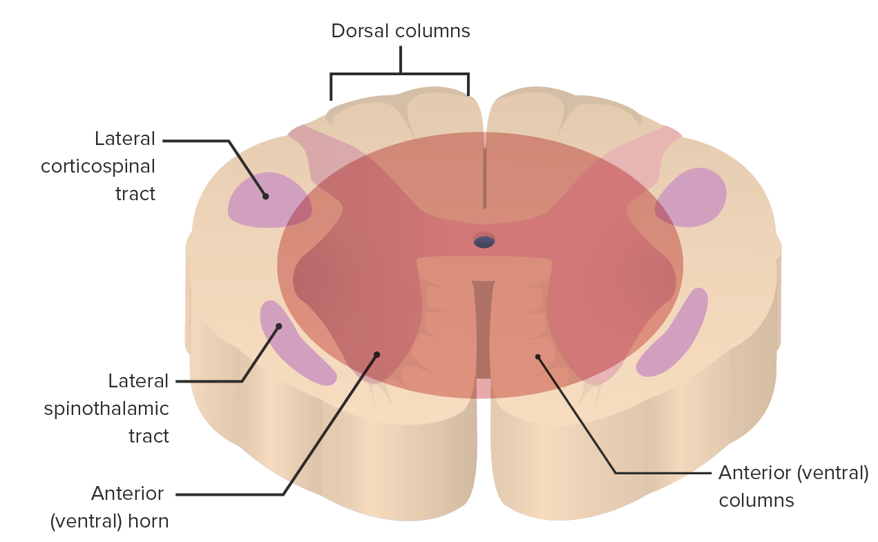
Affected areas of the spinal cordSpinal cordThe spinal cord is the major conduction pathway connecting the brain to the body; it is part of the CNS. In cross section, the spinal cord is divided into an H-shaped area of gray matter (consisting of synapsing neuronal cell bodies) and a surrounding area of white matter (consisting of ascending and descending tracts of myelinated axons). Spinal Cord: Anatomy by central cord syndrome:
Medial aspect of the CSTs/ anterior hornAnterior hornOne of three central columns of the spinal cord. It is composed of gray matter spinal laminae VIII and ix.Brown-Séquard Syndromegray matterGray matterRegion of central nervous system that appears darker in color than the other type, white matter. It is composed of neuronal cell bodies; neuropil; glial cells and capillaries but few myelinated nerve fibers.Cerebral Cortex: Anatomy → weakness in the arms > legs
Axonal disruption in the white matterWhite MatterThe region of central nervous system that appears lighter in color than the other type, gray matter. It mainly consists of myelinated nerve fibers and contains few neuronal cell bodies or dendrites.Brown-Séquard Syndrome as the fibers pass from the dorsal to the ventral hornVentral hornOne of three central columns of the spinal cord. It is composed of gray matter spinal laminae VIII and ix.Brown-Séquard Syndrome → loss of deep tendon reflexesDeep Tendon ReflexesNeurological Examination
Both somatic and visceral motorMotorNeurons which send impulses peripherally to activate muscles or secretory cells.Nervous System: Histology nuclei are affected.
Location of lesion in central cord syndrome (CSS): Rough depiction of the area affected by CSS (affects lateral spinothalamic tracts (STTs) and lateral corticospinal tracts (CSTs) to varying degrees)
Image by Lecturio.
Clinical Presentation and Diagnosis
Understanding the structures affected by a central cord lesion is key to their correlationCorrelationDetermination of whether or not two variables are correlated. This means to study whether an increase or decrease in one variable corresponds to an increase or decrease in the other variable.Causality, Validity, and Reliability with clinical signs and symptoms. Diagnosis of CCS is by clinical exam and diagnostic imaging.
Clinical presentation
History:
Site of injury: impact with the neckNeckThe part of a human or animal body connecting the head to the rest of the body.Peritonsillar Abscess hyperextended
Types of injuries that cause cervical fractures:
Blunt trauma from a motorMotorNeurons which send impulses peripherally to activate muscles or secretory cells.Nervous System: Histology vehicle accident
Penetrating trauma from gunshot or knife
Falling trauma: downstairs or from a significant height (e.g., off a roof or a ladder)
Inflammatory/demyelinating (MSMSMultiple sclerosis (MS) is a chronic inflammatory autoimmune disease that leads to demyelination of the nerves in the CNS. Young women are more predominantly affected by this most common demyelinating condition.Multiple Sclerosis)
Postinfectious inflammationInflammationInflammation is a complex set of responses to infection and injury involving leukocytes as the principal cellular mediators in the body’s defense against pathogenic organisms. Inflammation is also seen as a response to tissue injury in the process of wound healing. The 5 cardinal signs of inflammation are pain, heat, redness, swelling, and loss of function. Inflammation (transverse myelitisTransverse myelitisInflammation which extends horizontally across the spinal cord, believed to be immune-mediated and triggered by infection; associated with signs and symptoms of motor, sensory, and/or autonomic dysfunction.Mononucleosis)
SensorySensoryNeurons which conduct nerve impulses to the central nervous system.Nervous System: Histology deficits: PainPainAn unpleasant sensation induced by noxious stimuli which are detected by nerve endings of nociceptive neurons.Pain: Types and Pathways and temperature sensation are absent in the distribution of 1 or several adjacent dermatomesDermatomesSpinal Cord: Anatomy but intact further above and below the lesion (spinothalamic fibers disrupted).
Preserved vibrationVibrationA continuing periodic change in displacement with respect to a fixed reference.Neurological Examination and position sensation (intact posterior columns)
No usual bladderBladderA musculomembranous sac along the urinary tract. Urine flows from the kidneys into the bladder via the ureters, and is held there until urination.Pyelonephritis and Perinephric Abscess symptoms but urinary retentionUrinary retentionInability to empty the urinary bladder with voiding (urination).Delirium may occur
Loss of deep tendon reflexesDeep Tendon ReflexesNeurological Examination at the level of the spinal cordSpinal cordThe spinal cord is the major conduction pathway connecting the brain to the body; it is part of the CNS. In cross section, the spinal cord is divided into an H-shaped area of gray matter (consisting of synapsing neuronal cell bodies) and a surrounding area of white matter (consisting of ascending and descending tracts of myelinated axons). Spinal Cord: Anatomy lesion
Diagnosis
Physical exam as above
Imaging
X-raysX-raysX-rays are high-energy particles of electromagnetic radiation used in the medical field for the generation of anatomical images. X-rays are projected through the body of a patient and onto a film, and this technique is called conventional or projectional radiography. X-rays of the cervical spineSpineThe human spine, or vertebral column, is the most important anatomical and functional axis of the human body. It consists of 7 cervical vertebrae, 12 thoracic vertebrae, and 5 lumbar vertebrae and is limited cranially by the skull and caudally by the sacrum.Vertebral Column: Anatomy may show:
CT: shows impingement of the spinal canalSpinal CanalThe cavity within the spinal column through which the spinal cord passes.Spinal Cord Injuries
MRI: gold standard for evaluating the spinal cordSpinal cordThe spinal cord is the major conduction pathway connecting the brain to the body; it is part of the CNS. In cross section, the spinal cord is divided into an H-shaped area of gray matter (consisting of synapsing neuronal cell bodies) and a surrounding area of white matter (consisting of ascending and descending tracts of myelinated axons). Spinal Cord: Anatomy and surrounding soft tissues
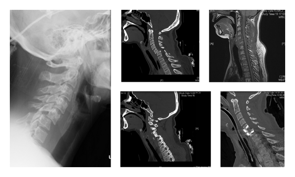
Imaging of a C4–5 fracture and dislocation: Conventional X-rays on admission showing fracture dislocation C4–5 (left); preoperative CT scan showing unilateral facet dislocation (middle); MRI after closed reduction (upper right); CT scan after anterior decompression and stabilization (lower right)
Image: “Figure 1: Conventional X-rays on admission showing fracture dislocation C4/5 (left); preoperative CT scan showing unilateral facet dislocation (middle); MRI after closed reduction (upper right); CT scan after anterior decompression and stabilization (lower right).” by Christian W. Müller et al. License: CC BY 3.0
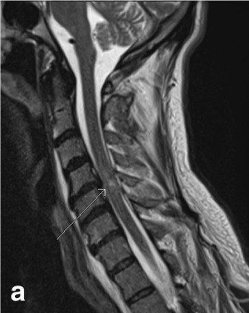
Sagittal MRI of a patient after acute spinal contusion: Notice the hyperintense lesion in the center of the spinal cord (white arrow), indicating likely edema or bleeding.
Image: “Fig. 7: Sagittal T2 weighted image (a) and axial gradient recalled echo (GRE) image (b) show the presence of nonhemorrhagic contusion in the spinal cord” by Kumar, Y., & Hayashi, D. License: CC BY 4.0
Management and Prognosis
Central cord syndrome has a good prognosisPrognosisA prediction of the probable outcome of a disease based on a individual’s condition and the usual course of the disease as seen in similar situations.Non-Hodgkin Lymphomas, although factors such as older age and more-severe neurologic injury at presentation are associated with a lower likelihood of neurologic recovery.
Management
Medical management:
Conservative treatment is the most common.
With acute severe trauma:
Treat hypotensionHypotensionHypotension is defined as low blood pressure, specifically < 90/60 mm Hg, and is most commonly a physiologic response. Hypotension may be mild, serious, or life threatening, depending on the cause. Hypotension due to neurogenic shockShockShock is a life-threatening condition associated with impaired circulation that results in tissue hypoxia. The different types of shock are based on the underlying cause: distributive (↑ cardiac output (CO), ↓ systemic vascular resistance (SVR)), cardiogenic (↓ CO, ↑ SVR), hypovolemic (↓ CO, ↑ SVR), obstructive (↓ CO), and mixed. Types of Shock.
BaclofenBaclofenA gamma-aminobutyric acid derivative that is a specific agonist of gaba-b receptors. It is used in the treatment of muscle spasticity, especially that due to spinal cord injuries. Its therapeutic effects result from actions at spinal and supraspinal sites, generally the reduction of excitatory transmission.Spasmolytics (muscle relaxant) for spasticitySpasticitySpinal Disk Herniation
Rehabilitation:
PT: for improving strength and range of motionRange of motionThe distance and direction to which a bone joint can be extended. Range of motion is a function of the condition of the joints, muscles, and connective tissues involved. Joint flexibility can be improved through appropriate muscle strength exercises.Examination of the Upper Limbs (ROM) of lower extremities
Occupational therapyOccupational TherapySkilled treatment that helps individuals achieve independence in all facets of their lives. It assists in the development of skills needed for independent living.Fetal Alcohol Spectrum Disorder: upper limb training
External fixation of the spineSpineThe human spine, or vertebral column, is the most important anatomical and functional axis of the human body. It consists of 7 cervical vertebrae, 12 thoracic vertebrae, and 5 lumbar vertebrae and is limited cranially by the skull and caudally by the sacrum.Vertebral Column: Anatomy: 4–6 weeks
Up to 75% of patientsPatientsIndividuals participating in the health care system for the purpose of receiving therapeutic, diagnostic, or preventive procedures.Clinician–Patient Relationship show some neurological improvement in functionality.
Surgical management:
Considered early on for:
Spinal instability
Ongoing spinal cordSpinal cordThe spinal cord is the major conduction pathway connecting the brain to the body; it is part of the CNS. In cross section, the spinal cord is divided into an H-shaped area of gray matter (consisting of synapsing neuronal cell bodies) and a surrounding area of white matter (consisting of ascending and descending tracts of myelinated axons). Spinal Cord: AnatomycompressionCompressionBlunt Chest Trauma with progressive neurologic deterioration
Involves procedures such as decompression laminectomyLaminectomyA surgical procedure that entails removing all (laminectomy) or part (laminotomy) of selected vertebral lamina to relieve pressure on the spinal cord and/or spinal nerve roots. Vertebral lamina is the thin flattened posterior wall of vertebral arch that forms the vertebral foramen through which pass the spinal cord and nerve roots.Neurosurgery
Complications of CCS
Autonomic dysreflexia: lack of a coordinated autonomic response with HR and blood pressure in spinal injuries above T6
Neurogenic bladderBladderA musculomembranous sac along the urinary tract. Urine flows from the kidneys into the bladder via the ureters, and is held there until urination.Pyelonephritis and Perinephric Abscess
PrognosisPrognosisA prediction of the probable outcome of a disease based on a individual’s condition and the usual course of the disease as seen in similar situations.Non-Hodgkin Lymphomas
VariableVariableVariables represent information about something that can change. The design of the measurement scales, or of the methods for obtaining information, will determine the data gathered and the characteristics of that data. As a result, a variable can be qualitative or quantitative, and may be further classified into subgroups.Types of Variables:
Functionality depends on the extent of the injury and rehabilitation.
Most patientsPatientsIndividuals participating in the health care system for the purpose of receiving therapeutic, diagnostic, or preventive procedures.Clinician–Patient Relationship recover the ability to walk.
Cervical fractures prolong recovery time.
BladderBladderA musculomembranous sac along the urinary tract. Urine flows from the kidneys into the bladder via the ureters, and is held there until urination.Pyelonephritis and Perinephric Abscess function usually returns 6–8 months after injury.
Differential Diagnosis
Ventral(anterior) cord syndrome (ACS): an injury to the anterior, or ventral, ⅔ of the spinal cordSpinal cordThe spinal cord is the major conduction pathway connecting the brain to the body; it is part of the CNS. In cross section, the spinal cord is divided into an H-shaped area of gray matter (consisting of synapsing neuronal cell bodies) and a surrounding area of white matter (consisting of ascending and descending tracts of myelinated axons). Spinal Cord: Anatomy (or incomplete cord syndrome) that spares the dorsal columnsDorsal ColumnsPosterior Cord Syndrome. The syndrome is caused by occlusion of the anterior spinal arteryAnterior Spinal ArteryAnterior Cord Syndrome or trauma causing disk herniationHerniationOmphalocele and boneBoneBone is a compact type of hardened connective tissue composed of bone cells, membranes, an extracellular mineralized matrix, and central bone marrow. The 2 primary types of bone are compact and spongy. Bones: Structure and Types fragments that disrupt the spinal cordSpinal cordThe spinal cord is the major conduction pathway connecting the brain to the body; it is part of the CNS. In cross section, the spinal cord is divided into an H-shaped area of gray matter (consisting of synapsing neuronal cell bodies) and a surrounding area of white matter (consisting of ascending and descending tracts of myelinated axons). Spinal Cord: Anatomy. Clinical manifestations are loss of motorMotorNeurons which send impulses peripherally to activate muscles or secretory cells.Nervous System: Histology and sensorySensoryNeurons which conduct nerve impulses to the central nervous system.Nervous System: Histology function below the level of injury. Diagnosis of ACS is by clinical exam and neuroimagingNeuroimagingNon-invasive methods of visualizing the central nervous system, especially the brain, by various imaging modalities.Febrile Infant with MRI. Management is directed at resolving the underlying cause.
Posterior cord syndromePosterior cord syndromePosterior cord syndrome (PCS) is an incomplete spinal cord syndrome affecting the dorsal columns, the corticospinal tracts (CSTs), and descending autonomic tracts to the bladder. Posterior cord syndrome is rare but has a diverse range of etiologies, including demyelinating disorders, degenerative spinal conditions, neoplastic causes, vascular abnormalities, and hereditary neurodegenerative disorders.Posterior Cord Syndrome (PCSPCSPosterior cord syndrome (PCS) is an incomplete spinal cord syndrome affecting the dorsal columns, the corticospinal tracts (csts), and descending autonomic tracts to the bladder. Posterior cord syndrome is rare but has a diverse range of etiologies, including demyelinating disorders, degenerative spinal conditions, neoplastic causes, vascular abnormalities, and hereditary neurodegenerative disorders.Posterior Cord Syndrome): an incomplete cord syndrome that affects the posterior aspect of the spinal cordSpinal cordThe spinal cord is the major conduction pathway connecting the brain to the body; it is part of the CNS. In cross section, the spinal cord is divided into an H-shaped area of gray matter (consisting of synapsing neuronal cell bodies) and a surrounding area of white matter (consisting of ascending and descending tracts of myelinated axons). Spinal Cord: Anatomy and is characterized by loss of vibrationVibrationA continuing periodic change in displacement with respect to a fixed reference.Neurological Examination and position senses below the level of injury. As a very rare condition, the status of PCSPCSPosterior cord syndrome (PCS) is an incomplete spinal cord syndrome affecting the dorsal columns, the corticospinal tracts (csts), and descending autonomic tracts to the bladder. Posterior cord syndrome is rare but has a diverse range of etiologies, including demyelinating disorders, degenerative spinal conditions, neoplastic causes, vascular abnormalities, and hereditary neurodegenerative disorders.Posterior Cord Syndrome as a separate clinical entity is still under debate in the literature and can overlap with CCS. Diagnosis is made clinically and supported by neuroimagingNeuroimagingNon-invasive methods of visualizing the central nervous system, especially the brain, by various imaging modalities.Febrile Infant. Management can be medical/rehabilitative or surgical if indicated.
Cruciate paralysis: a rare neurological condition that affects the cervicomedullary junction. The condition presents with bilateral upper limb paresisParesisA general term referring to a mild to moderate degree of muscular weakness, occasionally used as a synonym for paralysis (severe or complete loss of motor function). In the older literature, paresis often referred specifically to paretic neurosyphilis. ‘general paresis’ and ‘general paralysis’ may still carry that connotation. Bilateral lower extremity paresis is referred to as paraparesis.Spinal Disk Herniation without the involvement of the lower limbs. The etiologies of cruciate paralysis include traumatic injuries, postsurgical complications, and metabolic disorders. Diagnosis and management are similar to other incomplete cord syndromes.
References
Ameer, MA, Tessler, J, & Gillis, CC. (2023). Central cord syndrome. In StatPearls. StatPearls Publishing. Retrieved July 22, 2025, from http://www.ncbi.nlm.nih.gov/books/NBK441932/
Ropper, AH, Samuels, MA, Klein, JP, & Prasad, S. (2019). Diseases of the spinal cord. Ropper, AH, et al. (Eds.). Adams and Victor’s Principles of Neurology (11th ed.). McGraw-Hill Education. Retrieved July 22, 2025, from http://accessmedicine.mhmedical.com/content.aspx?aid=1162599484
Martínez-Palacios, K., Rubiano, A. M., Demetriades, A. K., & Vásquez-García, S. (2025). Traumatic central cord syndrome: An integrated neurosurgical and neurocritical care perspective. Brain and Spine, 5, 104281. https://doi.org/10.1016/j.bas.2025.104281
Create your free account or log in to continue reading!