A femoral herniaHerniaProtrusion of tissue, structure, or part of an organ through the bone, muscular tissue, or the membrane by which it is normally contained. Hernia may involve tissues such as the abdominal wall or the respiratory diaphragm. Hernias may be internal, external, congenital, or acquired.Abdominal Hernias is an uncommon type of groinGroinThe external junctural region between the lower part of the abdomen and the thigh.Male Genitourinary ExaminationherniaHerniaProtrusion of tissue, structure, or part of an organ through the bone, muscular tissue, or the membrane by which it is normally contained. Hernia may involve tissues such as the abdominal wall or the respiratory diaphragm. Hernias may be internal, external, congenital, or acquired.Abdominal Hernias in which intra-abdominal contents herniate under the inguinal ligament and through the femoral ring into the femoral canal. More common in adults than in children, femoral hernias usually present with swellingSwellingInflammation that protrudes into the femoral triangle (inferiorly to the inguinal ligament and medial to the femoral vein). Although uncommon, femoral hernias are frequently associated with complications, secondary to the small size of the canal, leading to herniaHerniaProtrusion of tissue, structure, or part of an organ through the bone, muscular tissue, or the membrane by which it is normally contained. Hernia may involve tissues such as the abdominal wall or the respiratory diaphragm. Hernias may be internal, external, congenital, or acquired.Abdominal HerniasincarcerationIncarcerationInguinal Canal: Anatomy and Hernias and/or strangulationStrangulationInguinal Canal: Anatomy and Hernias.
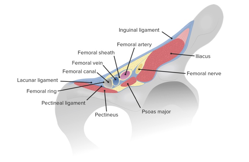
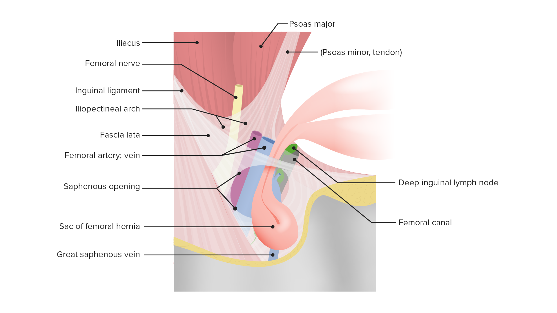
The femoral ring is theproximal or abdominal/pelvic opening of the femoral canal.
Boundaries:
Anterior: inguinal ligament
Posterior: pectineal ligament and muscle
Medial: lacunar ligament
Lateral: medial border of the femoral vein
Boundaries of the femoral ring and canal in relation to the femoral vessels and nerve
Image by Lecturio.
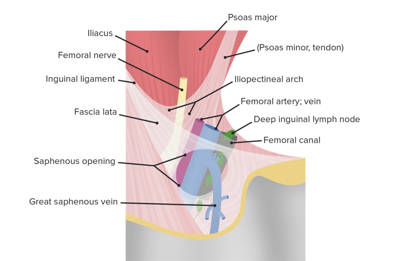
Femoral canal
The femoral canal is a cylindrical space, making up the medial compartment enclosed within the femoral sheath.
Same boundaries as the femoral ring
Extends 1–2 cm into the thighThighThe thigh is the region of the lower limb found between the hip and the knee joint. There is a single bone in the thigh called the femur, which is surrounded by large muscles grouped into 3 fascial compartments. Thigh: Anatomy
The femoral sheath is a fasciaFasciaLayers of connective tissue of variable thickness. The superficial fascia is found immediately below the skin; the deep fascia invests muscles, nerves, and other organs.Cellulitis that encloses the contents of the femoral triangle (except the femoral nerve), each within its own compartment.
Closeup of the femoral triangle featuring the location of the femoral canal in relation to the neighboring structures of the inner thigh
Image by Lecturio.
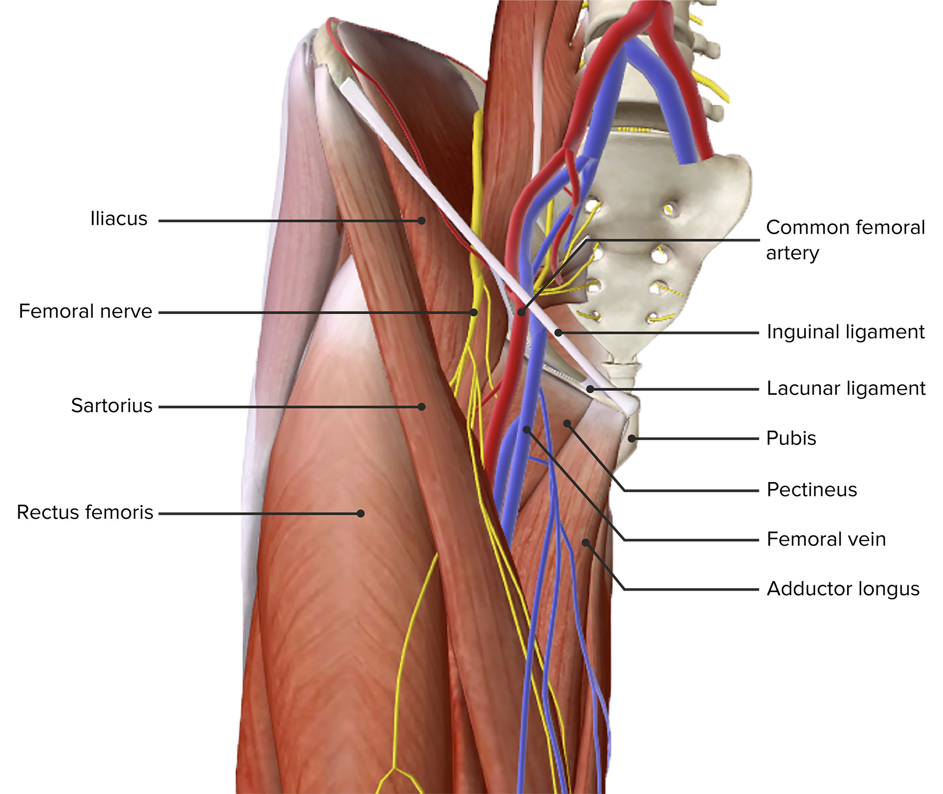
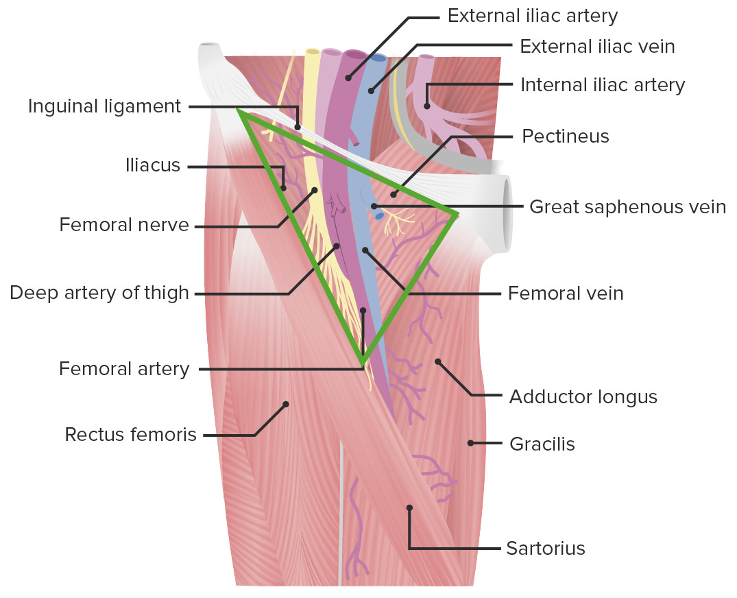
Femoral triangle
The femoral triangle is located on the medial aspect of the anterior thighThighThe thigh is the region of the lower limb found between the hip and the knee joint. There is a single bone in the thigh called the femur, which is surrounded by large muscles grouped into 3 fascial compartments. Thigh: Anatomy.
Roof: fasciaFasciaLayers of connective tissue of variable thickness. The superficial fascia is found immediately below the skin; the deep fascia invests muscles, nerves, and other organs.Cellulitis lata, superficial fasciaFasciaLayers of connective tissue of variable thickness. The superficial fascia is found immediately below the skin; the deep fascia invests muscles, nerves, and other organs.Cellulitis, and skinSkinThe skin, also referred to as the integumentary system, is the largest organ of the body. The skin is primarily composed of the epidermis (outer layer) and dermis (deep layer). The epidermis is primarily composed of keratinocytes that undergo rapid turnover, while the dermis contains dense layers of connective tissue.Skin: Structure and Functions
FasciaFasciaLayers of connective tissue of variable thickness. The superficial fascia is found immediately below the skin; the deep fascia invests muscles, nerves, and other organs.Cellulitis lata overlies the femoral triangle; the saphenous opening here allows entrance of lymphatic vesselsLymphatic VesselsTubular vessels that are involved in the transport of lymph and lymphocytes.Lymphatic Drainage System: Anatomy and the greater saphenous vein; the femoral herniaHerniaProtrusion of tissue, structure, or part of an organ through the bone, muscular tissue, or the membrane by which it is normally contained. Hernia may involve tissues such as the abdominal wall or the respiratory diaphragm. Hernias may be internal, external, congenital, or acquired.Abdominal Hernias protrudes through this opening
Anterior view of the thigh, featuring the femoral triangle
Image by Lecturio.
Anterior view of the thigh, featuring the femoral triangle with its borders and contents
Image by Lecturio.
Femoral herniaHerniaProtrusion of tissue, structure, or part of an organ through the bone, muscular tissue, or the membrane by which it is normally contained. Hernia may involve tissues such as the abdominal wall or the respiratory diaphragm. Hernias may be internal, external, congenital, or acquired.Abdominal Hernias
The femoral herniaHerniaProtrusion of tissue, structure, or part of an organ through the bone, muscular tissue, or the membrane by which it is normally contained. Hernia may involve tissues such as the abdominal wall or the respiratory diaphragm. Hernias may be internal, external, congenital, or acquired.Abdominal Hernias is the protrusion of intra-abdominal contents under the inguinal ligament, through the femoral ring, and into the femoral canal, producing a “bulge” or swellingSwellingInflammation in the femoral triangle.
Closeup of the femoral triangle featuring the location of a femoral hernia
Image by Lecturio.
Mnemonics
To remember the borders of the femoral triangle—SAIL:
S: Sartorius muscle
A: Adductor longus muscle
IL: Inguinal Ligament
To remember the contents of the femoral triangle (from lateral to medial)—NAVEL:
More common in women, with a female-to-male ratio of 3:1
Represents < 5% of all hernias
Etiology
Any condition that increases intra-abdominal pressure and enlarges/weakens the ligamentous structures of the pelvisPelvisThe pelvis consists of the bony pelvic girdle, the muscular and ligamentous pelvic floor, and the pelvic cavity, which contains viscera, vessels, and multiple nerves and muscles. The pelvic girdle, composed of 2 “hip” bones and the sacrum, is a ring-like bony structure of the axial skeleton that links the vertebral column with the lower extremities.Pelvis: Anatomy (e.g., the femoral ring)
Risk factors:
Increased intra-abdominal pressure
ObesityObesityObesity is a condition associated with excess body weight, specifically with the deposition of excessive adipose tissue. Obesity is considered a global epidemic. Major influences come from the western diet and sedentary lifestyles, but the exact mechanisms likely include a mixture of genetic and environmental factors. Obesity
ConstipationConstipationConstipation is common and may be due to a variety of causes. Constipation is generally defined as bowel movement frequency < 3 times per week. Patients who are constipated often strain to pass hard stools. The condition is classified as primary (also known as idiopathic or functional constipation) or secondary, and as acute or chronic. Constipation
Chronic cough
Recurrent, regularRegularInsulin, or violent vomitingVomitingThe forcible expulsion of the contents of the stomach through the mouth.Hypokalemia (e.g., eating disorder)
Prostatic hypertrophyHypertrophyGeneral increase in bulk of a part or organ due to cell enlargement and accumulation of fluids and secretions, not due to tumor formation, nor to an increase in the number of cells (hyperplasia).Cellular Adaptation (causes straining during micturition)
Enlarged/weakened femoral ring
Female genderGenderGender Dysphoria (wider pelvisPelvisThe pelvis consists of the bony pelvic girdle, the muscular and ligamentous pelvic floor, and the pelvic cavity, which contains viscera, vessels, and multiple nerves and muscles. The pelvic girdle, composed of 2 “hip” bones and the sacrum, is a ring-like bony structure of the axial skeleton that links the vertebral column with the lower extremities.Pelvis: Anatomy)
Advanced age
Multiparity
Previous surgical repair of inguinal herniasInguinal HerniasAn abdominal hernia with an external bulge in the groin region. It can be classified by the location of herniation. Indirect inguinal hernias occur through the internal inguinal ring. Direct inguinal hernias occur through defects in the abdominal wall (transversalis fascia) in Hesselbach’s triangle. The former type is commonly seen in children and young adults; the latter in adults.Inguinal Canal: Anatomy and Hernias
Clinical Presentation
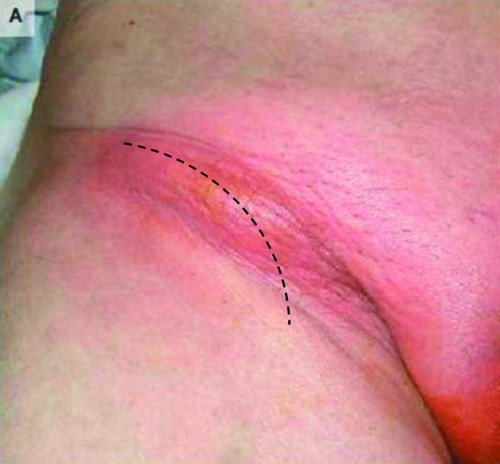
Globular, subcutaneous swellingSwellingInflammation or “bulge” in the groinGroinThe external junctural region between the lower part of the abdomen and the thigh.Male Genitourinary Examination that may or may not be tender
Located inferior to the inguinal ligament and medial to the femoral vein
Preoperative frontal view of a patient with a femoral hernia that demonstrates a red, round bulge in the groin area. The black dotted line shows how the curved low inguinal incision was performed.
Image: “De Garengeot’s hernia in a 60-year-old woman: a case report.
” by Konofaos P, Spartalis E, Smirnis A, Kontzoglou K, Kouraklis G. License: CC BY 2.0
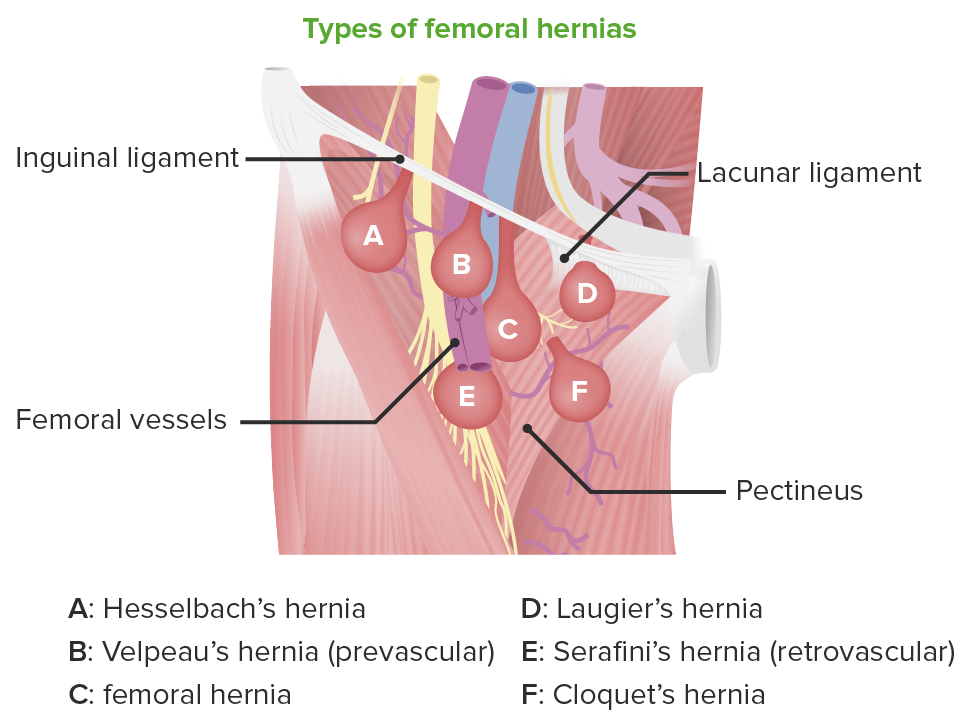
Types of femoral hernias
Depending on the location and contents of the protrusion, several subtypes of femoral hernias have been described:
Serafini’s herniaHerniaProtrusion of tissue, structure, or part of an organ through the bone, muscular tissue, or the membrane by which it is normally contained. Hernia may involve tissues such as the abdominal wall or the respiratory diaphragm. Hernias may be internal, external, congenital, or acquired.Abdominal Hernias: The hernial sac lies behind the femoral vessels.
Velpeau’s herniaHerniaProtrusion of tissue, structure, or part of an organ through the bone, muscular tissue, or the membrane by which it is normally contained. Hernia may involve tissues such as the abdominal wall or the respiratory diaphragm. Hernias may be internal, external, congenital, or acquired.Abdominal Hernias (or prevascular): The hernial sac lies in front of the femoral vessels, which can lead to this type being mistaken for a femoral aneurysmAneurysmAn aneurysm is a bulging, weakened area of a blood vessel that causes an abnormal widening of its diameter > 1.5 times the size of the native vessel. Aneurysms occur more often in arteries than in veins and are at risk of dissection and rupture, which can be life-threatening. Thoracic Aortic Aneurysms.
Laugier’s herniaHerniaProtrusion of tissue, structure, or part of an organ through the bone, muscular tissue, or the membrane by which it is normally contained. Hernia may involve tissues such as the abdominal wall or the respiratory diaphragm. Hernias may be internal, external, congenital, or acquired.Abdominal Hernias: The hernial sac transverses the lacunar ligament or the pectineal ligament of Cooper.
Hesselbach’s herniaHerniaProtrusion of tissue, structure, or part of an organ through the bone, muscular tissue, or the membrane by which it is normally contained. Hernia may involve tissues such as the abdominal wall or the respiratory diaphragm. Hernias may be internal, external, congenital, or acquired.Abdominal Hernias: The neckNeckThe part of a human or animal body connecting the head to the rest of the body.Peritonsillar Abscess of the sac lies lateral to the femoral vessels.
Cloquet’s herniaHerniaProtrusion of tissue, structure, or part of an organ through the bone, muscular tissue, or the membrane by which it is normally contained. Hernia may involve tissues such as the abdominal wall or the respiratory diaphragm. Hernias may be internal, external, congenital, or acquired.Abdominal Hernias: The hernial sac descends deep to the femoral vessels through the pectineal fasciaFasciaLayers of connective tissue of variable thickness. The superficial fascia is found immediately below the skin; the deep fascia invests muscles, nerves, and other organs.Cellulitis.
De Garengeot’s herniaHerniaProtrusion of tissue, structure, or part of an organ through the bone, muscular tissue, or the membrane by which it is normally contained. Hernia may involve tissues such as the abdominal wall or the respiratory diaphragm. Hernias may be internal, external, congenital, or acquired.Abdominal Hernias: The hernial contents include the vermiform appendixAppendixA worm-like blind tube extension from the cecum.Colon, Cecum, and Appendix: Anatomy, which often leads to incarcerationIncarcerationInguinal Canal: Anatomy and Hernias of the herniaHerniaProtrusion of tissue, structure, or part of an organ through the bone, muscular tissue, or the membrane by which it is normally contained. Hernia may involve tissues such as the abdominal wall or the respiratory diaphragm. Hernias may be internal, external, congenital, or acquired.Abdominal Hernias and requires an appendectomyAppendectomyAppendectomy is an invasive surgical procedure performed with the goal of resecting and extracting the vermiform appendix through either an open or a laparoscopic approach. The most common indication is acute appendicitis.Appendectomy as part of the surgical repair.
Types of femoral hernias
Image by Lecturio.
Complications
IncarcerationIncarcerationInguinal Canal: Anatomy and Hernias:HerniaHerniaProtrusion of tissue, structure, or part of an organ through the bone, muscular tissue, or the membrane by which it is normally contained. Hernia may involve tissues such as the abdominal wall or the respiratory diaphragm. Hernias may be internal, external, congenital, or acquired.Abdominal Hernias becomes irreducible.
Cardinal signs of inflammationInflammationInflammation is a complex set of responses to infection and injury involving leukocytes as the principal cellular mediators in the body’s defense against pathogenic organisms. Inflammation is also seen as a response to tissue injury in the process of wound healing. The 5 cardinal signs of inflammation are pain, heat, redness, swelling, and loss of function. Inflammation (erythemaErythemaRedness of the skin produced by congestion of the capillaries. This condition may result from a variety of disease processes.Chalazion, swellingSwellingInflammation, painPainAn unpleasant sensation induced by noxious stimuli which are detected by nerve endings of nociceptive neurons.Pain: Types and Pathways, warm to touch)
NauseaNauseaAn unpleasant sensation in the stomach usually accompanied by the urge to vomit. Common causes are early pregnancy, sea and motion sickness, emotional stress, intense pain, food poisoning, and various enteroviruses.Antiemetics, vomitingVomitingThe forcible expulsion of the contents of the stomach through the mouth.Hypokalemia, and severe abdominal painAbdominal PainAcute Abdomen
Signs of mechanical bowel obstructionBowel obstructionAny impairment, arrest, or reversal of the normal flow of intestinal contents toward the anal canal.Ascaris/Ascariasis (colicky abdominal painAbdominal PainAcute Abdomen, nauseaNauseaAn unpleasant sensation in the stomach usually accompanied by the urge to vomit. Common causes are early pregnancy, sea and motion sickness, emotional stress, intense pain, food poisoning, and various enteroviruses.Antiemetics, bilious or fecal vomitingVomitingThe forcible expulsion of the contents of the stomach through the mouth.Hypokalemia, constipationConstipationConstipation is common and may be due to a variety of causes. Constipation is generally defined as bowel movement frequency < 3 times per week. Patients who are constipated often strain to pass hard stools. The condition is classified as primary (also known as idiopathic or functional constipation) or secondary, and as acute or chronic. Constipation, abdominal distension)
Signs of peritonitisPeritonitisInflammation of the peritoneum lining the abdominal cavity as the result of infectious, autoimmune, or chemical processes. Primary peritonitis is due to infection of the peritoneal cavity via hematogenous or lymphatic spread and without intra-abdominal source. Secondary peritonitis arises from the abdominal cavity itself through rupture or abscess of intra-abdominal organs.Penetrating Abdominal Injury, followed by paralytic ileusParalytic ileusSmall Bowel Obstruction (due to intestinal perforationIntestinal perforationPerforated viscus or GI perforation represents a condition in which the integrity of the GI wall is lost with subsequent leakage of enteric contents into the peritoneal cavity, resulting in peritonitis. The causes of perforated viscus include trauma, bowel ischemia, infections, or ulcerative conditions, all of which ultimately lead to a full-thickness disruption of the intestinal wall.Perforated Viscus)
If palpationPalpationApplication of fingers with light pressure to the surface of the body to determine consistency of parts beneath in physical diagnosis; includes palpation for determining the outlines of organs.Dermatologic Examination is difficult (e.g., obesityObesityObesity is a condition associated with excess body weight, specifically with the deposition of excessive adipose tissue. Obesity is considered a global epidemic. Major influences come from the western diet and sedentary lifestyles, but the exact mechanisms likely include a mixture of genetic and environmental factors. Obesity), diagnosis can be confirmed by an ultrasound, computed tomography (CT), or magnetic resonance imaging (MRI) of the groinGroinThe external junctural region between the lower part of the abdomen and the thigh.Male Genitourinary Examination area, which will show signs of small bowel obstructionBowel obstructionAny impairment, arrest, or reversal of the normal flow of intestinal contents toward the anal canal.Ascaris/Ascariasis.
Differential diagnoses
Inguinal herniaHerniaProtrusion of tissue, structure, or part of an organ through the bone, muscular tissue, or the membrane by which it is normally contained. Hernia may involve tissues such as the abdominal wall or the respiratory diaphragm. Hernias may be internal, external, congenital, or acquired.Abdominal Hernias
AneurysmAneurysmAn aneurysm is a bulging, weakened area of a blood vessel that causes an abnormal widening of its diameter > 1.5 times the size of the native vessel. Aneurysms occur more often in arteries than in veins and are at risk of dissection and rupture, which can be life-threatening. Thoracic Aortic Aneurysms of the femoral artery
Dilation of the saphenous vein
AbscessAbscessAccumulation of purulent material in tissues, organs, or circumscribed spaces, usually associated with signs of infection.Chronic Granulomatous Disease of the psoas musclePsoas muscleA powerful flexor of the thigh at the hip joint (psoas major) and a weak flexor of the trunk and lumbar spinal column (psoas minor). Psoas is derived from the greek ‘psoa.Pyelonephritis and Perinephric Abscess
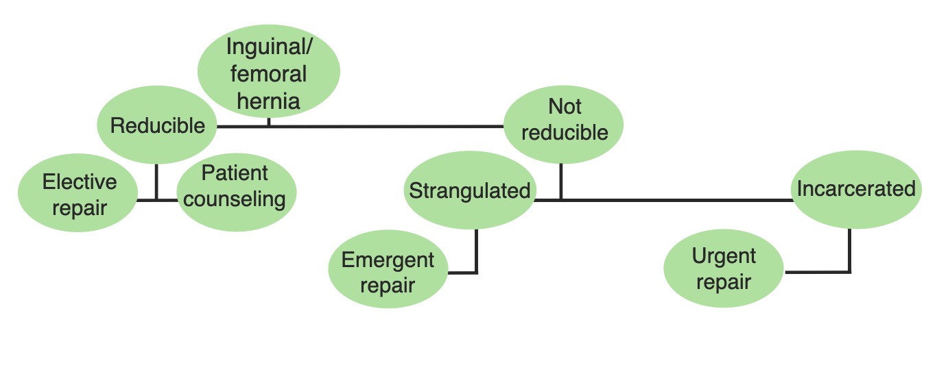
Management
Due to the high risk of complications because of the small size of the canal/ring, all femoral hernias should be surgically repaired.
Non-complicated hernias: early elective surgical repair with mesh hernioplasty
Complicated hernias: herniorrhaphy within 4 hours of the onset of symptoms in order to prevent bowel ischemiaBowel ischemiaMesenteric ischemia is a rare, life-threatening condition caused by inadequate blood flow through the mesenteric vessels, which results in ischemia and necrosis of the intestinal wall. Mesenteric ischemia can be either acute or chronic.Mesenteric Ischemia and necrosisNecrosisThe death of cells in an organ or tissue due to disease, injury or failure of the blood supply.Ischemic Cell Damage
Schematic diagram of the difference in location between direct inguinal hernias, indirect inguinal hernias, and femoral hernias
Image by Lecturio.
Ultrasonography of an incarcerated femoral hernia showing the oedematous hernia sac, above the femoral vessels
Image: “Incarcerated femoral hernia containing ipsilateral fallopian tube” by Atmatzidis S, Chatzimavroudis G, Dragoumis D, Atmatzidis K. License: CC BY 3.0
Schematic diagram of the management options for femoral hernias, depending on their state of reducibility.
Image by Lecturio.
Clinical Relevance
Femoral vascular access: The femoral artery and vein are easily accessed within the femoral triangle for interventional procedures.
The following conditions are included in the differential diagnoses of femoral hernias:
Inguinal herniasInguinal HerniasAn abdominal hernia with an external bulge in the groin region. It can be classified by the location of herniation. Indirect inguinal hernias occur through the internal inguinal ring. Direct inguinal hernias occur through defects in the abdominal wall (transversalis fascia) in Hesselbach’s triangle. The former type is commonly seen in children and young adults; the latter in adults.Inguinal Canal: Anatomy and Hernias: a protrusion of intra-abdominal contents through the deep inguinal ringDeep inguinal ringInguinal Canal: Anatomy and Hernias, producing an indirect inguinal herniaHerniaProtrusion of tissue, structure, or part of an organ through the bone, muscular tissue, or the membrane by which it is normally contained. Hernia may involve tissues such as the abdominal wall or the respiratory diaphragm. Hernias may be internal, external, congenital, or acquired.Abdominal Hernias, or through a weakness in the posterior wall of the inguinal canalInguinal canalThe tunnel in the lower anterior abdominal wall through which the spermatic cord, in the male; round ligament, in the female; nerves; and vessels pass. Its internal end is at the deep inguinal ring and its external end is at the superficial inguinal ring.Inguinal Canal: Anatomy and Hernias, producing a direct inguinal herniaHerniaProtrusion of tissue, structure, or part of an organ through the bone, muscular tissue, or the membrane by which it is normally contained. Hernia may involve tissues such as the abdominal wall or the respiratory diaphragm. Hernias may be internal, external, congenital, or acquired.Abdominal Hernias. Presents with similar clinical manifestations as the femoral herniaHerniaProtrusion of tissue, structure, or part of an organ through the bone, muscular tissue, or the membrane by which it is normally contained. Hernia may involve tissues such as the abdominal wall or the respiratory diaphragm. Hernias may be internal, external, congenital, or acquired.Abdominal Hernias; however, inguinal herniasInguinal HerniasAn abdominal hernia with an external bulge in the groin region. It can be classified by the location of herniation. Indirect inguinal hernias occur through the internal inguinal ring. Direct inguinal hernias occur through defects in the abdominal wall (transversalis fascia) in Hesselbach’s triangle. The former type is commonly seen in children and young adults; the latter in adults.Inguinal Canal: Anatomy and Hernias are located above the inguinal ligament. In some cases, the 2 types may be indistinguishable during physical examination.
AneurysmAneurysmAn aneurysm is a bulging, weakened area of a blood vessel that causes an abnormal widening of its diameter > 1.5 times the size of the native vessel. Aneurysms occur more often in arteries than in veins and are at risk of dissection and rupture, which can be life-threatening. Thoracic Aortic Aneurysms of the femoral artery: an arterial dilation caused by weakness of the wall of the femoral artery, located in the medial aspect of the thighThighThe thigh is the region of the lower limb found between the hip and the knee joint. There is a single bone in the thigh called the femur, which is surrounded by large muscles grouped into 3 fascial compartments. Thigh: Anatomy. AneurysmAneurysmAn aneurysm is a bulging, weakened area of a blood vessel that causes an abnormal widening of its diameter > 1.5 times the size of the native vessel. Aneurysms occur more often in arteries than in veins and are at risk of dissection and rupture, which can be life-threatening. Thoracic Aortic Aneurysms of the femoral artery is the 2nd-most common peripheral aneurysmAneurysmAn aneurysm is a bulging, weakened area of a blood vessel that causes an abnormal widening of its diameter > 1.5 times the size of the native vessel. Aneurysms occur more often in arteries than in veins and are at risk of dissection and rupture, which can be life-threatening. Thoracic Aortic Aneurysms. May present as painless, pulsatile swellingSwellingInflammation with a palpable thrill and a continuous murmur at the mid-inguinal point. Femoral aneurysms can rupture, which may cause life-threatening, uncontrollable bleeding.
References
Drake, R.L., Vogl, A.W., & Mitchell, A.W.M. (2014). Gray’s Anatomy for Students (3rd ed.). Philadelphia, PA: Churchill Livingstone.