The foot is the terminal portion of the lower limb, whose primary function is to bear weight and facilitate locomotion. The foot comprises 26 bones, including the tarsal bones, metatarsal bones, and phalangesPhalangesBones that make up the skeleton of the fingers, consisting of two for the thumb, and three for each of the other fingers.Hand: Anatomy. The bones of the foot form longitudinal and transverse arches and are supported by various muscles, ligaments, and tendons that allow for flexibility as well as dynamic and static support. The foot has 3 primary arches and multiple ligaments that are essential to its structure. The arches are important in absorbing weight during standing, walking, and running and the ability to adapt to uneven terrain during locomotion.
The 26 bones of the foot are divided into 3 groups: tarsals, metatarsals, and phalangesPhalangesBones that make up the skeleton of the fingers, consisting of two for the thumb, and three for each of the other fingers.Hand: Anatomy.
Tarsal bones (7):
Calcaneus:
Largest and strongest
Acts as a lever for the strong muscles of the legLegThe lower leg, or just “leg” in anatomical terms, is the part of the lower limb between the knee and the ankle joint. The bony structure is composed of the tibia and fibula bones, and the muscles of the leg are grouped into the anterior, lateral, and posterior compartments by extensions of fascia.Leg: Anatomy, specifically the muscles of the posterior compartment
Articulates with the talusTalusThe second largest of the tarsal bones. It articulates with the tibia and fibula to form the ankle joint.Ankle Joint: Anatomy at the superior portion, forming the subtalar jointSubtalar JointFormed by the articulation of the talus with the calcaneus.Ankle Joint: Anatomy
Articulates with the cuboid anteriorly
TalusTalusThe second largest of the tarsal bones. It articulates with the tibia and fibula to form the ankle joint.Ankle Joint: Anatomy:
2nd largest tarsal boneBoneBone is a compact type of hardened connective tissue composed of bone cells, membranes, an extracellular mineralized matrix, and central bone marrow. The 2 primary types of bone are compact and spongy. Bones: Structure and Types
The majority of the talar surface is covered with articular cartilageCartilageCartilage is a type of connective tissue derived from embryonic mesenchyme that is responsible for structural support, resilience, and the smoothness of physical actions. Perichondrium (connective tissue membrane surrounding cartilage) compensates for the absence of vasculature in cartilage by providing nutrition and support. Cartilage: Histology.
No tendons or muscles insert to or originate from the talusTalusThe second largest of the tarsal bones. It articulates with the tibia and fibula to form the ankle joint.Ankle Joint: Anatomy.
Articulates with the tibiaTibiaThe second longest bone of the skeleton. It is located on the medial side of the lower leg, articulating with the fibula laterally, the talus distally, and the femur proximally.Knee Joint: Anatomy, fibulaFibulaThe bone of the lower leg lateral to and smaller than the tibia. In proportion to its length, it is the most slender of the long bones.Leg: Anatomy, navicular, and calcaneus bones
Cuboid:
Anterior to the calcaneus, on the lateral side of the foot
Articulates with the calcaneus, lateral cuneiform, 4th–5th metatarsals, and occasionally the navicular
Navicular:
Anterior to the talusTalusThe second largest of the tarsal bones. It articulates with the tibia and fibula to form the ankle joint.Ankle Joint: Anatomy on the medial aspect of the foot
Articulates with the talusTalusThe second largest of the tarsal bones. It articulates with the tibia and fibula to form the ankle joint.Ankle Joint: Anatomy, medial, middle, and lateral cuneiforms, and occasionally with the cuboid
Cuneiforms (3):
Medial, middle (intermediate), and lateral
Configuration creates a keystone effect that contributes to the stability of the foot
Metatarsal bones (5):
The basesBasesUsually a hydroxide of lithium, sodium, potassium, rubidium or cesium, but also the carbonates of these metals, ammonia, and the amines.Acid-Base Balance articulate with the tarsal bones proximally:
1st–3rd metatarsals articulate with the cuneiforms
3rd–5th metatarsals articulate with the cuboid
The distal heads articulate with the proximal phalangesPhalangesBones that make up the skeleton of the fingers, consisting of two for the thumb, and three for each of the other fingers.Hand: Anatomy.
The 1st metatarsal is the shortest and strongest.
The 2nd metatarsal is the longest.
PhalangesPhalangesBones that make up the skeleton of the fingers, consisting of two for the thumb, and three for each of the other fingers.Hand: Anatomy (14):
Small bones of the digits
Each toe has 3 phalangesPhalangesBones that make up the skeleton of the fingers, consisting of two for the thumb, and three for each of the other fingers.Hand: Anatomy: proximal, middle, and distal
The exception is the hallux, which has only a proximal and a distal phalanx.
A common variant is a fused middle and distal phalange of the 5th digit.
Other bones of the foot:
Sesamoid bones:
2 sesamoid bones are usually present at the plantar area of the 1st metatarsal phalangeal joint, within the tendon of the flexor hallucis brevis.
Increase the mechanical advantage of the 1st digit
Common accessory ossicles or accessory bones of the foot:
Os trigonum:found at the posterior aspect of the talusTalusThe second largest of the tarsal bones. It articulates with the tibia and fibula to form the ankle joint.Ankle Joint: Anatomy
Os navicular(accessory navicular):medial aspect of the navicular
Os peroneum:accessory boneBoneBone is a compact type of hardened connective tissue composed of bone cells, membranes, an extracellular mineralized matrix, and central bone marrow. The 2 primary types of bone are compact and spongy. Bones: Structure and Types within the peroneus longus tendon
Bipartite sesamoid: Sesamoids of the 1st digit fail to ossify, resulting in a fibrousFibrousFibrocystic Change union.
Mnemonic
From superior to inferior and from medial to lateral in a right foot: Talus, Calcaneus, Navicular, Medial cuneiform, Intermediate or middle cuneiform, Lateral cuneiform, Cuboid
The Cab in New Mexico Is a Land Cruiser
The Cub Needs MILC
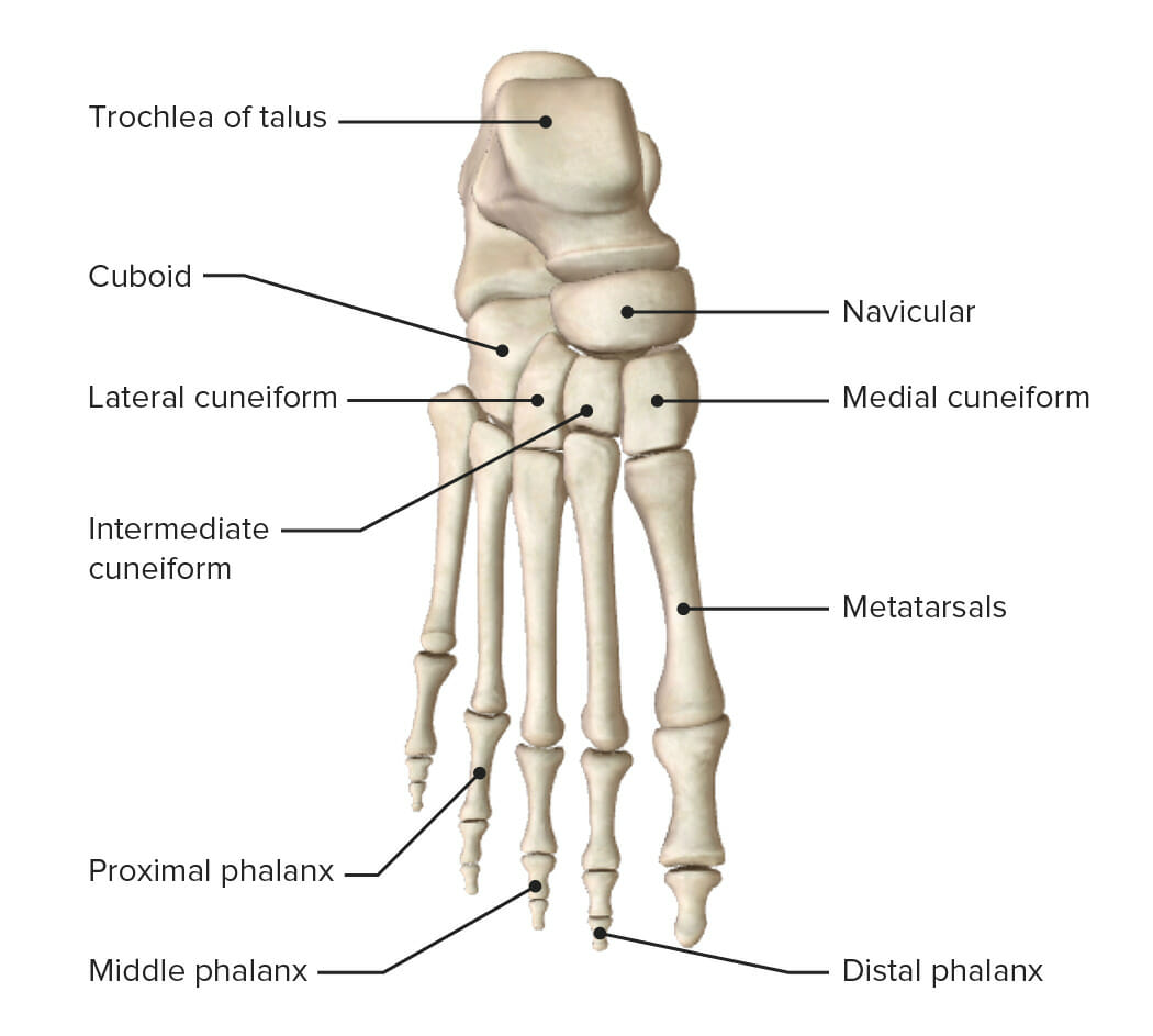
Superior view of the right foot featuring the bones of the foot and the tarsus
Image by Lecturio.
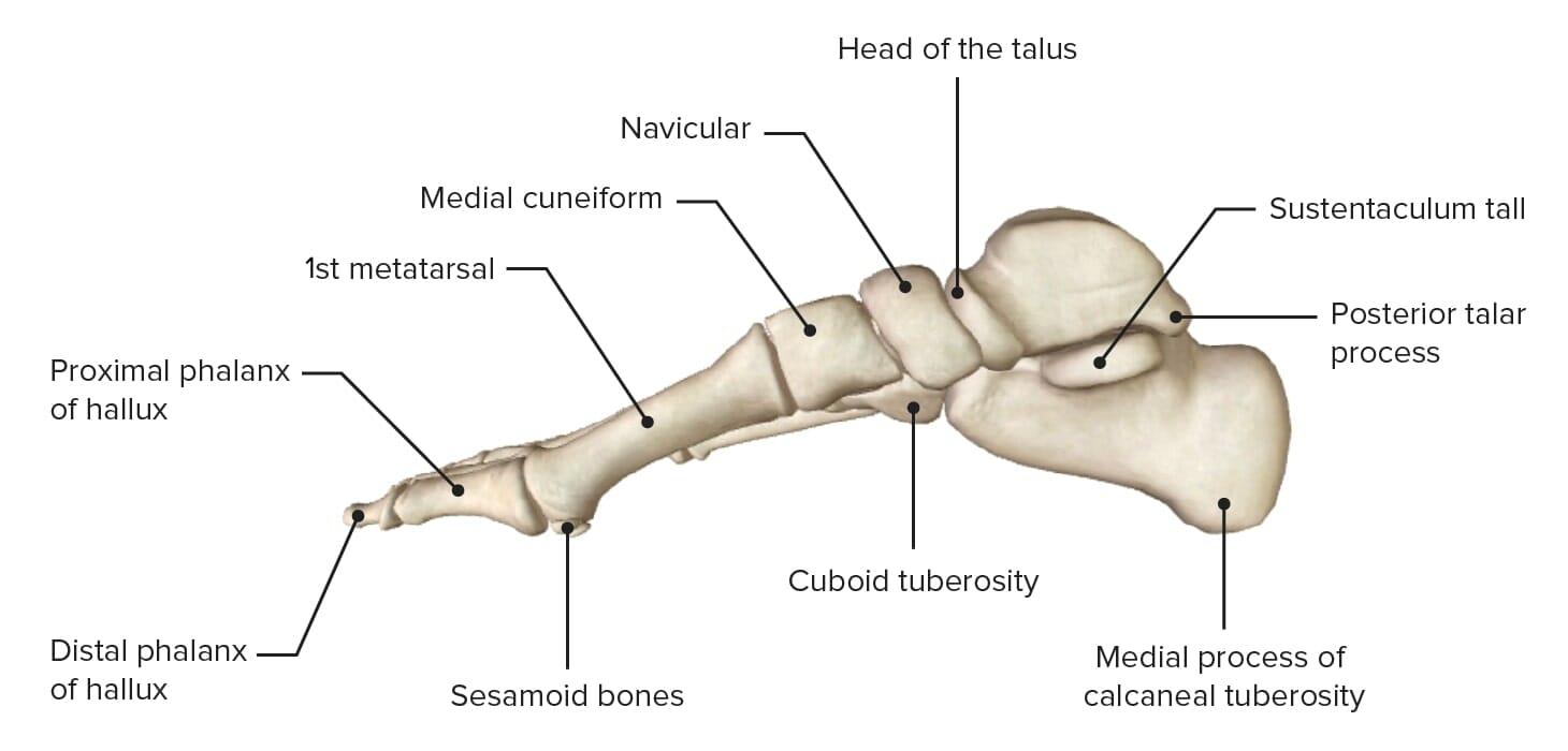
Medial view of the right foot featuring the bones of the foot and the tarsus
Image by Lecturio.
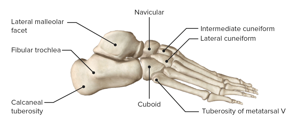
Lateral view of the right foot featuring the bones of the foot and the tarsus
Image by Lecturio.
Joints of the foot
The joints of the foot, from proximal to distal, include the following articulations.
Composed of 3 alternating convex–concave areas on each of the talusTalusThe second largest of the tarsal bones. It articulates with the tibia and fibula to form the ankle joint.Ankle Joint: Anatomy and calcaneus bones
Supporting ligaments:
Anterior, posterior, lateral, and medial talocalcaneal ligaments
Interosseous talocalcaneal ligament (the strongest of this joint, lies within the sinus tarsi or tunnel between the talusTalusThe second largest of the tarsal bones. It articulates with the tibia and fibula to form the ankle joint.Ankle Joint: Anatomy and calcaneus)
Talocalcaneonavicular jointTalocalcaneonavicular jointAnkle Joint: Anatomy: articular capsuleCapsuleAn envelope of loose gel surrounding a bacterial cell which is associated with the virulence of pathogenic bacteria. Some capsules have a well-defined border, whereas others form a slime layer that trails off into the medium. Most capsules consist of relatively simple polysaccharides but there are some bacteria whose capsules are made of polypeptides.Bacteroides, dorsal talonavicular ligament, and plantar calcaneonavicular ligament
Calcaneocuboid jointCalcaneocuboid jointAnkle Joint: Anatomy: bifurcate (Y-shaped) ligament superiorly, long plantar ligament inferiorly, and short plantar ligament
1st–3rd metatarsals articulate with the cuneiforms
3rd–5th metatarsals articulate with the cuboid
Supporting ligaments:
Dorsal and plantar tarsometatarsal ligaments
Interosseous cuneometatarsal ligaments
The strongest of these is the Lisfranc ligament, which extends from the 2nd metatarsal to the lateral aspect of medial cuneiform.
Function:
Minimal gliding movement
Primarily stability
Metatarsophalangeal joints:
Type: condyloid joints
Components: articulations between the metatarsal heads and the base of the proximal phalangesPhalangesBones that make up the skeleton of the fingers, consisting of two for the thumb, and three for each of the other fingers.Hand: Anatomy of the digits
Supporting ligaments:
A capsuleCapsuleAn envelope of loose gel surrounding a bacterial cell which is associated with the virulence of pathogenic bacteria. Some capsules have a well-defined border, whereas others form a slime layer that trails off into the medium. Most capsules consist of relatively simple polysaccharides but there are some bacteria whose capsules are made of polypeptides.Bacteroides encloses each joint.
The foot has 3 primary arches and multiple supporting ligaments.
Plantar arches
The plantar arches function to distribute and absorb the body weight, provide the foot with elasticityElasticityResistance and recovery from distortion of shape.Skeletal Muscle Contraction and resilience during locomotion, adapt to uneven surfaces, and protect the neurovasculature on the plantar surface.
Medial longitudinal arch:
Formed by the calcaneus, talusTalusThe second largest of the tarsal bones. It articulates with the tibia and fibula to form the ankle joint.Ankle Joint: Anatomy, navicular, cuneiforms, and 1st–3rd metatarsals
Generally the highest arch of the foot
Supported by the intrinsic muscles of the foot
Lateral longitudinal arch:
Formed by the calcaneus, cuboid, and 4th–5th metatarsals
Transverse arch:
Formed by the cuboid, cuneiforms, and basesBasesUsually a hydroxide of lithium, sodium, potassium, rubidium or cesium, but also the carbonates of these metals, ammonia, and the amines.Acid-Base Balance of the 1st–4th metatarsal bones
Forms the medial to lateral midfoot curvature
Medial view of the foot featuring the arches of the foot
Image by Lecturio.
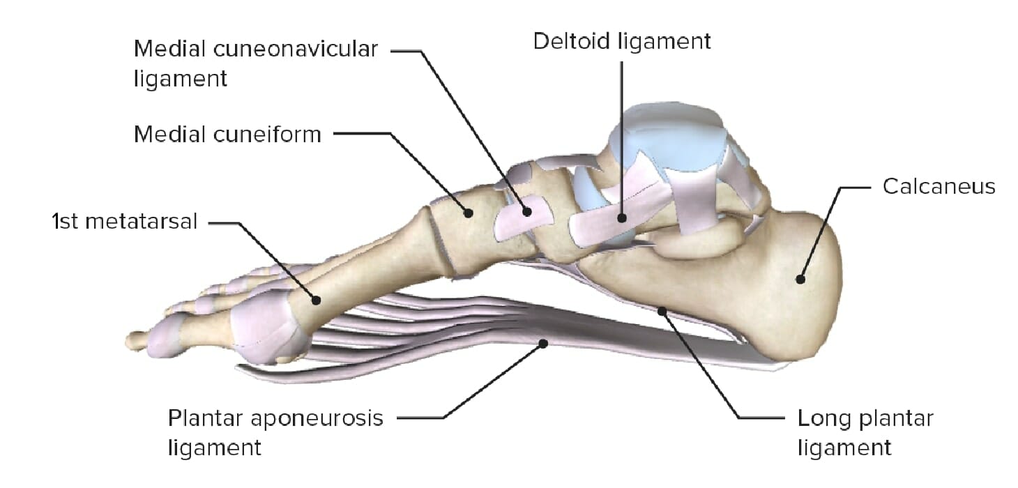
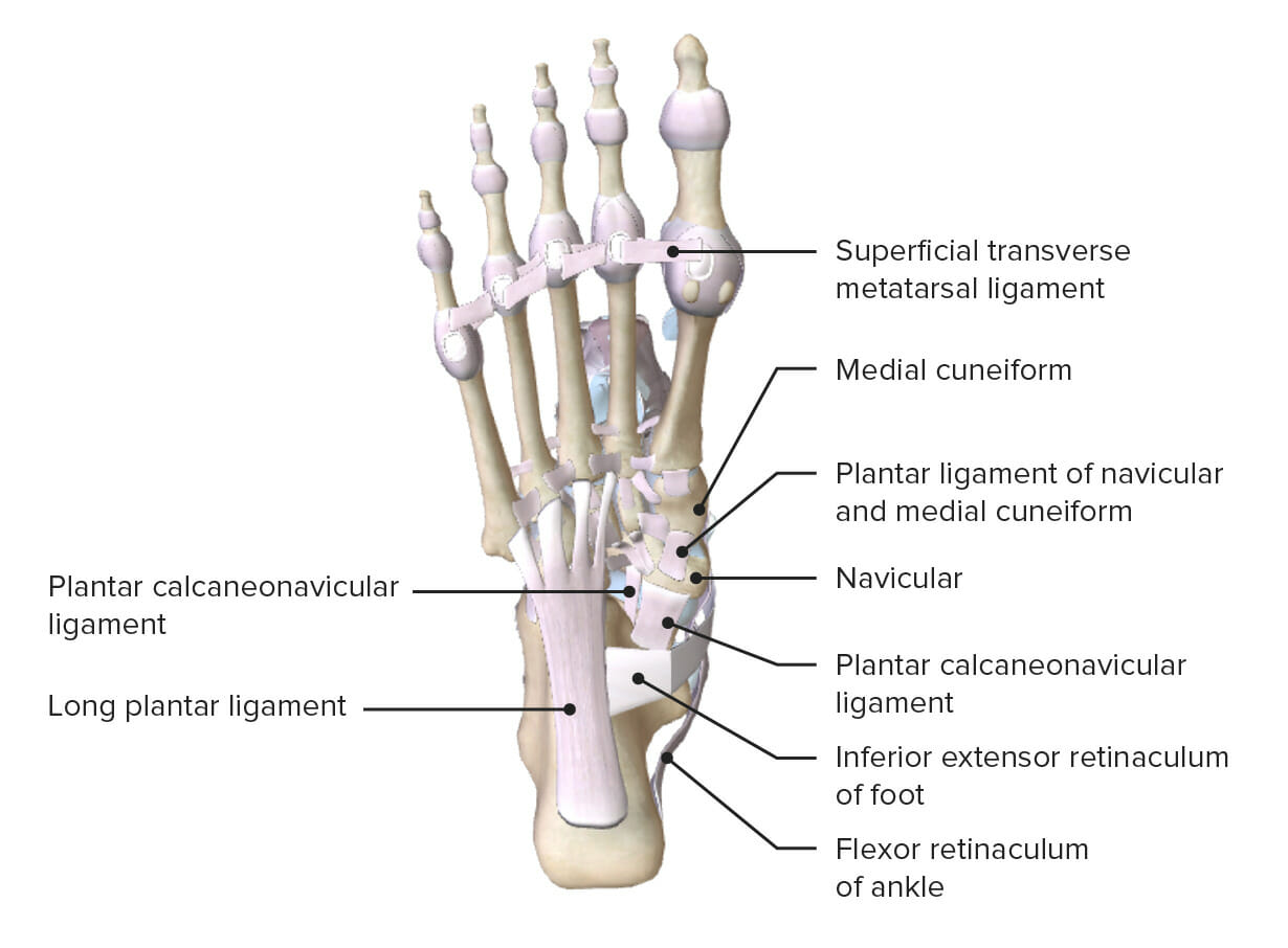
Plantar ligaments
The plantar ligaments are essential in the maintenance of the functional integrity of the arches on the sole of the foot.
Long plantar ligament:
Longest and strongest ligament in the body
Supports the longitudinal arches
Connects the calcaneus and the cuboid boneBoneBone is a compact type of hardened connective tissue composed of bone cells, membranes, an extracellular mineralized matrix, and central bone marrow. The 2 primary types of bone are compact and spongy. Bones: Structure and Types/base of 5th metatarsal
Converts the cuboid groove into a canal for the fibularis longus tendon
Short plantar ligament:
Deep to the long plantar ligament
Plantar calcaneonavicular ligament:
Also known as the spring ligament
Runs from the sustentaculum tali to the plantar surface of the navicular boneBoneBone is a compact type of hardened connective tissue composed of bone cells, membranes, an extracellular mineralized matrix, and central bone marrow. The 2 primary types of bone are compact and spongy. Bones: Structure and Types
Other ligaments that contribute to the structural integrity of the arches:
Plantar cuneonavicular ligament
Plantar intercuneiform ligaments
Plantar cuboideonavicular ligament
Plantar cuneocuboid ligaments
Plantar view of the foot featuring the numerous plantar ligaments
Image by Lecturio.
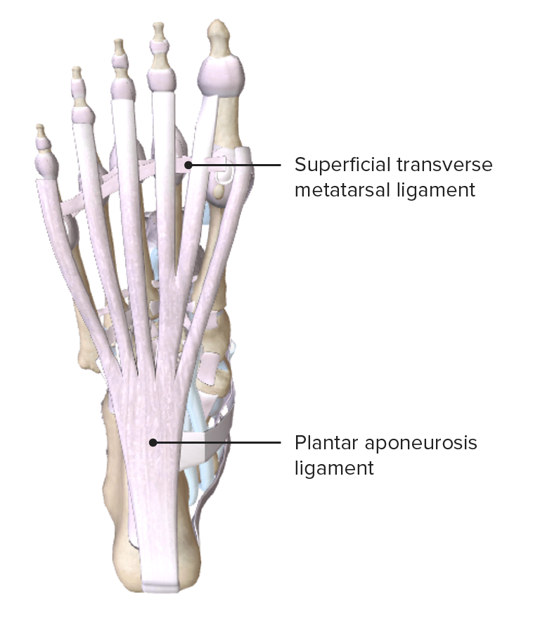
Plantar fasciaFasciaLayers of connective tissue of variable thickness. The superficial fascia is found immediately below the skin; the deep fascia invests muscles, nerves, and other organs.Cellulitis or plantar aponeurosis
Thick band of connective tissueConnective tissueConnective tissues originate from embryonic mesenchyme and are present throughout the body except inside the brain and spinal cord. The main function of connective tissues is to provide structural support to organs. Connective tissues consist of cells and an extracellular matrix.Connective Tissue: Histology that supports the bony arches of the foot
Extends from the calcaneal tuberosity to the proximal phalangesPhalangesBones that make up the skeleton of the fingers, consisting of two for the thumb, and three for each of the other fingers.Hand: Anatomy
Divides the foot into lateral, medial, and central compartments through septa of the plantar aponeurosis
Plantar fascia or aponeurosis: Note how the deep fascia is continuous with the
plantar fascia, which is thickened centrally as the toughened plantar aponeurosis. The aponeurosis has bands and intermuscular septa that divide the sole of the foot into 3 compartments: medial, lateral, and central.
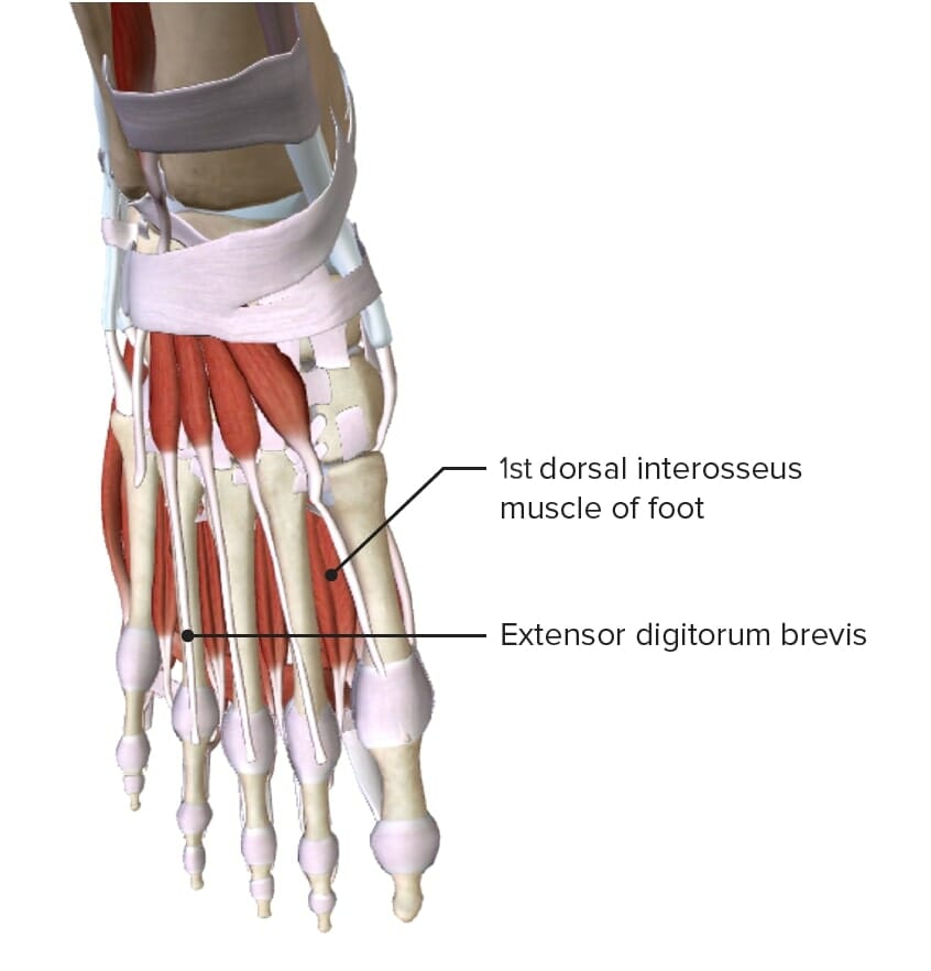
Aside from the tendons of the extrinsic muscles from the anterior compartment of the legLegThe lower leg, or just “leg” in anatomical terms, is the part of the lower limb between the knee and the ankle joint. The bony structure is composed of the tibia and fibula bones, and the muscles of the leg are grouped into the anterior, lateral, and posterior compartments by extensions of fascia.Leg: Anatomy (extensor hallucis longusExtensor hallucis longusLeg: Anatomy, extensor digitorumExtensor digitorumForearm: Anatomy, tibialis anteriorTibialis anteriorLeg: Anatomy, and peroneus tertiusPeroneus tertiusLeg: Anatomy), which pass under the extensor retinaculum, only 2 intrinsic muscles exist on the dorsum of the foot:
The intrinsic muscles of the plantar surface, or sole, of the foot both originate and insert within the foot. These muscles produce the fine movements of the digits and support the arches of the foot during standing, walking, and running. Commonly, the plantar muscles of the foot are organized into 4 layers, from superficial to deep:
Opponens digiti minimiOpponens digiti minimiHand: Anatomy (variableVariableVariables represent information about something that can change. The design of the measurement scales, or of the methods for obtaining information, will determine the data gathered and the characteristics of that data. As a result, a variable can be qualitative or quantitative, and may be further classified into subgroups.Types of Variables)
Middle phalangesPhalangesBones that make up the skeleton of the fingers, consisting of two for the thumb, and three for each of the other fingers.Hand: Anatomy of digits 2–5
Flexes the proximal phalangesPhalangesBones that make up the skeleton of the fingers, consisting of two for the thumb, and three for each of the other fingers.Hand: Anatomy
Extends the middle and distal phalangesPhalangesBones that make up the skeleton of the fingers, consisting of two for the thumb, and three for each of the other fingers.Hand: Anatomy of digits 2–5
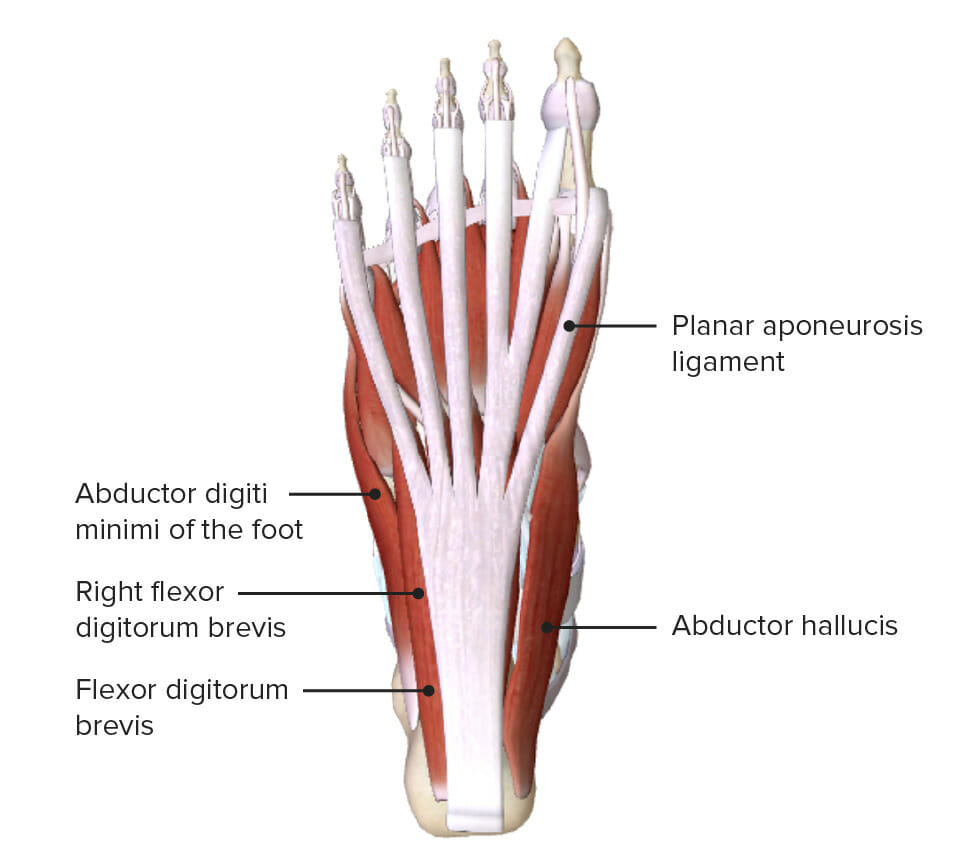
2nd most superficial layer of the muscles of the sole of the foot
Image by Lecturio.
Table: 3rd most superficial muscle layer of the foot
Opponens digiti minimiOpponens digiti minimiHand: Anatomy (variableVariableVariables represent information about something that can change. The design of the measurement scales, or of the methods for obtaining information, will determine the data gathered and the characteristics of that data. As a result, a variable can be qualitative or quantitative, and may be further classified into subgroups.Types of Variables)
1st: medial surface of the proximal phalanx of the 2nd digit
2nd-4th: lateral surface of the proximal phalanx of digits 2–4
Abducts digits 2–4 and flexes metatarsophalangeal joints
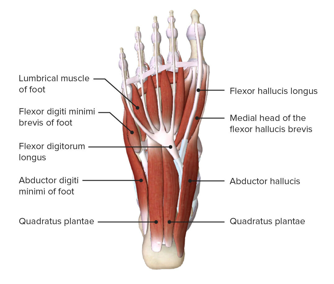
Deepest layer of the muscles of the sole of the foot
Image by Lecturio.
Alternatively, the intrinsic muscles of the plantar surface, or sole, of the foot can be divided into 3 groups, using the medial, lateral and central compartments of the foot. These compartments are formed by the deep fasciaFasciaLayers of connective tissue of variable thickness. The superficial fascia is found immediately below the skin; the deep fascia invests muscles, nerves, and other organs.Cellulitis or plantar aponeurosis.
Opponens digiti minimiOpponens digiti minimiHand: Anatomy (variableVariableVariables represent information about something that can change. The design of the measurement scales, or of the methods for obtaining information, will determine the data gathered and the characteristics of that data. As a result, a variable can be qualitative or quantitative, and may be further classified into subgroups.Types of Variables)
Primarily from branches of the tibial nerveTibial NerveThe medial terminal branch of the sciatic nerve. The tibial nerve fibers originate in lumbar and sacral spinal segments (L4 to S2). They supply motor and sensory innervation to parts of the calf and foot.Popliteal Fossa: Anatomy and the deep fibular nerve.
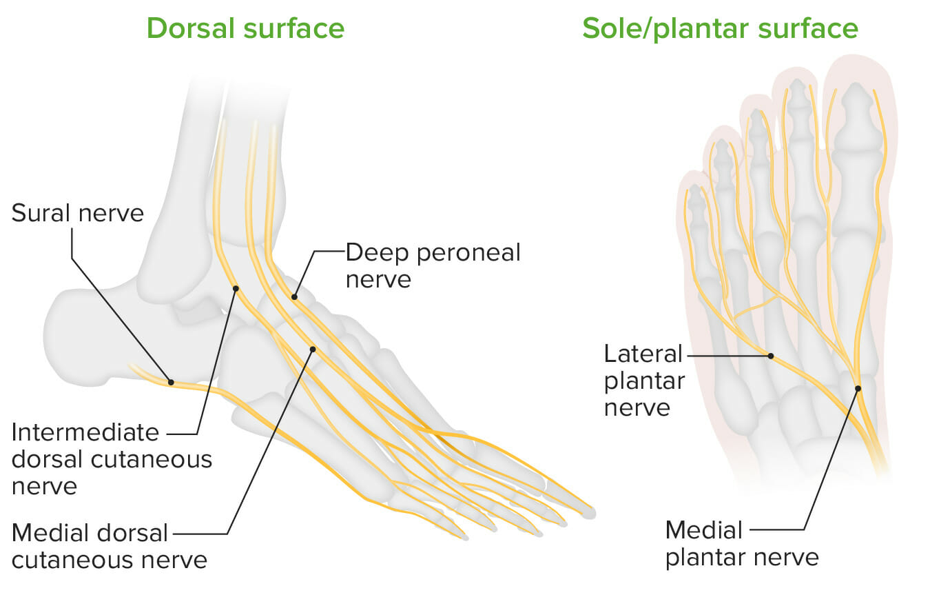
Innervation of the plantar surface:
The tibial nerveTibial NerveThe medial terminal branch of the sciatic nerve. The tibial nerve fibers originate in lumbar and sacral spinal segments (L4 to S2). They supply motor and sensory innervation to parts of the calf and foot.Popliteal Fossa: Anatomy branches into the medial and lateral plantar nerves at the ankle within the tarsal tunnel.
Superficial fibular or peroneal nervePeroneal nerveThe lateral of the two terminal branches of the sciatic nerve. The peroneal (or fibular) nerve provides motor and sensory innervation to parts of the leg and foot.Popliteal Fossa: Anatomy:
Originates from the common fibular nerveCommon Fibular NerveThe lateral of the two terminal branches of the sciatic nerve. The peroneal (or fibular) nerve provides motor and sensory innervation to parts of the leg and foot.Popliteal Fossa: Anatomy
Supplies the skinSkinThe skin, also referred to as the integumentary system, is the largest organ of the body. The skin is primarily composed of the epidermis (outer layer) and dermis (deep layer). The epidermis is primarily composed of keratinocytes that undergo rapid turnover, while the dermis contains dense layers of connective tissue.Skin: Structure and Functions of the dorsum of the foot, excluding digits 1, 2, and 5
Deep fibular or peroneal nervePeroneal nerveThe lateral of the two terminal branches of the sciatic nerve. The peroneal (or fibular) nerve provides motor and sensory innervation to parts of the leg and foot.Popliteal Fossa: Anatomy:
SensorySensoryNeurons which conduct nerve impulses to the central nervous system.Nervous System: Histology innervation to the 1st web space (1st and 2nd digits)
Originates from the femoral nerveFemoral NerveA nerve originating in the lumbar spinal cord (usually L2 to L4) and traveling through the lumbar plexus to provide motor innervation to extensors of the thigh and sensory innervation to parts of the thigh, lower leg, and foot, and to the hip and knee joints.Femoral Region and Hernias: Anatomy
Supplies the skinSkinThe skin, also referred to as the integumentary system, is the largest organ of the body. The skin is primarily composed of the epidermis (outer layer) and dermis (deep layer). The epidermis is primarily composed of keratinocytes that undergo rapid turnover, while the dermis contains dense layers of connective tissue.Skin: Structure and Functions of the medial side of the ankle jointAnkle jointThe ankle is a hinged synovial joint formed between the articular surfaces of the distal tibia, distal fibula, and talus. The ankle primarily allows plantar flexion and dorsiflexion of the foot. and foot
Supplies the distal aspect of the 1st metatarsal
Sural nerve:
Lateral dorsal cutaneous
Lateral calcaneal
Innervation of the dorsal and plantar portions of the foot
Image by Lecturio.
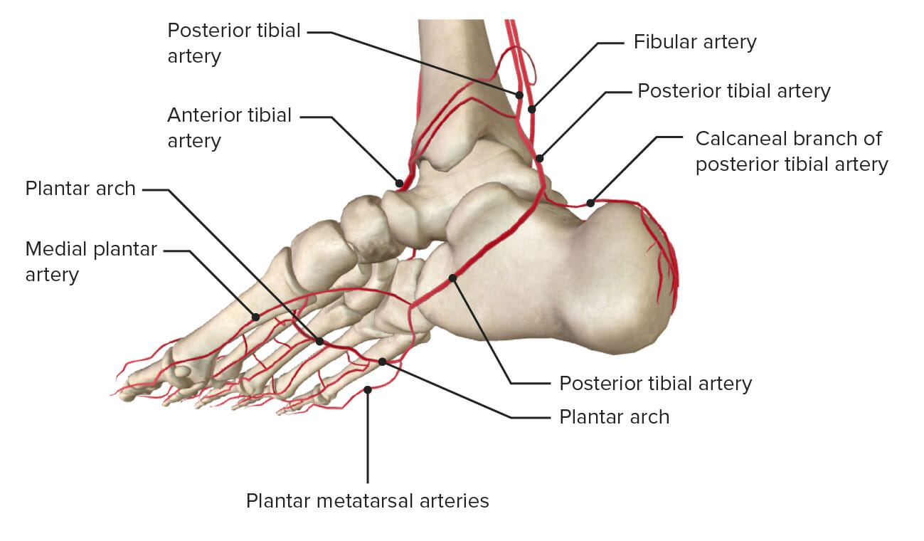
Blood supply
Posterior tibial artery:
Origin:
The most proximal posterior tibial artery
The largest terminal branch of the popliteal arteryPopliteal ArteryThe continuation of the femoral artery coursing through the popliteal fossa; it divides into the anterior and posterior tibial arteries.Popliteal Fossa: Anatomy
Enters the foot by passing behind the medial malleolusMedial malleolusAnkle Joint: Anatomy of the tibiaTibiaThe second longest bone of the skeleton. It is located on the medial side of the lower leg, articulating with the fibula laterally, the talus distally, and the femur proximally.Knee Joint: Anatomy, where it provides branches to the ankle jointAnkle jointThe ankle is a hinged synovial joint formed between the articular surfaces of the distal tibia, distal fibula, and talus. The ankle primarily allows plantar flexion and dorsiflexion of the foot.
The terminal branches are the lateral and medial plantar arteriesArteriesArteries are tubular collections of cells that transport oxygenated blood and nutrients from the heart to the tissues of the body. The blood passes through the arteries in order of decreasing luminal diameter, starting in the largest artery (the aorta) and ending in the small arterioles. Arteries are classified into 3 types: large elastic arteries, medium muscular arteries, and small arteries and arterioles. Arteries: Histology.
Supplies the posterior compartment of the legLegThe lower leg, or just “leg” in anatomical terms, is the part of the lower limb between the knee and the ankle joint. The bony structure is composed of the tibia and fibula bones, and the muscles of the leg are grouped into the anterior, lateral, and posterior compartments by extensions of fascia.Leg: Anatomy via the tarsal tunnel
Supplies the entire plantar surface of the foot via the medial plantar and lateral plantar arteriesArteriesArteries are tubular collections of cells that transport oxygenated blood and nutrients from the heart to the tissues of the body. The blood passes through the arteries in order of decreasing luminal diameter, starting in the largest artery (the aorta) and ending in the small arterioles. Arteries are classified into 3 types: large elastic arteries, medium muscular arteries, and small arteries and arterioles. Arteries: Histology:
Medial plantar artery: supplies medial aspect of the 1st metatarsal and the 1st digit
Lateral plantar artery: supplies the majority of the sole of the foot and gives rise to the deep plantar arch
Oblique view of the plantar aspect of the foot showing the branches of the posterior tibial artery
Image by Lecturio.
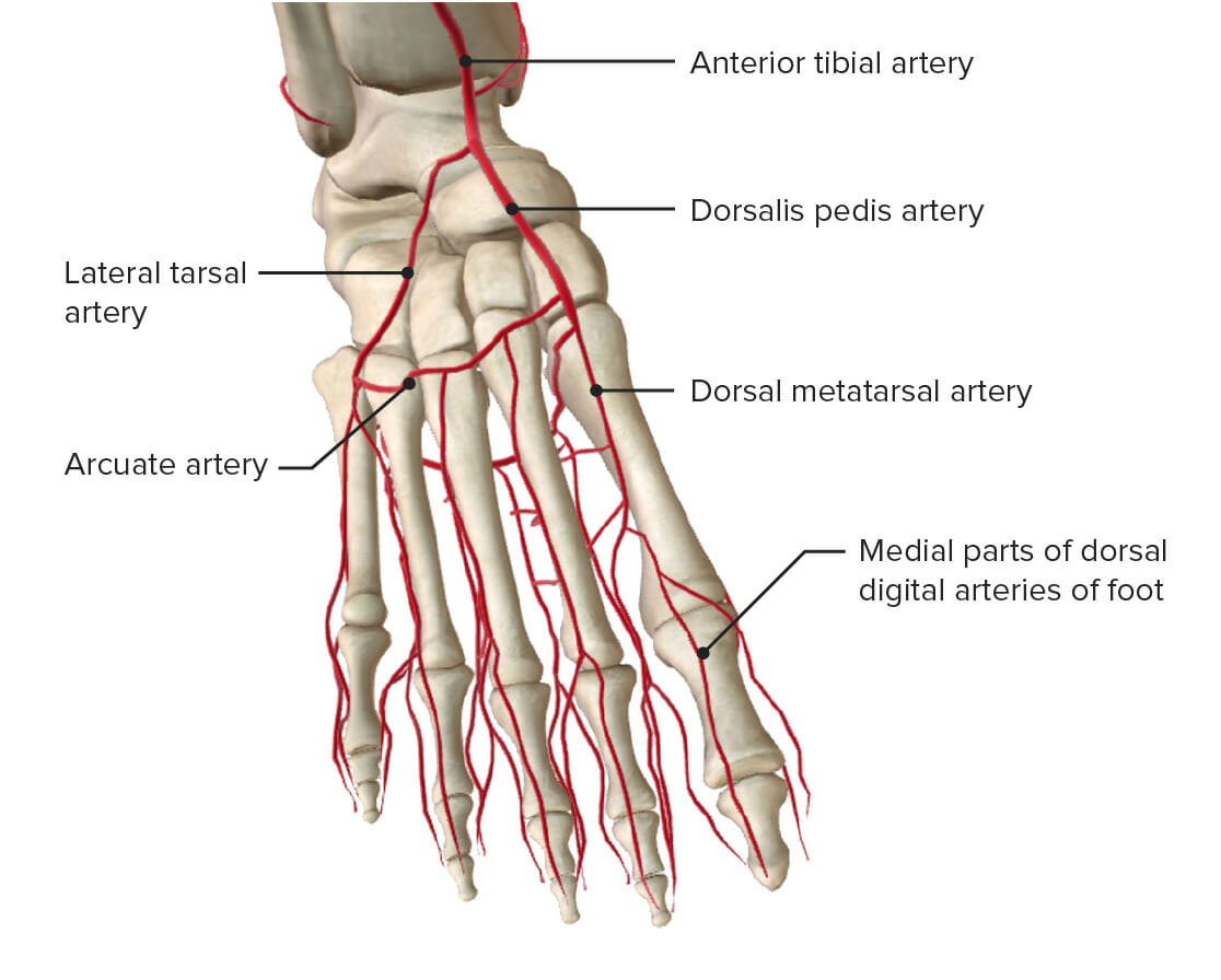
Anterior tibial artery:
Origin:
A terminal branch of the popliteal arteryPopliteal ArteryThe continuation of the femoral artery coursing through the popliteal fossa; it divides into the anterior and posterior tibial arteries.Popliteal Fossa: Anatomy
Passes in front of the ankle jointAnkle jointThe ankle is a hinged synovial joint formed between the articular surfaces of the distal tibia, distal fibula, and talus. The ankle primarily allows plantar flexion and dorsiflexion of the foot. and becomes the dorsalis pedis artery in the foot
Reaches the 1st intermetatarsal space and branches into:
Lateral tarsal artery → arcuate artery
1st dorsal metatarsal artery
Forms an anastomosis with the lateral plantar artery → deep plantar arch
Medial to the extensor digitorum longusExtensor digitorum longusLeg: Anatomy and deep peroneal nervePeroneal nerveThe lateral of the two terminal branches of the sciatic nerve. The peroneal (or fibular) nerve provides motor and sensory innervation to parts of the leg and foot.Popliteal Fossa: Anatomy
Anterior view of the dorsal foot demonstrating the anterior tibial artery to the dorsalis pedis artery and its branches
Image by Lecturio.
Venous drainage
Venous drainage of the foot begins with the digital veinsVeinsVeins are tubular collections of cells, which transport deoxygenated blood and waste from the capillary beds back to the heart. Veins are classified into 3 types: small veins/venules, medium veins, and large veins. Each type contains 3 primary layers: tunica intima, tunica media, and tunica adventitia. Veins: Histology, which run proximally to form both a deep plantar venous arch and a dorsal venous arch. These veinsVeinsVeins are tubular collections of cells, which transport deoxygenated blood and waste from the capillary beds back to the heart. Veins are classified into 3 types: small veins/venules, medium veins, and large veins. Each type contains 3 primary layers: tunica intima, tunica media, and tunica adventitia. Veins: Histology drain into the legLegThe lower leg, or just “leg” in anatomical terms, is the part of the lower limb between the knee and the ankle joint. The bony structure is composed of the tibia and fibula bones, and the muscles of the leg are grouped into the anterior, lateral, and posterior compartments by extensions of fascia.Leg: Anatomy via the anterior and posterior tibial veinsVeinsVeins are tubular collections of cells, which transport deoxygenated blood and waste from the capillary beds back to the heart. Veins are classified into 3 types: small veins/venules, medium veins, and large veins. Each type contains 3 primary layers: tunica intima, tunica media, and tunica adventitia. Veins: Histology and the peroneal vein.
Foot deformitiesFoot deformitiesFoot deformities in children include congenital or acquired malformations of the feet. Two common examples are talipes equinovarus, commonly known as clubfoot, and metatarsus adductus, also called metatarsus varus. Foot Deformities
Lisfranc injury: fractureFractureA fracture is a disruption of the cortex of any bone and periosteum and is commonly due to mechanical stress after an injury or accident. Open fractures due to trauma can be a medical emergency. Fractures are frequently associated with automobile accidents, workplace injuries, and trauma.Overview of Bone Fractures/dislocation of the tarsal–metatarsal articulations at the junction of the midfoot and forefoot. This injury commonly occurs when there is indirect loading on a plantar-flexed foot or with a crush injuryCrush injuryExcessive compression of parts of the body that causes muscle swelling, fracture, and/or neurological disturbances in the affected areas. Crush injury with systemic manifestations is referred to as crush syndrome.Crush Syndrome. The Lisfranc ligament, which is found between the medial cuneiform and the base of the 2nd metatarsal boneBoneBone is a compact type of hardened connective tissue composed of bone cells, membranes, an extracellular mineralized matrix, and central bone marrow. The 2 primary types of bone are compact and spongy. Bones: Structure and Types, is disrupted. This type of injury commonly needs a CT scan for detection, as it may be missed with an x-rayX-rayPenetrating electromagnetic radiation emitted when the inner orbital electrons of an atom are excited and release radiant energy. X-ray wavelengths range from 1 pm to 10 nm. Hard x-rays are the higher energy, shorter wavelength x-rays. Soft x-rays or grenz rays are less energetic and longer in wavelength. The short wavelength end of the x-ray spectrum overlaps the gamma rays wavelength range. The distinction between gamma rays and x-rays is based on their radiation source.Pulmonary Function Tests. A Lisfranc injury may lead to chronic injury.
PesPESRemoval of plasma and replacement with various fluids, e.g., fresh frozen plasma, plasma protein fractions (ppf), albumin preparations, dextran solutions, saline. Used in treatment of autoimmune diseases, immune complex diseases, diseases of excess plasma factors, and other conditions.Thrombotic Thrombocytopenic Purpura cavus: excessively arched foot or “claw foot.” PesPESRemoval of plasma and replacement with various fluids, e.g., fresh frozen plasma, plasma protein fractions (ppf), albumin preparations, dextran solutions, saline. Used in treatment of autoimmune diseases, immune complex diseases, diseases of excess plasma factors, and other conditions.Thrombotic Thrombocytopenic Purpura cavus is usually caused by neurologic disorders, resulting in plantar hyperflexion of the 1st metatarsal.
Plantar metatarsophalangeal sprain or turf toe: sprain or disruption of the plantar stabilizers of the 1st metatarsal phalangeal joint.
Plantar fasciitisPlantar fasciitisInflammation of the plantar fascia (aponeurosis) on the bottom of the foot causing heel pain. The etiology of plantar fasciitis remains controversial but is likely to involve a biomechanical imbalance. Though often presenting along with heel spur, they do not appear to be causally related.Ankle and Foot Pain: common degenerative condition of the proximal plantar fasciaFasciaLayers of connective tissue of variable thickness. The superficial fascia is found immediately below the skin; the deep fascia invests muscles, nerves, and other organs.Cellulitis. Presentation is with heel painPainAn unpleasant sensation induced by noxious stimuli which are detected by nerve endings of nociceptive neurons.Pain: Types and Pathways with the 1st step in the morning or with prolonged standing.
Tarsal coalitions: union of ≥ 2 tarsal bones. Tarsal coalitions occur most commonly between the talusTalusThe second largest of the tarsal bones. It articulates with the tibia and fibula to form the ankle joint.Ankle Joint: Anatomy and calcaneus or the navicular and calcaneus and are usually asymptomatic until adolescence.