Definition, Epidemiology, and Etiology
Definition
- Benign breast masses
- Composed of fibrous and glandular tissue
Epidemiology
- Most common in women < 35 years of age (peak incidence: 15–35 years)
- Most common benign tumor of the breast, and the most common tumor in young women
- Accounts for half of all breast biopsies
- Only a slightly increased risk of developing malignant breast cancer, with a somewhat higher risk if “complex” features are present:
- Cysts > 0.3 cm
- Sclerosing adenosis
- Epithelial calcifications
- Papillary apocrine change
Etiology
- Unknown
- Unlikely to be hereditary
- Negative correlation with tobacco smoking
- A relationship has been established with estrogen:
- Increases in size during pregnancy, lactation, cyclically with menstruation, and with the use of oral contraceptives
- Regresses after menopause
Clinical Presentation
- Generally asymptomatic
- Solitary lesion in 80% of cases
- 20% of cases consist of multiple fibroadenomas occurring in the same breast or bilaterally.
- Small, spherical, well-defined, mobile mass with a rubbery or firm consistency
- Diameter of 3–10 cm that causes breast asymmetry (usually < 3 cm)
- Usually found in the upper outer quadrant of the breast
- Cyclic changes in size related to the menstrual cycle due to sensitivity to estrogen
- Other types of fibroadenomas:
- Juvenile fibroadenomas
- Usually occur in African American patients aged 10–18 years
- Rapid increase in size
- May cause skin ulceration
- Giant fibroadenomas
- Histologically typical fibroadenomas > 10 cm in size
- Excision is recommended
- Complex fibroadenomas
- Proliferative pathologic changes
- Associated with a slightly increased risk of cancer
- Management is controversial
- Juvenile fibroadenomas
Diagnosis and Management
Diagnosis
- Ultrasonography:
- Well-defined mass with smooth and regular boundaries and a weak echo signal
- 25% of cases have irregular borders → biopsy to confirm benignancy
- Mammogram or magnetic resonance imaging (MRI): regular boundaries and popcorn-like calcifications
- Fine needle aspiration (method of choice for biopsy):
- Fibrous and glandular tissue with clusters of spindle cells
- Antler horn clusters
- Honeycomb sheets
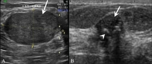
A: Breast ultrasonography in a 25-year-old patient shows a homogenous, hypoechoic, gently lobulated lesion (arrow), suggestive of a fibroadenoma.
Image: “Pictorial essay: Breast USG” by Advanced Radiology Centre, Mumbai, India. License: CC BY 2.0
B: Ultrasonography shows a degenerating fibroadenoma (arrow) with coarse calcifications (arrowhead) and posterior shadowing from the calcific foci.Preoperative imaging, histology, absoption- and phase-contrast CT
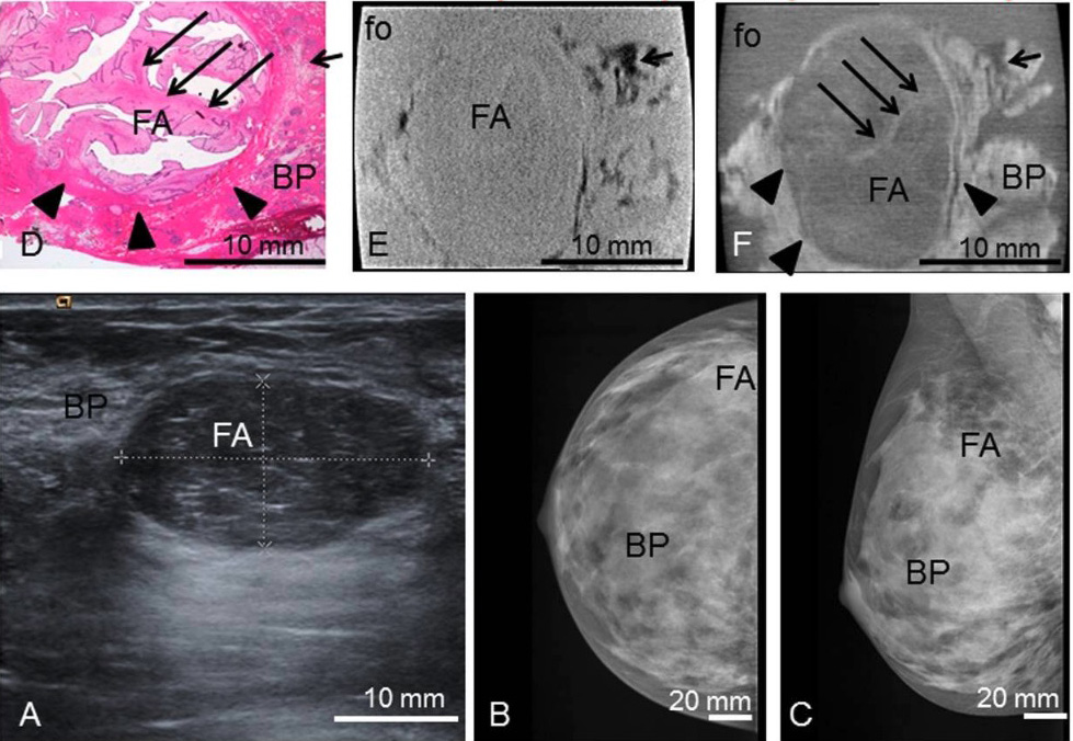
Image: “Typical features of breast fibroadenomas” by Institute for Clinical Radiology, Ludwig-Maximilians-University Hospital, Munich, Germany. License: CC BY 4.0
Preoperative ultrasonography (A) with typical benign imaging characteristics of the fibroadenoma (FA) (oval form, smooth margins, posterior enhancement);
preoperative craniocaudal (B) and mediolateral oblique (C) mammography projections show the fibroadenoma (FA) partially hidden within the dense breast parenchyma (BP) (ACR IV).
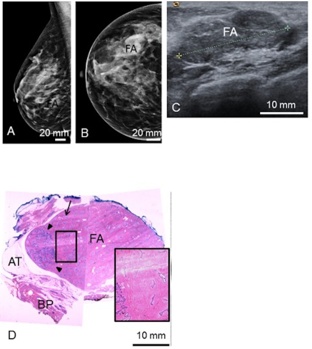
Representative histological slice (D) and corresponding absorption- (E) and phase-contrast CT (F) image. The black sharp lines in the corners of (E) and (F) correspond to the walls of the plastic container. The ducts are artificially torn open in (D) due to cutting and staining procedures.Preoperative mediolateral-oblique (A) and craniocaudal (B) mammogram and ultrasonography (C) show the fibroadenoma (FA).
Image: “Visualizing typical features of breast fibroadenomas” by Institute for Clinical Radiology, Ludwig-Maximilians-University Hospital, Munich, Germany. License: CC BY 4.0, edited by Lecturio.
A representative histological slice (D) shows the FA, surrounded by adipose tissue (AT) and breast parenchyma (BP).
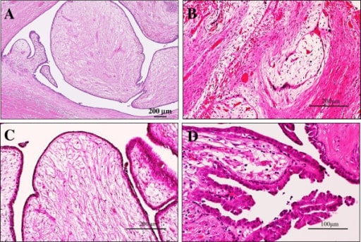
(D) The black rectangle shows a zoomed view of a polygonal sclerotic frame filled with basophilic branched ducts; 1 linear duct is indicated by the arrow.Microscopic examination of the intraductal fibroadenoma or intraductal phyllodes tumor of the breast
Image: “Intraductal fibroadenoma” by Department of Pathology and Cell Biology, School of Medicine, University of Occupational and Environmental Health, 1-1 Iseigaoka, Yahatanishi-ku, Kitakyushu 807-8555, Japan. License: CC BY 2.0
A: Low-power view shows that its polypoid parts are composed of leaf-like processes with a hypocellular and prominent myxoid stroma protruding into cystic spaces, reminiscent of intracanalicular type fibroadenoma or benign phyllodes tumor features (H&E stains). Bar = 200 μm.
B: In some areas, there are foci of a typical intracanalicular variant of fibroadenoma, in which duct lumens are compressed by the proliferating myxoid stroma (H&E stains). Bar = 200 μm.
C: On high-power view, those stromal cells show no significant atypia, but the covering hyperplastic epithelial components are also bland looking in 2 cell layers. Mitotic figures are very rarely seen (H&E stains). Bar = 200 μm.
D: In others, tiny foci of benign intraductal papilloma with characteristic delicate fibrovascular stalks are rarely seen (H&E stains). Bar = 100 μm.
Management
- Regular check-ups in cases confirmed to be benign (“wait-and-see” approach)
- Cryoablation for small but symptomatic fibroadenomas
- Surgical resection for cases of juvenile and giant fibroadenomas, irregular boundaries, and patients with personal or family history of breast cancer
Differential Diagnosis
The following conditions are differential diagnoses of fibroadenomas:
- Galactocele: retention cyst within the mammary gland containing milk, which presents as a palpable, firm mass in the subareolar region.
- Breast abscess: accumulation of pus within the mammary gland. Usually associated with lactational mastitis. Presents as a unilateral and fluctuant mass within a painful, erythematous, and edematous breast.
- Intraductal papilloma: benign mass with a papillary configuration of breast stroma and epithelium within a breast duct, which presents as a solitary mass found close to or behind the nipple, with bloody nipple discharge.
- Phyllodes tumor: uncommon fibroepithelial tumor similar to a fibroadenoma but requiring complete surgical excision with clean borders because of its malignant potential. The tumors are categorized into benign, borderline, and malignant types. Histologically, these tumors have stromal overgrowth with increased mitotic activity, with the borderline and malignant tumors showing infiltrative borders.
- Fibrocystic changes of the breast: refers to the common changes that happen to breast tissue as a woman ages, causing a “lumpy-bumpy” breast in the more pronounced expressions of the condition. These changes can be non-proliferative (cysts, fibrosis, adenosis) or proliferative (epithelial proliferation, sclerosing adenosis, papilloma), with or without atypia. Atypical hyperplasia has a moderately increased risk of carcinoma.
- Mastitis: inflammation of the mammary gland tissue; can be lactational or non-lactational. Mastitis is most common in women of childbearing age.
- Fat necrosis of the breast: usually due to breast injury, which often goes unnoticed. As the damaged breast tissue is repaired, it is replaced by scar tissue. However, some fat cells may have a different response and form what are known as oily cysts.
- Breast cancer: malignant tumor of the mammary gland. The 2 most common histological types of breast cancer are ductal carcinoma and lobular carcinoma.
References
- Vinay K, Abbas A, Aster J. Robbins & Cotran Pathologic Basis of Disease. 2020, 10th Edition, Elsevier.
- Sabel MS. Overview of benign breast disease. UpToDate. Retrieved on August 28, 2020, from https://www.uptodate.com/contents/overview-of-benign-breast-disease#H14
- Rohan, T. E., & Miller, A. B. (1999). A cohort study of cigarette smoking and risk of fibroadenoma. Journal of Epidemiology and Biostatistics, 4(4), 297–302.