Achieve Mastery of Medical Concepts

Study for medical school and boards with Lecturio

Testicular Torsion

Testicular torsion is the sudden rotation Rotation Motion of an object in which either one or more points on a line are fixed. It is also the motion of a particle about a fixed point. X-rays of the testicle, specifically the spermatic cord Spermatic Cord Either of a pair of tubular structures formed by ductus deferens; arteries; veins; lymphatic vessels; and nerves. The spermatic cord extends from the deep inguinal ring through the inguinal canal to the testis in the scrotum. Testicles: Anatomy, around its axis in the inguinal canal Inguinal canal The tunnel in the lower anterior abdominal wall through which the spermatic cord, in the male; round ligament, in the female; nerves; and vessels pass. Its internal end is at the deep inguinal ring and its external end is at the superficial inguinal ring. Inguinal Canal: Anatomy and Hernias or below. The acute rotation Rotation Motion of an object in which either one or more points on a line are fixed. It is also the motion of a particle about a fixed point. X-rays results in compromised blood flow Blood flow Blood flow refers to the movement of a certain volume of blood through the vasculature over a given unit of time (e.g., mL per minute). Vascular Resistance, Flow, and Mean Arterial Pressure to and from the testicle, which puts the testicle at risk for necrosis Necrosis The death of cells in an organ or tissue due to disease, injury or failure of the blood supply. Ischemic Cell Damage. Quick diagnosis and intervention is key to saving the affected testicle. Emergent surgical exploration with subsequent orchidopexy is required. Ultrasound or manual detorsion should not delay definitive care.

Last updated: Sep 29, 2022

Editorial responsibility: Stanley Oiseth, Lindsay Jones, Evelin Maza

Overview

Definition

  • Sudden rotation Rotation Motion of an object in which either one or more points on a line are fixed. It is also the motion of a particle about a fixed point. X-rays of the testicle, specifically the spermatic cord Spermatic Cord Either of a pair of tubular structures formed by ductus deferens; arteries; veins; lymphatic vessels; and nerves. The spermatic cord extends from the deep inguinal ring through the inguinal canal to the testis in the scrotum. Testicles: Anatomy, around its axis
  • A urological emergency

Epidemiology

  • Can occur at any age 
  • Peak incidence Incidence The number of new cases of a given disease during a given period in a specified population. It also is used for the rate at which new events occur in a defined population. It is differentiated from prevalence, which refers to all cases in the population at a given time. Measures of Disease Frequency
    • Neonatal period 
    • Boys 12–18 years old
  • Yearly incidence Incidence The number of new cases of a given disease during a given period in a specified population. It also is used for the rate at which new events occur in a defined population. It is differentiated from prevalence, which refers to all cases in the population at a given time. Measures of Disease Frequency: 3.8 per 100,000 boys < 18 years old
  • Cryptorchidism Cryptorchidism Cryptorchidism is one of the most common congenital anomalies in young boys. Typically, this asymptomatic condition presents during a routine well-child examination where 1 or both testicles are not palpable in the scrotum. Cryptorchidism increases the risk of testicular torsion.

Pathophysiology and Clinical Presentation

Pathogenesis

Testicular torsion

Testicular torsion:
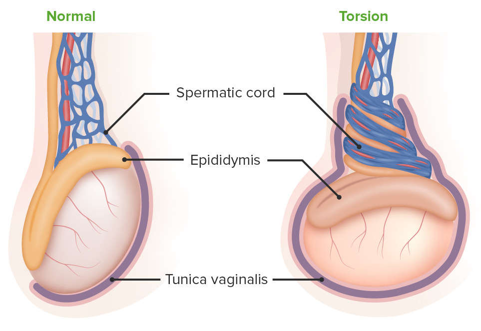
The left image depicts a normal testicle. Testicular torsion is seen on the right: The testicle lies horizontally, creating an increased risk of torsion of the spermatic vessels.

Image by Lecturio.

Clinical presentation

  • Newborn Newborn An infant during the first 28 days after birth. Physical Examination of the Newborn
    • Hard scrotal mass Mass Three-dimensional lesion that occupies a space within the breast Imaging of the Breast that does not transilluminate
    • Discolored or bruised hemiscrotum with swelling Swelling Inflammation 
    • Acute tenderness on exam
  • Older patients Patients Individuals participating in the health care system for the purpose of receiving therapeutic, diagnostic, or preventive procedures. Clinician–Patient Relationship
    • Acute-onset, severe, constant testicular or scrotal pain Pain An unpleasant sensation induced by noxious stimuli which are detected by nerve endings of nociceptive neurons. Pain: Types and Pathways (typically < 12-hour duration)
    • Associated nausea Nausea An unpleasant sensation in the stomach usually accompanied by the urge to vomit. Common causes are early pregnancy, sea and motion sickness, emotional stress, intense pain, food poisoning, and various enteroviruses. Antiemetics and vomiting Vomiting The forcible expulsion of the contents of the stomach through the mouth. Hypokalemia 
    • No clear inciting factor, but history of strenuous activity or trauma reported
    • Children may awaken at night with scrotal pain Pain An unpleasant sensation induced by noxious stimuli which are detected by nerve endings of nociceptive neurons. Pain: Types and Pathways from cremasteric contraction.

Diagnosis and Management

Clinical findings

  • Physical exam: 
    • Edematous, tender, indurated testicle/ scrotum Scrotum A cutaneous pouch of skin containing the testicles and spermatic cords. Testicles: Anatomy 
    • Affected testicle lies horizontally.
    • High-riding testicle due to spermatic cord Spermatic Cord Either of a pair of tubular structures formed by ductus deferens; arteries; veins; lymphatic vessels; and nerves. The spermatic cord extends from the deep inguinal ring through the inguinal canal to the testis in the scrotum. Testicles: Anatomy shortening
  • Key physical exam maneuvers: 
    • Cremasteric reflex Cremasteric Reflex Male Genitourinary Examination (elevation of testicle when stroking upper inner thigh Thigh The thigh is the region of the lower limb found between the hip and the knee joint. There is a single bone in the thigh called the femur, which is surrounded by large muscles grouped into 3 fascial compartments. Thigh: Anatomy) is usually absent.
    • Prehn sign: Lifting the scrotum Scrotum A cutaneous pouch of skin containing the testicles and spermatic cords. Testicles: Anatomy relieves pain Pain An unpleasant sensation induced by noxious stimuli which are detected by nerve endings of nociceptive neurons. Pain: Types and Pathways in epididymitis Epididymitis Epididymitis and orchitis are characterized by acute inflammation of the epididymis and the testicle, respectively, due to viral or bacterial infections. Patients typically present with gradually worsening testicular pain and scrotal swelling along with systemic symptoms such as fever, depending on severity. Epididymitis and Orchitis and increases pain Pain An unpleasant sensation induced by noxious stimuli which are detected by nerve endings of nociceptive neurons. Pain: Types and Pathways in torsion. 
  • Scrotal ultrasound: 
    • Should not delay definitive management 
    • Color Doppler Doppler Ultrasonography applying the doppler effect, with frequency-shifted ultrasound reflections produced by moving targets (usually red blood cells) in the bloodstream along the ultrasound axis in direct proportion to the velocity of movement of the targets, to determine both direction and velocity of blood flow. Ultrasound (Sonography) will demonstrate decreased testicular vascular perfusion.

Treatment

  • Time is essential in testicle preservation:
    • Within 4–6 hours: 95% viability
    • After 12 hours: 20%–60% viability
    • After 24 hours: 0%–20% viability
  • Emergency surgical exploration of the affected testicle with reduction (untwisting) and bilateral orchidopexy (fixation of testicle to scrotal wall) indicated
  • Manual detorsion:
    • Bedside procedure is attempted if emergency operative care is not readily available.
    • Grasp the affected testicle and rotate from medial to lateral direction (“open book technique”). 
    • May provide prompt relief of pain Pain An unpleasant sensation induced by noxious stimuli which are detected by nerve endings of nociceptive neurons. Pain: Types and Pathways prior to going to the operating room
Testicular torsion surgery

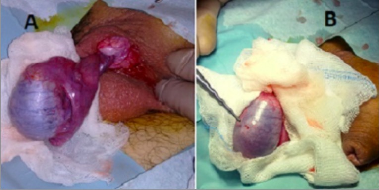
Intraoperative scrotal exploration for testicular torsion:
A: blue discolored testicle devoid of blood supply and twisted spermatic cord
B: prior orchiopexy sutures denoting recurrent testicular torsion

Image: “Intraoperative image of emergent scrotal exploration” by Department of Urology, University General Hospital of Heraklion, Heraklion, Crete, Greece. License: CC BY 2.0
Manual testicular detorsion

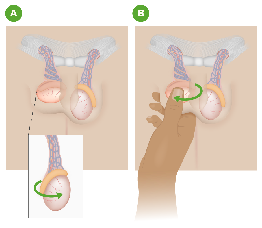
Manual testicular detorsion:
Image A depicts a right testicular torsion.
Image B illustrates the manual detorsion method of grasping and rotating the affected testicle with the “open book technique” from the medial to lateral position.

Image by Lecturio.

Differential Diagnosis

  • Torsion of appendix Appendix A worm-like blind tube extension from the cecum. Colon, Cecum, and Appendix: Anatomy testis ( Müllerian duct Müllerian duct A pair of ducts near the wolffian ducts in a developing embryo. In the male embryo, they degenerate with the appearance of testicular anti-mullerian hormone. In the absence of anti-mullerian hormone, mullerian ducts give rise to the female reproductive tract, including the oviducts; uterus; cervix; and vagina. Uterus, Cervix, and Fallopian Tubes: Anatomy remnant) or appendix Appendix A worm-like blind tube extension from the cecum. Colon, Cecum, and Appendix: Anatomy epididymis Epididymis The convoluted cordlike structure attached to the posterior of the testis. Epididymis consists of the head (caput), the body (corpus), and the tail (cauda). A network of ducts leaving the testis joins into a common epididymal tubule proper which provides the transport, storage, and maturation of spermatozoa. Testicles: Anatomy ( Wolffian duct Wolffian duct A pair of excretory ducts of the middle kidneys of an embryo, also called mesonephric ducts. In higher vertebrates, wolffian ducts persist in the male forming vas deferens, but atrophy into vestigial structures in the female. Kidneys: Anatomy remnant): sudden onset of testicular pain Pain An unpleasant sensation induced by noxious stimuli which are detected by nerve endings of nociceptive neurons. Pain: Types and Pathways similar to testicular torsion. However, the testicle itself is not tender and the pain Pain An unpleasant sensation induced by noxious stimuli which are detected by nerve endings of nociceptive neurons. Pain: Types and Pathways is focused at the superior pole of the testicle depicting a “blue dot sign” (inflamed appendage visualized through the scrotal skin Skin The skin, also referred to as the integumentary system, is the largest organ of the body. The skin is primarily composed of the epidermis (outer layer) and dermis (deep layer). The epidermis is primarily composed of keratinocytes that undergo rapid turnover, while the dermis contains dense layers of connective tissue. Skin: Structure and Functions). Doppler Doppler Ultrasonography applying the doppler effect, with frequency-shifted ultrasound reflections produced by moving targets (usually red blood cells) in the bloodstream along the ultrasound axis in direct proportion to the velocity of movement of the targets, to determine both direction and velocity of blood flow. Ultrasound (Sonography) ultrasound shows normal flow Flow Blood flows through the heart, arteries, capillaries, and veins in a closed, continuous circuit. Flow is the movement of volume per unit of time. Flow is affected by the pressure gradient and the resistance fluid encounters between 2 points. Vascular resistance is the opposition to flow, which is caused primarily by blood friction against vessel walls. Vascular Resistance, Flow, and Mean Arterial Pressure to the testicle and torsion of the appendage. Management is supportive with analgesics and bedrest. 
  • Intermittent testicular torsion: acute, sudden-onset testicular pain Pain An unpleasant sensation induced by noxious stimuli which are detected by nerve endings of nociceptive neurons. Pain: Types and Pathways with scrotal swelling Swelling Inflammation and rapid resolution. The cycle Cycle The type of signal that ends the inspiratory phase delivered by the ventilator Invasive Mechanical Ventilation may go on for several hours or days but must be worked up in the same manner as a suspected testicular torsion. Clinical diagnosis includes a physical exam and Doppler Doppler Ultrasonography applying the doppler effect, with frequency-shifted ultrasound reflections produced by moving targets (usually red blood cells) in the bloodstream along the ultrasound axis in direct proportion to the velocity of movement of the targets, to determine both direction and velocity of blood flow. Ultrasound (Sonography) scrotal ultrasound for radiographic evidence of halted blood flow Blood flow Blood flow refers to the movement of a certain volume of blood through the vasculature over a given unit of time (e.g., mL per minute). Vascular Resistance, Flow, and Mean Arterial Pressure to the testicles Testicles The testicles, also known as the testes or the male gonads, are a pair of egg-shaped glands suspended within the scrotum. The testicles have multiple layers: an outer tunica vaginalis, an intermediate tunica albuginea, and an innermost tunica vasculosa. The testicles are composed of testicular lobules and seminiferous tubules. Testicles: Anatomy.  
  • Epididymitis Epididymitis Epididymitis and orchitis are characterized by acute inflammation of the epididymis and the testicle, respectively, due to viral or bacterial infections. Patients typically present with gradually worsening testicular pain and scrotal swelling along with systemic symptoms such as fever, depending on severity. Epididymitis and Orchitis and orchitis Orchitis Inflammation of a testis. It has many features of epididymitis, such as swollen scrotum; pain; pyuria; and fever. It is usually related to infections in the urinary tract, which likely spread to the epididymis and then the testis through either the vas deferens or the lymphatics of the spermatic cord. Epididymitis and Orchitis: inflammatory process of the epididymis Epididymis The convoluted cordlike structure attached to the posterior of the testis. Epididymis consists of the head (caput), the body (corpus), and the tail (cauda). A network of ducts leaving the testis joins into a common epididymal tubule proper which provides the transport, storage, and maturation of spermatozoa. Testicles: Anatomy or testicle causing gradual-onset pain Pain An unpleasant sensation induced by noxious stimuli which are detected by nerve endings of nociceptive neurons. Pain: Types and Pathways and swelling Swelling Inflammation. Condition commonly presents with dysuria Dysuria Painful urination. It is often associated with infections of the lower urinary tract. Urinary tract infections (UTIs), urinary frequency, discharge, and fevers. Diagnosis consists of physical exam, history of gradual-onset pain Pain An unpleasant sensation induced by noxious stimuli which are detected by nerve endings of nociceptive neurons. Pain: Types and Pathways (vs. acute with torsion), and urinalysis Urinalysis Examination of urine by chemical, physical, or microscopic means. Routine urinalysis usually includes performing chemical screening tests, determining specific gravity, observing any unusual color or odor, screening for bacteriuria, and examining the sediment microscopically. Urinary Tract Infections (UTIs) in Children/culture. Sexually transmitted disease Sexually Transmitted Disease Sexually transmitted diseases (STDs) are infections that spread either by vaginal intercourse, anal sex, or oral sex. Symptoms and signs may include vaginal discharge, penile discharge, dysuria, skin lesions (e.g., warts, ulcers) on or around the genitals, and pelvic pain. Some infections can lead to infertility and chronic debilitating disease. Sexually Transmitted Infections (STIs) testing or Doppler Doppler Ultrasonography applying the doppler effect, with frequency-shifted ultrasound reflections produced by moving targets (usually red blood cells) in the bloodstream along the ultrasound axis in direct proportion to the velocity of movement of the targets, to determine both direction and velocity of blood flow. Ultrasound (Sonography) scrotal ultrasound should be considered based on the clinical scenario. Treatment is antibiotics, analgesics, and scrotal support.
  • Immunoglobulin A ( IgA IgA Represents 15-20% of the human serum immunoglobulins, mostly as the 4-chain polymer in humans or dimer in other mammals. Secretory iga is the main immunoglobulin in secretions. Immunoglobulins: Types and Functions) vasculitis Vasculitis Inflammation of any one of the blood vessels, including the arteries; veins; and rest of the vasculature system in the body. Systemic Lupus Erythematosus: systemic vasculitis Vasculitis Inflammation of any one of the blood vessels, including the arteries; veins; and rest of the vasculature system in the body. Systemic Lupus Erythematosus syndrome with nonthrombocytopenic purpura, arthralgia Arthralgia Pain in the joint. Rheumatic Fever, renal disease, abdominal pain Abdominal Pain Acute Abdomen, and sometimes scrotal pain Pain An unpleasant sensation induced by noxious stimuli which are detected by nerve endings of nociceptive neurons. Pain: Types and Pathways. The onset of scrotal pain Pain An unpleasant sensation induced by noxious stimuli which are detected by nerve endings of nociceptive neurons. Pain: Types and Pathways may be acute or gradual. Torsion should be suspected if the patient does not exhibit any other sequelae of vasculitis Vasculitis Inflammation of any one of the blood vessels, including the arteries; veins; and rest of the vasculature system in the body. Systemic Lupus Erythematosus; otherwise, treatment is supportive.  
  • Inguinal canal Inguinal canal The tunnel in the lower anterior abdominal wall through which the spermatic cord, in the male; round ligament, in the female; nerves; and vessels pass. Its internal end is at the deep inguinal ring and its external end is at the superficial inguinal ring. Inguinal Canal: Anatomy and Hernias and hernias: Incarcerated inguinal hernias Inguinal Hernias An abdominal hernia with an external bulge in the groin region. It can be classified by the location of herniation. Indirect inguinal hernias occur through the internal inguinal ring. Direct inguinal hernias occur through defects in the abdominal wall (transversalis fascia) in Hesselbach’s triangle. The former type is commonly seen in children and young adults; the latter in adults. Inguinal Canal: Anatomy and Hernias will cause acute-onset inguinal or scrotal pain Pain An unpleasant sensation induced by noxious stimuli which are detected by nerve endings of nociceptive neurons. Pain: Types and Pathways. Physical exam reveals inguinal swelling Swelling Inflammation, pain Pain An unpleasant sensation induced by noxious stimuli which are detected by nerve endings of nociceptive neurons. Pain: Types and Pathways, and sometimes bowel sounds in the scrotum Scrotum A cutaneous pouch of skin containing the testicles and spermatic cords. Testicles: Anatomy due to herniated bowel. Ultrasound may be necessary for a definitive diagnosis if the presentation is unclear. Management consists of attempted hernia Hernia Protrusion of tissue, structure, or part of an organ through the bone, muscular tissue, or the membrane by which it is normally contained. Hernia may involve tissues such as the abdominal wall or the respiratory diaphragm. Hernias may be internal, external, congenital, or acquired. Abdominal Hernias reduction or, in the case of incarceration Incarceration Inguinal Canal: Anatomy and Hernias, immediate operative care. 
  • Varicocele Varicocele A condition characterized by the dilated tortuous veins of the spermatic cord with a marked left-sided predominance. Adverse effect on male fertility occurs when varicocele leads to an increased scrotal (and testicular) temperature and reduced testicular volume. Varicocele, Hydrocele, and Spermatocele, hydrocele Hydrocele Accumulation of serous fluid between the layers of membrane (tunica vaginalis) covering the testis in the scrotum. Varicocele, Hydrocele, and Spermatocele, and spermatocele Spermatocele A cystic dilation of the epididymis, usually in the head portion (caput epididymis). The cyst fluid contains dead spermatozoa and can be easily differentiated from testicular hydrocele and other testicular lesions. Varicocele, Hydrocele, and Spermatocele: scrotal conditions that are usually asymptomatic or associated with dull, aching sensation. Key physical exam findings include: “bag of worms” for varicocele Varicocele A condition characterized by the dilated tortuous veins of the spermatic cord with a marked left-sided predominance. Adverse effect on male fertility occurs when varicocele leads to an increased scrotal (and testicular) temperature and reduced testicular volume. Varicocele, Hydrocele, and Spermatocele, tense scrotum Scrotum A cutaneous pouch of skin containing the testicles and spermatic cords. Testicles: Anatomy and large swelling Swelling Inflammation for hydrocele Hydrocele Accumulation of serous fluid between the layers of membrane (tunica vaginalis) covering the testis in the scrotum. Varicocele, Hydrocele, and Spermatocele, and cyst-like mass Mass Three-dimensional lesion that occupies a space within the breast Imaging of the Breast of the epididymis Epididymis The convoluted cordlike structure attached to the posterior of the testis. Epididymis consists of the head (caput), the body (corpus), and the tail (cauda). A network of ducts leaving the testis joins into a common epididymal tubule proper which provides the transport, storage, and maturation of spermatozoa. Testicles: Anatomy for spermatocele Spermatocele A cystic dilation of the epididymis, usually in the head portion (caput epididymis). The cyst fluid contains dead spermatozoa and can be easily differentiated from testicular hydrocele and other testicular lesions. Varicocele, Hydrocele, and Spermatocele. Management consists of conservative care or surgical excision based on the patient’s level of discomfort. 

References

  1. Brenner, J.S. (2020). Causes of scrotal pain in children and adolescents. UpToDate. Retrieved January 23, 2021, from https://www.uptodate.com/contents/causes-of-scrotal-pain-in-children-and-adolescents
  2. Hittelman, A.B. (2020). Neonatal testicular torsion. UpToDate. Retrieved January 25, 2021, from https://www.uptodate.com/contents/neonatal-testicular-torsion
  3. Sharp, V., Kieran, K., Arlen, A.  (2013) Testicular torsion: Diagnosis, Evaluation and Management. Am Fam Physician. 15;88(12):835-840.

Create your free account or log in to continue reading!

Sign up now and get free access to Lecturio with concept pages, medical videos, and questions for your medical education.

User Reviews

Details