The shoulder complex comprises the glenohumeral joint, sternoclavicular joint, acromioclavicular joint, and the scapulothoracic articulation, and connects the upper limb to the trunk. This group of joints consists of the clavicle Clavicle A bone on the ventral side of the shoulder girdle, which in humans is commonly called the collar bone. Clavicle Fracture, scapula, and humerus Humerus Bone in humans and primates extending from the shoulder joint to the elbow joint. Arm: Anatomy bones, multiple muscles and supporting ligaments, cartilage Cartilage Cartilage is a type of connective tissue derived from embryonic mesenchyme that is responsible for structural support, resilience, and the smoothness of physical actions. Perichondrium (connective tissue membrane surrounding cartilage) compensates for the absence of vasculature in cartilage by providing nutrition and support. Cartilage: Histology, and bursae. The muscles ensure the mobility and stability of the shoulder and upper limb and are divided into 3 groups: anterior axioappendicular, posterior axioappendicular, and scapulohumeral muscles.
Last updated: Dec 15, 2025
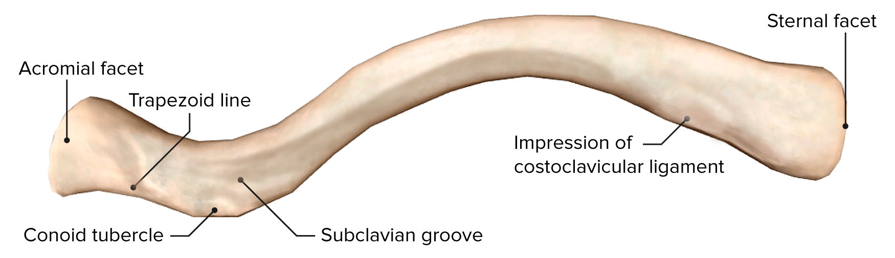
Inferior view of the right clavicle
Image by BioDigital, edited by Lecturio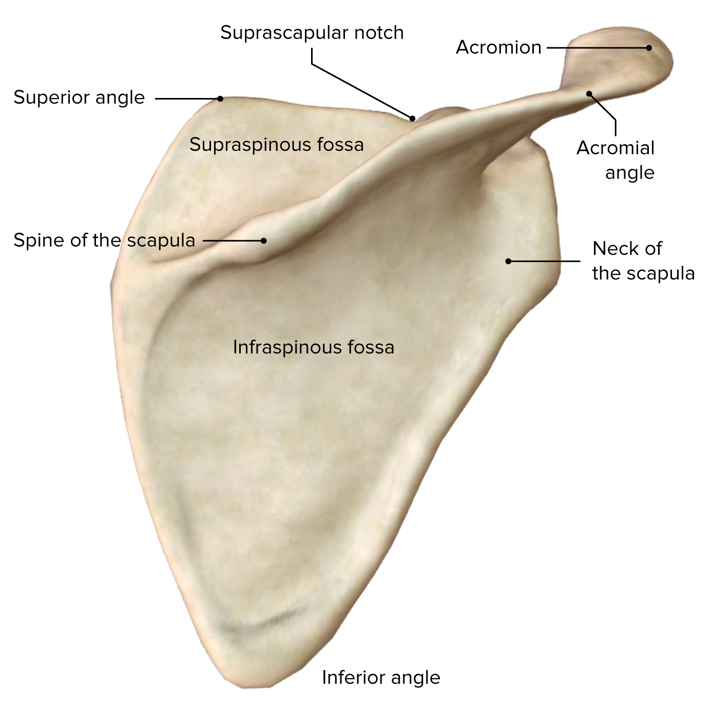
Posterior view of the right scapula
Image by BioDigital, edited by Lecturio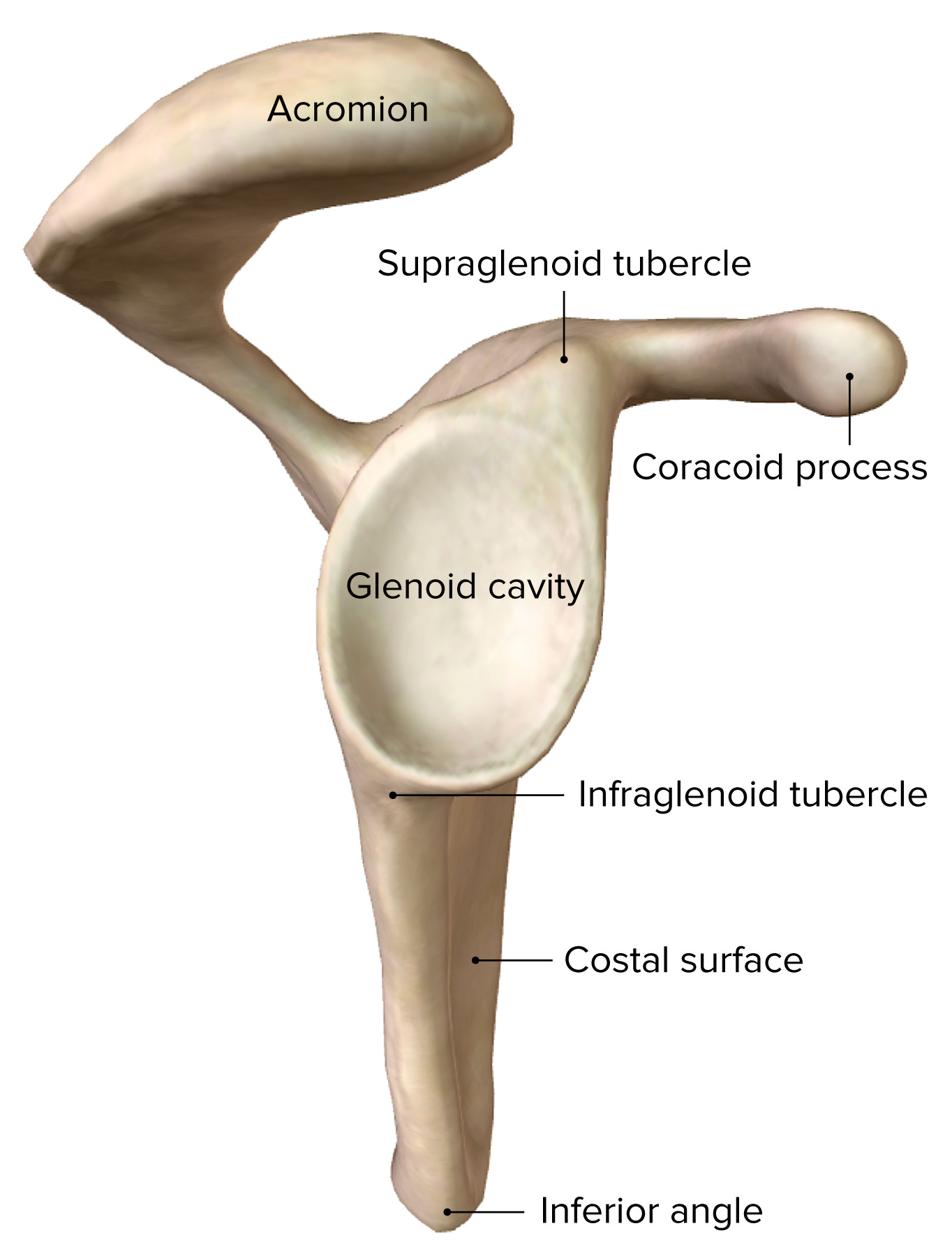
Lateral view of the right scapula
Image by BioDigital, edited by Lecturio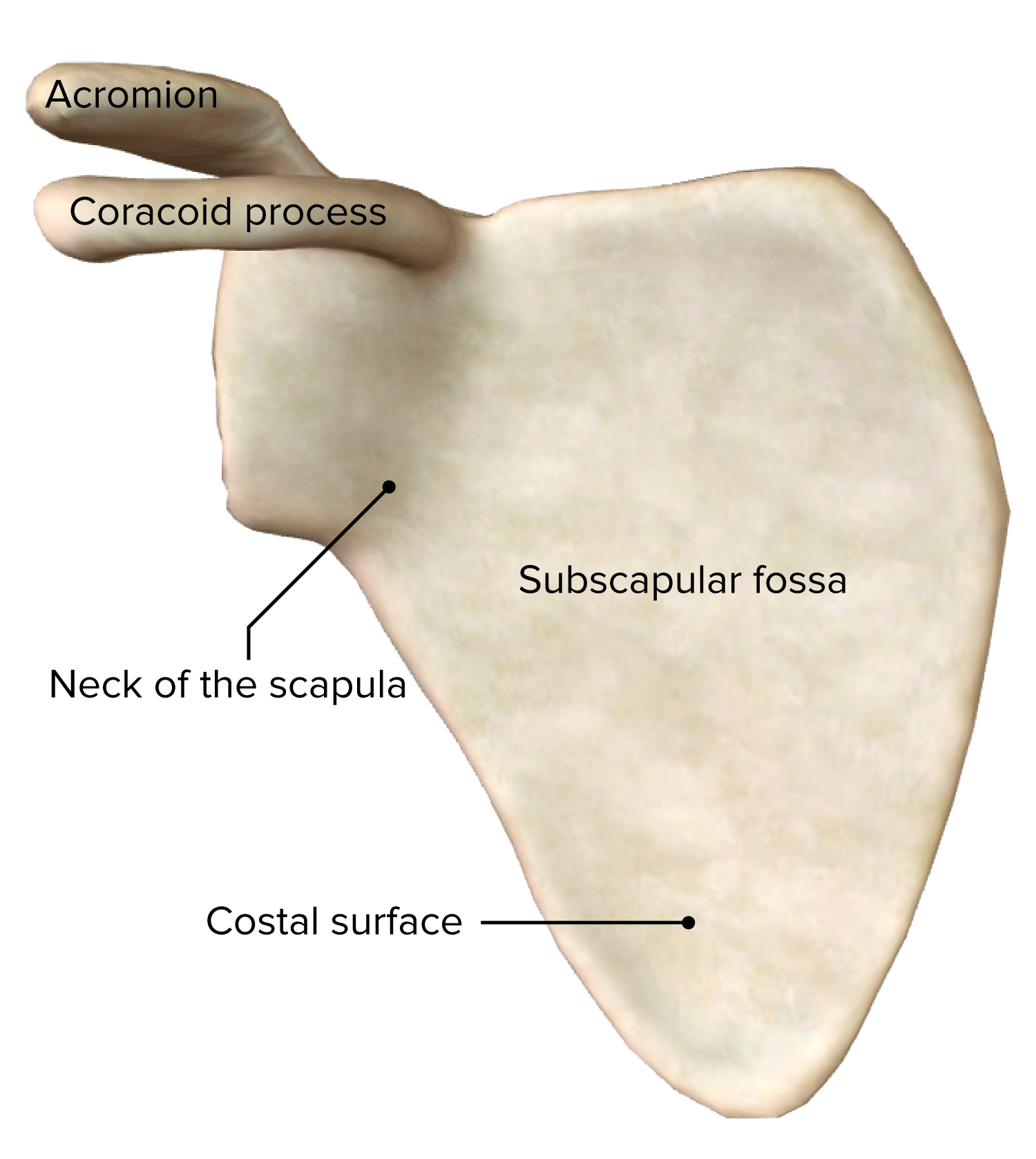
Anterior view of the right scapula
Image by BioDigital, edited by Lecturio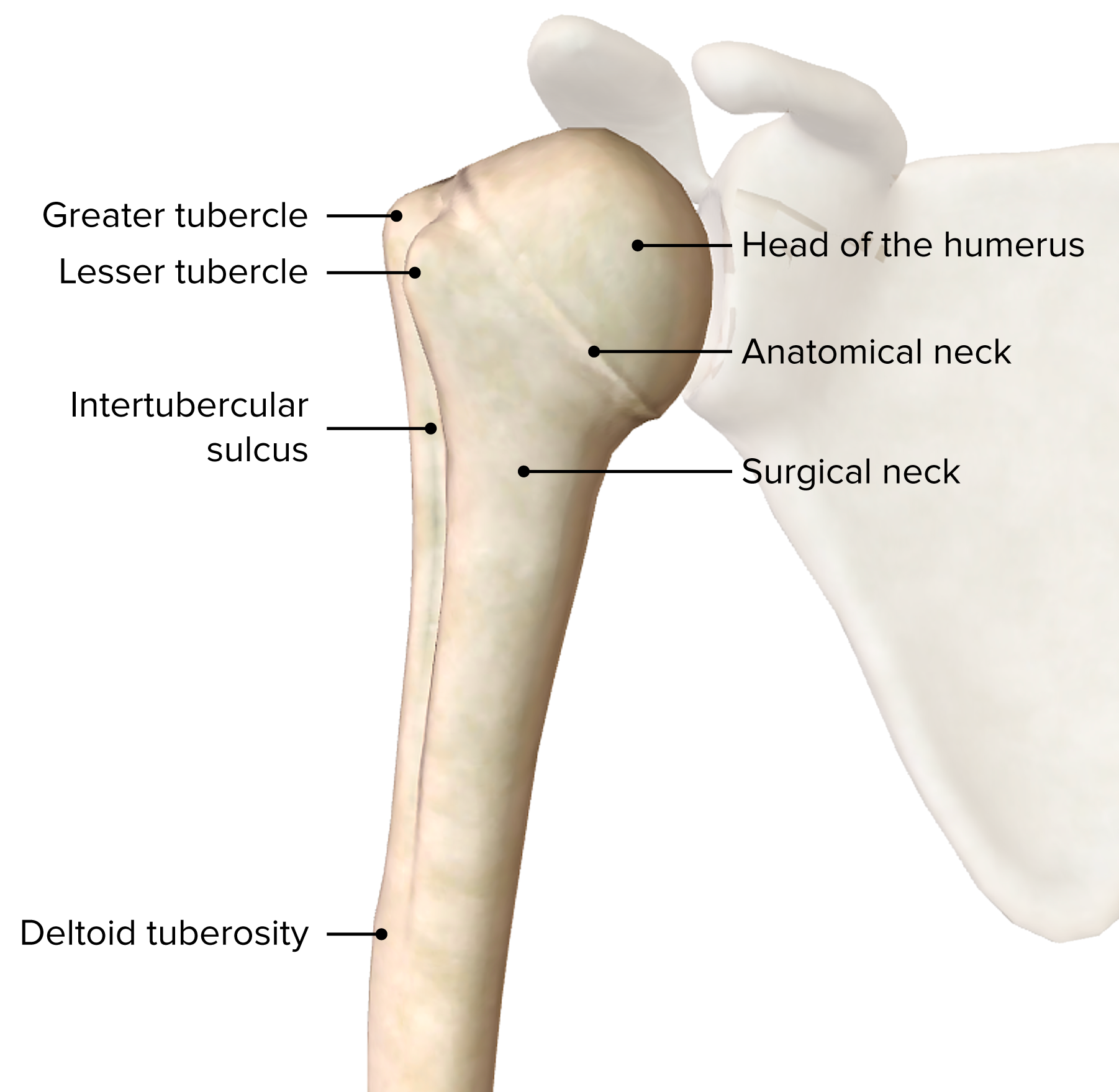
Anterior view of the proximal half of the right humerus bone and its bony landmarks
Image by BioDigital, edited by Lecturio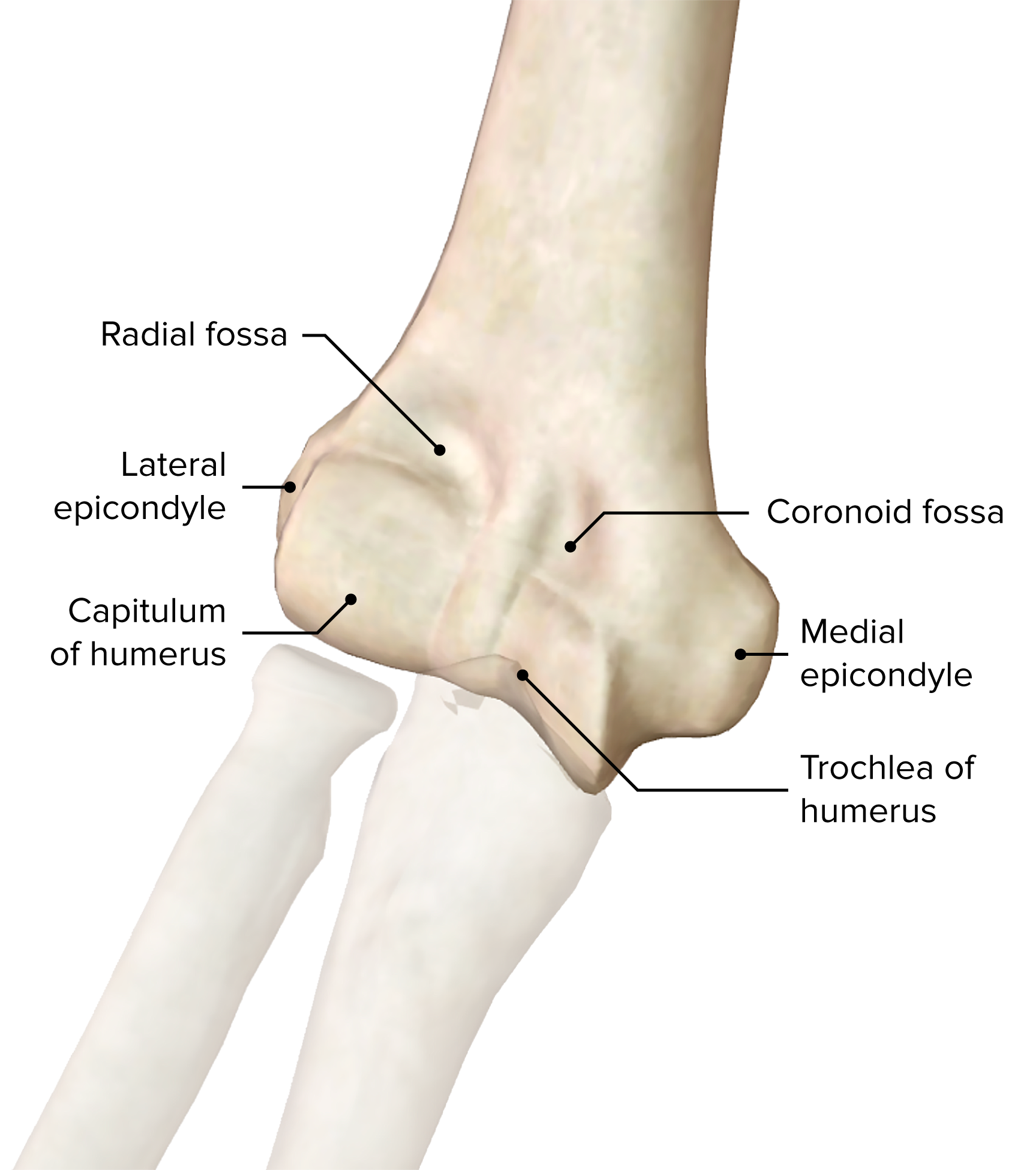
Anterior view of the distal end of the right humerus bone and its bony landmarks
Image by BioDigital, edited by Lecturio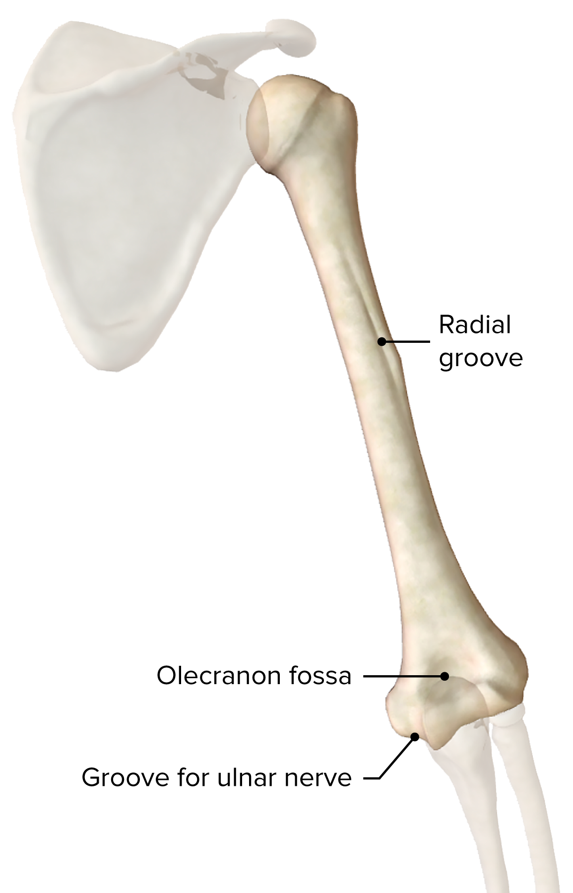
Posterior view of the right humerus bone and its bony landmarks
Image by BioDigital, edited by Lecturio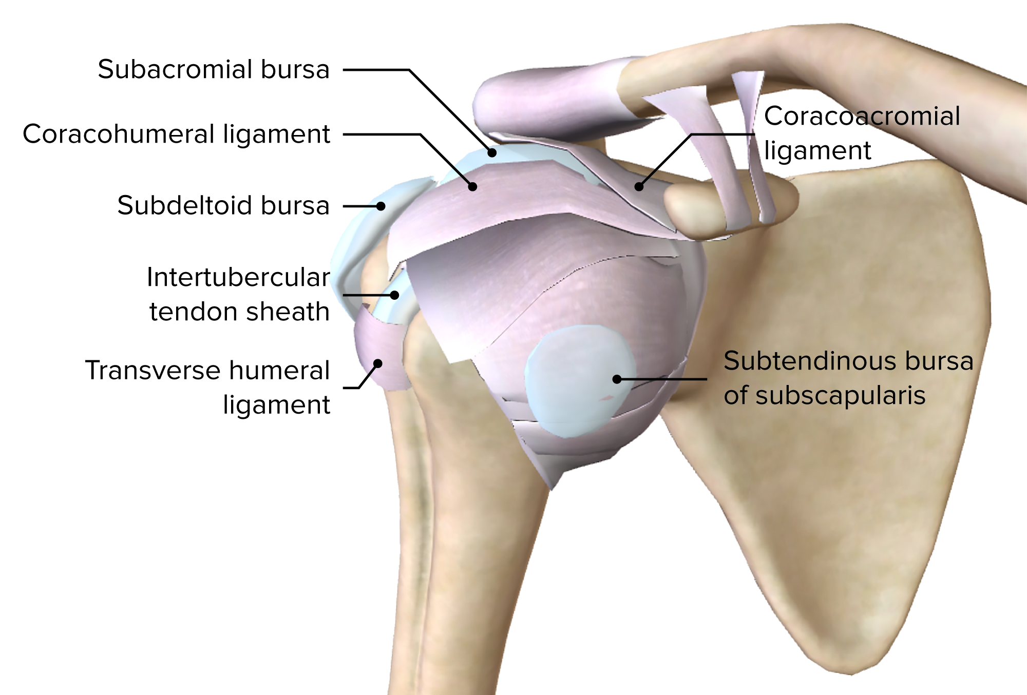
Anterior view of the superficial layer of ligaments and bursa of the right glenohumeral joint
Image by BioDigital, edited by Lecturio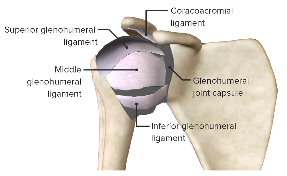
Anterior view of the deeper ligaments of the right glenohumeral joint
Image by BioDigital, edited by Lecturio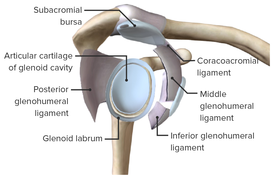
Lateral view of the right scapula, featuring the articular surfaces of the glenohumeral joint, the glenoid labrum and glenoid cavity
Image by BioDigital, edited by Lecturio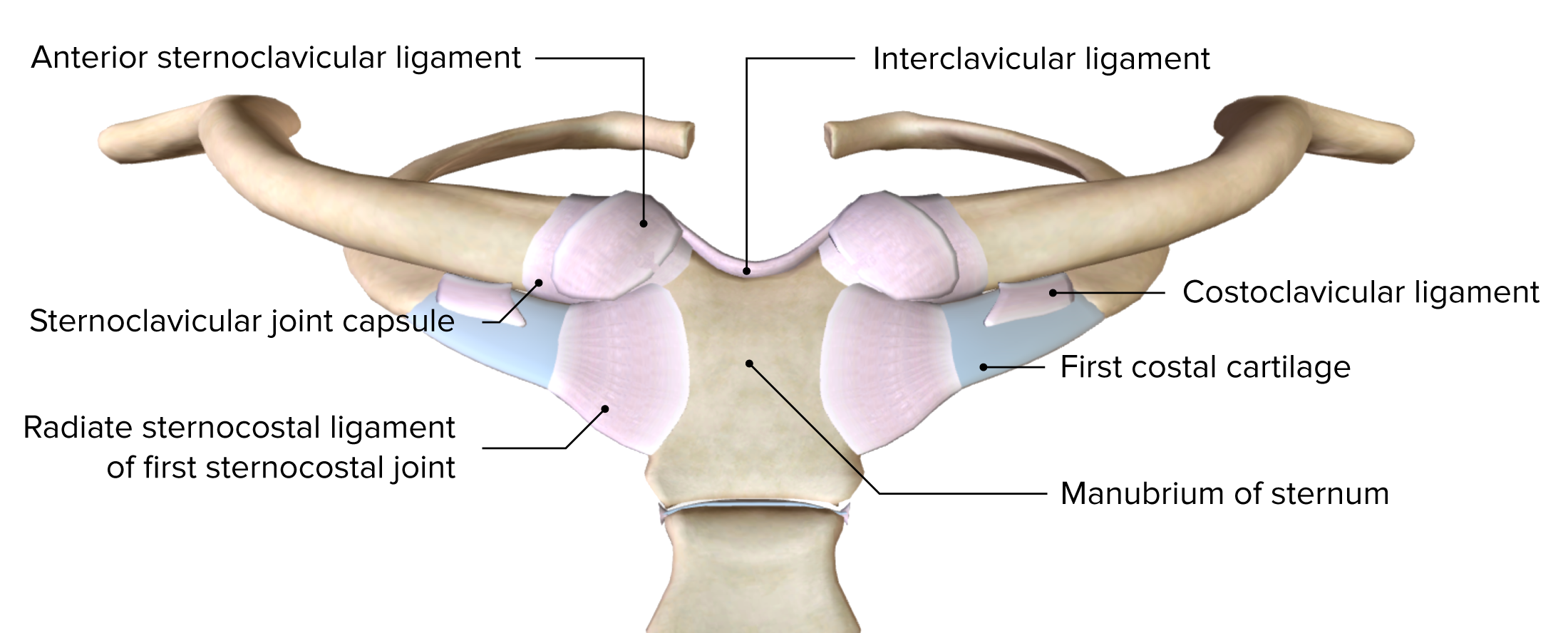
Anterior view of the sternoclavicular joint and the first sternocostal joint
Image by BioDigital, edited by Lecturio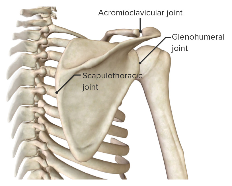
Posterior view of the scapular region, highlighting the scapulothoracic pseudo-joint
Image by BioDigital, edited by LecturioThe anterior axioappendicular muscles function to move the pectoral girdle, stabilize the clavicle Clavicle A bone on the ventral side of the shoulder girdle, which in humans is commonly called the collar bone. Clavicle Fracture, and move the upper arm Upper Arm The arm, or “upper arm” in common usage, is the region of the upper limb that extends from the shoulder to the elbow joint and connects inferiorly to the forearm through the cubital fossa. It is divided into 2 fascial compartments (anterior and posterior). Arm: Anatomy.
| Muscle | Origin | Insertion | Nerve supply | Function |
|---|---|---|---|---|
| Pectoralis major |
|
Lateral lip of intertubercular sulcus Intertubercular sulcus Arm: Anatomy | Lateral and medial pectoral nerves (C5, C6: clavicular head; C7, C8: sternocostal head) |
|
| Pectoralis minor | Ribs Ribs A set of twelve curved bones which connect to the vertebral column posteriorly, and terminate anteriorly as costal cartilage. Together, they form a protective cage around the internal thoracic organs. Chest Wall: Anatomy 3–5 | Coracoid process of scapula | Medial pectoral nerves (C8, T1) | Stabilizes scapula and pulls anteriorly and inferiorly |
| Subclavius Subclavius Muscles of the Neck: Anatomy | Rib 1 and sternum Sternum A long, narrow, and flat bone commonly known as breastbone occurring in the midsection of the anterior thoracic segment or chest region, which stabilizes the rib cage and serves as the point of origin for several muscles that move the arms, head, and neck. Chest Wall: Anatomy junction | Middle 3rd of clavicle Clavicle A bone on the ventral side of the shoulder girdle, which in humans is commonly called the collar bone. Clavicle Fracture | Subclavian nerve (C5) | Stabilizes and depresses clavicle Clavicle A bone on the ventral side of the shoulder girdle, which in humans is commonly called the collar bone. Clavicle Fracture |
| Serratus anterior | External surface of ribs Ribs A set of twelve curved bones which connect to the vertebral column posteriorly, and terminate anteriorly as costal cartilage. Together, they form a protective cage around the internal thoracic organs. Chest Wall: Anatomy 1–8 | Medial border of scapula | Long thoracic nerve Long thoracic nerve Axilla and Brachial Plexus: Anatomy (C6, C7) | Protracts scapula; holds scapula against posterior thoracic wall |
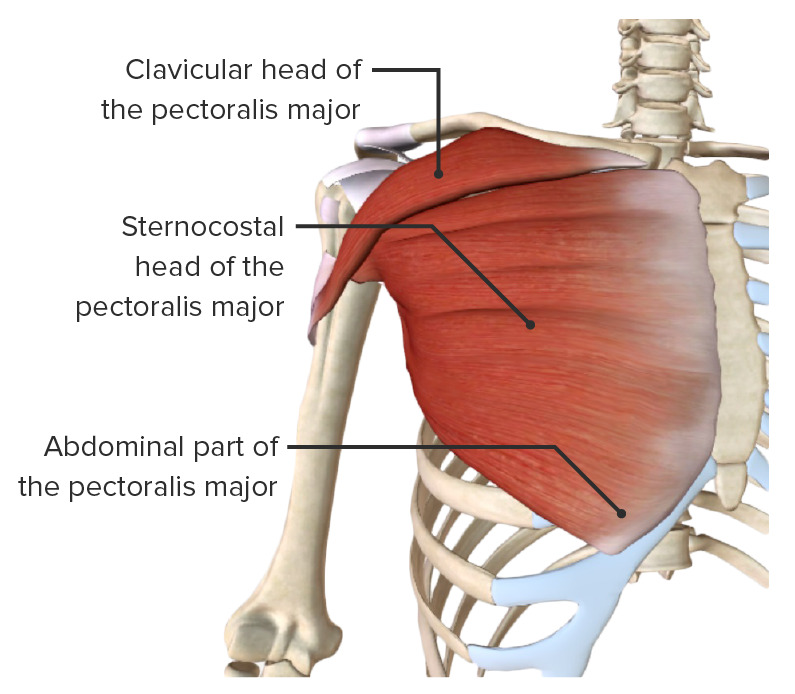
Pectoralis major muscle
Image by BioDigital, edited by Lecturio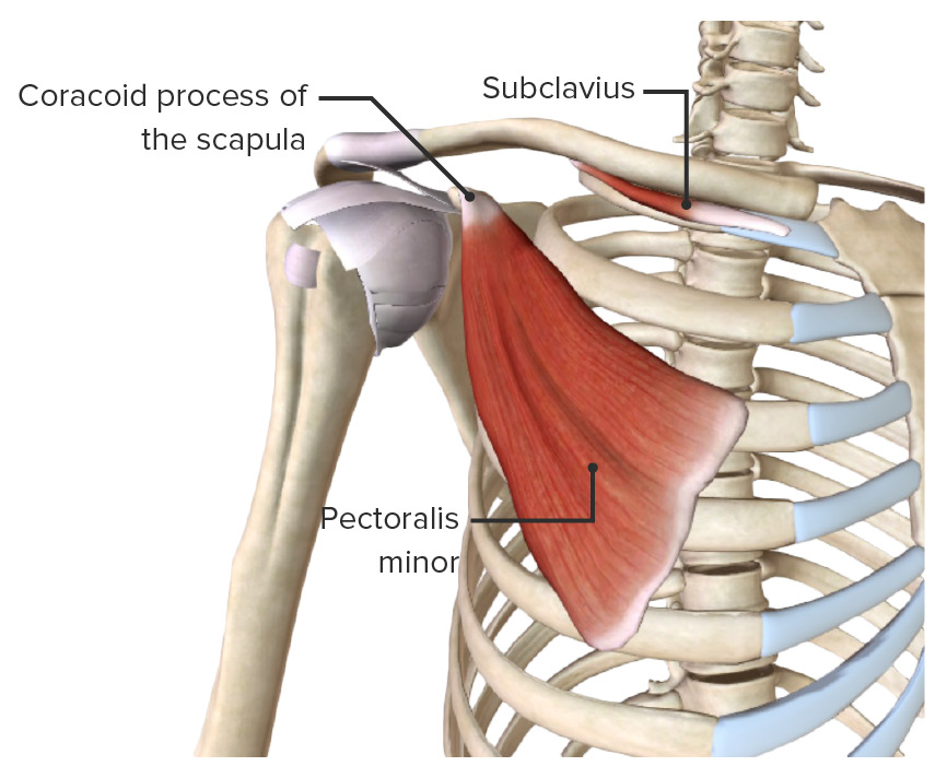
Pectoralis minor and subclavius muscles
Image by BioDigital, edited by Lecturio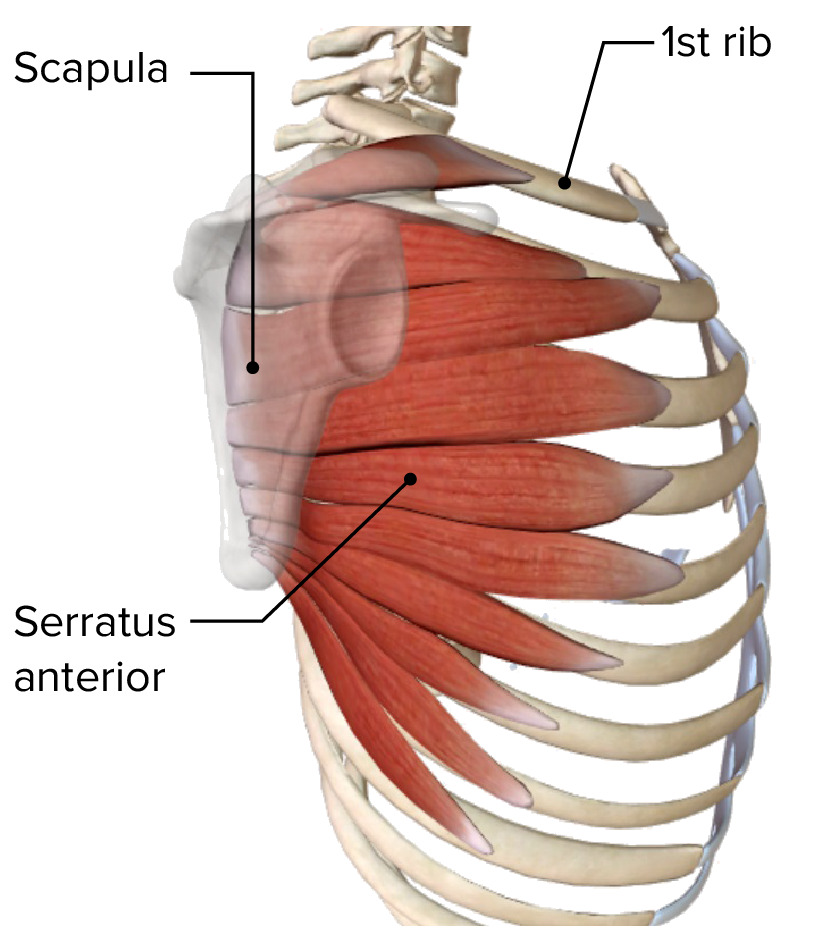
Serratus anterior muscle
Image by BioDigital, edited by LecturioThe posterior axioappendicular muscles stabilize and move the scapula and consist of a superficial and a deep layer.
| Muscle | Origin | Insertion | Nerve supply | Function |
|---|---|---|---|---|
| Trapezius |
|
Lateral 3rd of clavicle Clavicle A bone on the ventral side of the shoulder girdle, which in humans is commonly called the collar bone. Clavicle Fracture and spine Spine The human spine, or vertebral column, is the most important anatomical and functional axis of the human body. It consists of 7 cervical vertebrae, 12 thoracic vertebrae, and 5 lumbar vertebrae and is limited cranially by the skull and caudally by the sacrum. Vertebral Column: Anatomy of scapula | Spinal accessory nerve Spinal accessory nerve The 11th cranial nerve which originates from neurons in the medulla and in the cervical spinal cord. It has a cranial root, which joins the vagus nerve (10th cranial) and sends motor fibers to the muscles of the larynx, and a spinal root, which sends motor fibers to the trapezius and the sternocleidomastoid muscles. The 12 Cranial Nerves: Overview and Functions (CN XI) and C3, C4 spinal nerves Spinal nerves The 31 paired peripheral nerves formed by the union of the dorsal and ventral spinal roots from each spinal cord segment. The spinal nerve plexuses and the spinal roots are also included. Spinal Cord: Anatomy for proprioception Proprioception Sensory functions that transduce stimuli received by proprioceptive receptors in joints, tendons, muscles, and the inner ear into neural impulses to be transmitted to the central nervous system. Proprioception provides sense of stationary positions and movements of one’s body parts, and is important in maintaining kinesthesia and postural balance. Neurological Examination |
|
| Latissimus dorsi | T6–12 spinous processes, thoracolumbar fascia Thoracolumbar fascia Posterior Abdominal Wall: Anatomy and iliac crest | Floor of intertubercular groove | Thoracodorsal nerve Thoracodorsal nerve Axilla and Brachial Plexus: Anatomy (C6, C7) | Extends, adducts, and medially rotates shoulder joint Shoulder joint The articulation between the head of the humerus and the glenoid cavity of the scapula. Examination of the Upper Limbs |
| Rhomboid |
|
|
Dorsal scapula (C5) |
|
| Levator scapulae | Transverse process of C1–C4 | Superior angle of scapula | Dorsal scapula (C5) |
|
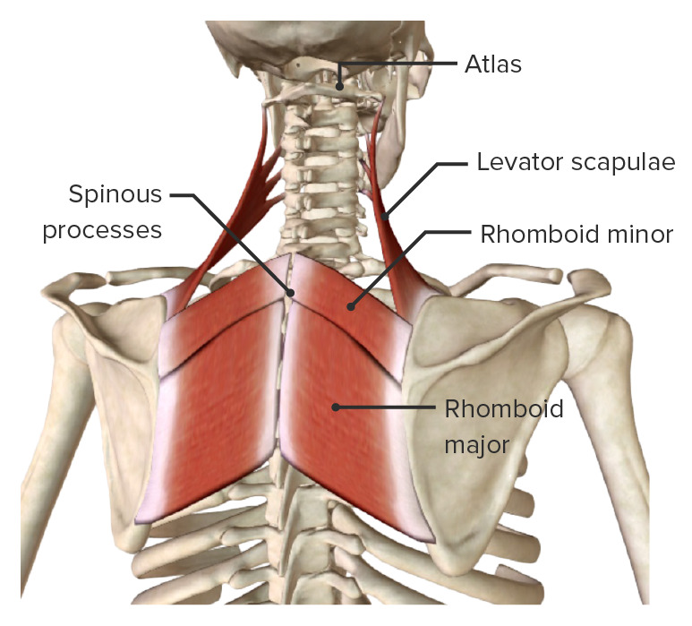
Rhomboid major, rhomboid minor, and levator scapulae muscles, the deep or intrinsic posterior axioappendicular muscles
Image by BioDigital, edited by Lecturio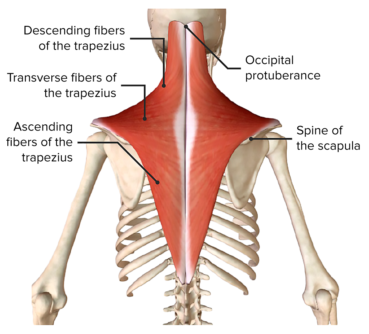
Trapezius muscle, part of the superficial or extrinsic posterior axioappendicular muscles
Image by BioDigital, edited by Lecturio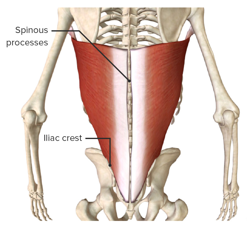
Latissimus dorsi muscle, part of the superficial or extrinsic posterior axioappendicular muscles
Image by BioDigital, edited by LecturioThe deep or intrinsic layer consists of 6 muscles:
The scapulohumeral muscles stabilize the glenohumeral joint by connecting the humerus Humerus Bone in humans and primates extending from the shoulder joint to the elbow joint. Arm: Anatomy to the scapula, including the rotator cuff muscles.
| Muscle | Origin | Insertion | Nerve supply | Function |
|---|---|---|---|---|
| Deltoid |
|
Deltoid tuberosity Deltoid tuberosity Arm: Anatomy of humerus Humerus Bone in humans and primates extending from the shoulder joint to the elbow joint. Arm: Anatomy | Axillary nerve Axillary nerve Axilla and Brachial Plexus: Anatomy (C5) |
|
| Teres major | Lateral border of scapula, inferior portion | Medial lip of intertubercular groove | Lower subscapular nerve Lower subscapular nerve Axilla and Brachial Plexus: Anatomy (C6) | Adducts and medially rotates shoulder joint Shoulder joint The articulation between the head of the humerus and the glenoid cavity of the scapula. Examination of the Upper Limbs |
| Coracobrachialis Coracobrachialis Arm: Anatomy | Coracoid process of scapula | Shaft of humerus Humerus Bone in humans and primates extending from the shoulder joint to the elbow joint. Arm: Anatomy, anteromedial | Musculocutaneous nerve Musculocutaneous Nerve A major nerve of the upper extremity. The fibers of the musculocutaneous nerve originate in the lower cervical spinal cord (usually C5 to C7), travel via the lateral cord of the brachial plexus, and supply sensory and motor innervation to the upper arm, elbow, and forearm. Axilla and Brachial Plexus: Anatomy (C5, C6, C7) | Flexes, adducts, and medially rotates the shoulder joint Shoulder joint The articulation between the head of the humerus and the glenoid cavity of the scapula. Examination of the Upper Limbs |
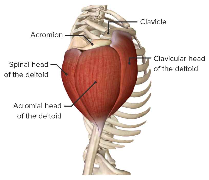
Deltoid muscle, part of the scapulohumeral muscles
Image by BioDigital, edited by Lecturio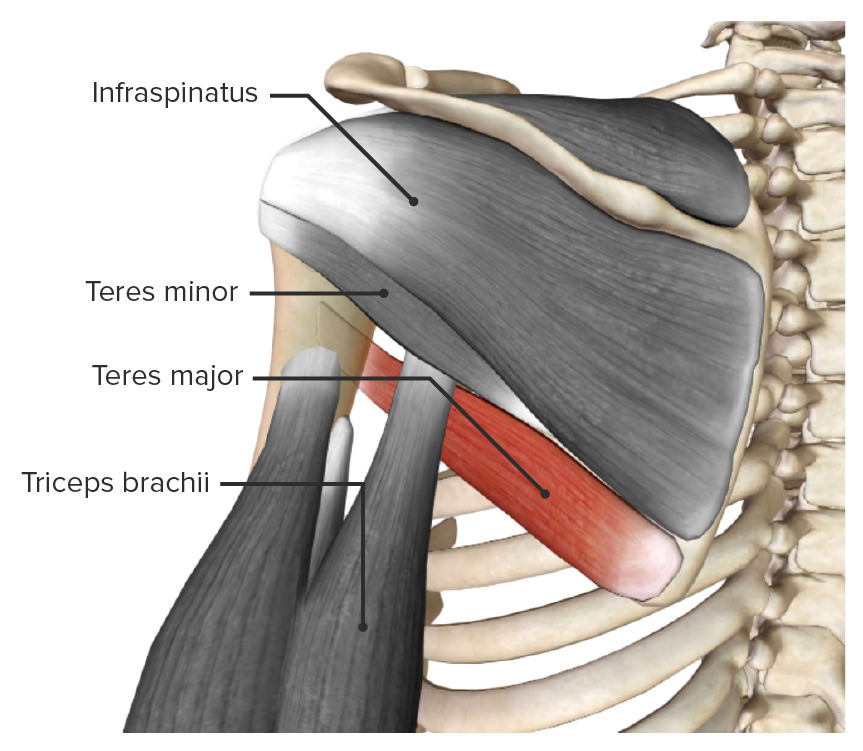
Teres major muscle (highlighted), part of the scapulohumeral muscles
Image by BioDigital, edited by Lecturio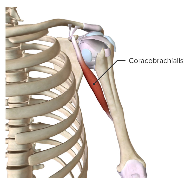
Coracobrachialis muscle (highlighted), part of the scapulohumeral muscles
Image by BioDigital, edited by LecturioRotator cuff muscles stabilize the shoulder joint Shoulder joint The articulation between the head of the humerus and the glenoid cavity of the scapula. Examination of the Upper Limbs and prevent detachment of the head of the humerus Head of The Humerus The upper rounded extremity of the humerus fitting into the glenoid cavity of the scapula. Arm: Anatomy from the glenoid cavity via “concavity compression Compression Blunt Chest Trauma,” especially during abduction Abduction Examination of the Upper Limbs of the arm Arm The arm, or “upper arm” in common usage, is the region of the upper limb that extends from the shoulder to the elbow joint and connects inferiorly to the forearm through the cubital fossa. It is divided into 2 fascial compartments (anterior and posterior). Arm: Anatomy.
Mnemonic
To recall the rotator cuff muscles, remember “SITS”:
S: Supraspinatus
I: Infraspinatus
T: Teres minor
S: Subscapularis
| Muscle | Origin | Insertion | Nerve supply | Function |
|---|---|---|---|---|
| Supraspinatus | Supraspinous fossa | Superior facet of greater tubercle of humerus Humerus Bone in humans and primates extending from the shoulder joint to the elbow joint. Arm: Anatomy | Suprascapular nerve Suprascapular nerve Axilla and Brachial Plexus: Anatomy (C5) | Initiates and assists deltoid in abduction Abduction Examination of the Upper Limbs |
| Infraspinatus | Infraspinous fossa | Middle facet of greater tubercle of humerus Humerus Bone in humans and primates extending from the shoulder joint to the elbow joint. Arm: Anatomy | Suprascapular nerve Suprascapular nerve Axilla and Brachial Plexus: Anatomy (C5) | Laterally rotates the shoulder joint Shoulder joint The articulation between the head of the humerus and the glenoid cavity of the scapula. Examination of the Upper Limbs, holds head of humerus Humerus Bone in humans and primates extending from the shoulder joint to the elbow joint. Arm: Anatomy in glenoid cavity |
| Teres minor | Lateral border of scapula, middle portion | Inferior facet of greater tubercle of humerus Humerus Bone in humans and primates extending from the shoulder joint to the elbow joint. Arm: Anatomy | Axillary nerve Axillary nerve Axilla and Brachial Plexus: Anatomy (C6) | Laterally rotates the shoulder joint Shoulder joint The articulation between the head of the humerus and the glenoid cavity of the scapula. Examination of the Upper Limbs, holds head of humerus Humerus Bone in humans and primates extending from the shoulder joint to the elbow joint. Arm: Anatomy in glenoid cavity |
| Subscapularis | Subscapular fossa | Lesser tubercle of humerus Humerus Bone in humans and primates extending from the shoulder joint to the elbow joint. Arm: Anatomy | Upper and lower subscapular nerves (C6) | Medially rotates and adducts shoulder joint Shoulder joint The articulation between the head of the humerus and the glenoid cavity of the scapula. Examination of the Upper Limbs, holds head of humerus Humerus Bone in humans and primates extending from the shoulder joint to the elbow joint. Arm: Anatomy in glenoid cavity |
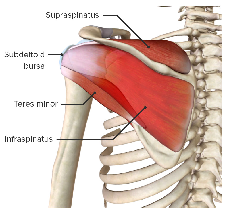
Posterior view of the scapular region and rotator cuff muscles (subscapularis missing)
Image by BioDigital, edited by Lecturio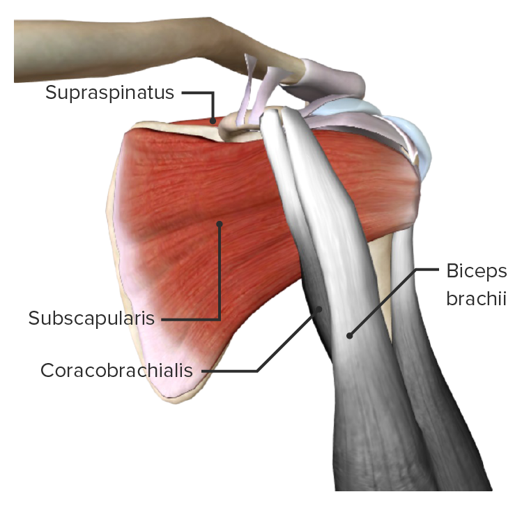
Anterior view of the scapular region, featuring the subscapularis muscle (part of the rotator cuff muscles)
Image by BioDigital, edited by Lecturio
Human right shoulder joint seen from lateral side, illustrating the scapula and humerus. Muscles shown are subscapularis muscle (shown at right), infraspinatus muscle (shown at upper left), and teres minor muscle (shown at bottom left).
Image: “Human right shoulder joint” by Young Lae, Moon M.D. Chair of 3D Based Medical Application Working group. Chairman and Professor of Orthopaedics, Chosun University Hospital, Korea. License: CC BY 3.0
Function of the supraspinatus muscle
Image: “Shoulder motion with rotator cuff” by Young Lae, Moon M.D. Chair of 3D Based Medical Application Working group. Chairman and Professor of Orthopaedics, Chosun University Hospital, Korea. License: CC BY 3.0The following conditions are common conditions associated with the shoulder: