Immunoassays are plate-based techniques that can detect and quantify many types of molecules through antibody-antigen reactions. An immunoassay typically involves an analyte, a targeted antibody, and labels. Classification of immunoassays is based on the type of label utilized, which includes enzymes (ELISA), light-emitting molecules/tracers (e.g., chemiluminescence and fluorescence immunoassays), and radioactive isotopes (radioimmunoassays). These specialized immunoassays are relatively sensitive, specific, inexpensive, and rapid, and are widely used in a clinical setting. Immunoassays are used in the diagnosis of infectious diseases, identification of tumor markers, allergy testing, and monitoring drug levels.
Immunoassays are plate-based assay techniques designed for detecting and quantifying peptides, proteinsProteinsLinear polypeptides that are synthesized on ribosomes and may be further modified, crosslinked, cleaved, or assembled into complex proteins with several subunits. The specific sequence of amino acids determines the shape the polypeptide will take, during protein folding, and the function of the protein.Energy Homeostasis, antibodiesAntibodiesImmunoglobulins (Igs), also known as antibodies, are glycoprotein molecules produced by plasma cells that act in immune responses by recognizing and binding particular antigens. The various Ig classes are IgG (the most abundant), IgM, IgE, IgD, and IgA, which differ in their biologic features, structure, target specificity, and distribution.Immunoglobulins: Types and Functions, and hormonesHormonesHormones are messenger molecules that are synthesized in one part of the body and move through the bloodstream to exert specific regulatory effects on another part of the body. Hormones play critical roles in coordinating cellular activities throughout the body in response to the constant changes in both the internal and external environments. Hormones: Overview and Types. The most crucial element of the detection strategy is a highly specific antibody-antigen reaction.
Components
Analyte: the molecule of interest (antigenAntigenSubstances that are recognized by the immune system and induce an immune reaction.Vaccination)
Antibody: carefully selected for the specific analyte
Labels:
Molecules that have the potential to conjugate with the antibody-antigen complex
Allow for detection and quantification
Types of immunoassays
The type of label defines the immunoassay being performed:
EnzymesEnzymesEnzymes are complex protein biocatalysts that accelerate chemical reactions without being consumed by them. Due to the body’s constant metabolic needs, the absence of enzymes would make life unsustainable, as reactions would occur too slowly without these molecules. Basics of Enzymes: ELISAELISAAn immunoassay utilizing an antibody labeled with an enzyme marker such as horseradish peroxidase. While either the enzyme or the antibody is bound to an immunosorbent substrate, they both retain their biologic activity; the change in enzyme activity as a result of the enzyme-antibody-antigen reaction is proportional to the concentration of the antigen and can be measured spectrophotometrically or with the naked eye. Many variations of the method have been developed.St. Louis Encephalitis Virus (enzyme-linked immunosorbent assay)
Specialized molecules/tracers (along with enzymesEnzymesEnzymes are complex protein biocatalysts that accelerate chemical reactions without being consumed by them. Due to the body’s constant metabolic needs, the absence of enzymes would make life unsustainable, as reactions would occur too slowly without these molecules. Basics of Enzymes) that have the property of light emission:
A washing step is performed in some assays to remove unbound antigenAntigenSubstances that are recognized by the immune system and induce an immune reaction.Vaccination/antibodiesAntibodiesImmunoglobulins (Igs), also known as antibodies, are glycoprotein molecules produced by plasma cells that act in immune responses by recognizing and binding particular antigens. The various Ig classes are IgG (the most abundant), IgM, IgE, IgD, and IgA, which differ in their biologic features, structure, target specificity, and distribution.Immunoglobulins: Types and Functions.
A substrateSubstrateA substance upon which the enzyme acts.Basics of Enzymes is added, which reacts with the label and results in:
Color change (ELISAELISAAn immunoassay utilizing an antibody labeled with an enzyme marker such as horseradish peroxidase. While either the enzyme or the antibody is bound to an immunosorbent substrate, they both retain their biologic activity; the change in enzyme activity as a result of the enzyme-antibody-antigen reaction is proportional to the concentration of the antigen and can be measured spectrophotometrically or with the naked eye. Many variations of the method have been developed.St. Louis Encephalitis Virus)
RadiationRadiationEmission or propagation of acoustic waves (sound), electromagnetic energy waves (such as light; radio waves; gamma rays; or x-rays), or a stream of subatomic particles (such as electrons; neutrons; protons; or alpha particles).Osteosarcoma emission (radioimmunoassay)
Changes/signals are detected to determine the presence/amount of antigenAntigenSubstances that are recognized by the immune system and induce an immune reaction.Vaccination in a sample. Detection methods may include:
Spectrophotometry (color change)
Luminometry (light emission)
Gamma counter (radiationRadiationEmission or propagation of acoustic waves (sound), electromagnetic energy waves (such as light; radio waves; gamma rays; or x-rays), or a stream of subatomic particles (such as electrons; neutrons; protons; or alpha particles).Osteosarcoma)
Variants of ELISAELISAAn immunoassay utilizing an antibody labeled with an enzyme marker such as horseradish peroxidase. While either the enzyme or the antibody is bound to an immunosorbent substrate, they both retain their biologic activity; the change in enzyme activity as a result of the enzyme-antibody-antigen reaction is proportional to the concentration of the antigen and can be measured spectrophotometrically or with the naked eye. Many variations of the method have been developed.St. Louis Encephalitis Virus
There are 4 major types of ELISAELISAAn immunoassay utilizing an antibody labeled with an enzyme marker such as horseradish peroxidase. While either the enzyme or the antibody is bound to an immunosorbent substrate, they both retain their biologic activity; the change in enzyme activity as a result of the enzyme-antibody-antigen reaction is proportional to the concentration of the antigen and can be measured spectrophotometrically or with the naked eye. Many variations of the method have been developed.St. Louis Encephalitis Virus, which are variations of the general immunoassay process:
Direct ELISAELISAAn immunoassay utilizing an antibody labeled with an enzyme marker such as horseradish peroxidase. While either the enzyme or the antibody is bound to an immunosorbent substrate, they both retain their biologic activity; the change in enzyme activity as a result of the enzyme-antibody-antigen reaction is proportional to the concentration of the antigen and can be measured spectrophotometrically or with the naked eye. Many variations of the method have been developed.St. Louis Encephalitis Virus:
The plate is coated with an antigenAntigenSubstances that are recognized by the immune system and induce an immune reaction.Vaccination.
IncubationIncubationThe amount time between exposure to an infectious agent and becoming symptomatic.Rabies Virus with a specific, labeled antibody
Indirect ELISAELISAAn immunoassay utilizing an antibody labeled with an enzyme marker such as horseradish peroxidase. While either the enzyme or the antibody is bound to an immunosorbent substrate, they both retain their biologic activity; the change in enzyme activity as a result of the enzyme-antibody-antigen reaction is proportional to the concentration of the antigen and can be measured spectrophotometrically or with the naked eye. Many variations of the method have been developed.St. Louis Encephalitis Virus:
The plate is coated with an antigenAntigenSubstances that are recognized by the immune system and induce an immune reaction.Vaccination.
IncubationIncubationThe amount time between exposure to an infectious agent and becoming symptomatic.Rabies Virus with a primary, unlabeled antibody (specific to the antigenAntigenSubstances that are recognized by the immune system and induce an immune reaction.Vaccination)
IncubationIncubationThe amount time between exposure to an infectious agent and becoming symptomatic.Rabies Virus again with a secondary, labeled antibody (specific to the primary antibody)
Sandwich ELISAELISAAn immunoassay utilizing an antibody labeled with an enzyme marker such as horseradish peroxidase. While either the enzyme or the antibody is bound to an immunosorbent substrate, they both retain their biologic activity; the change in enzyme activity as a result of the enzyme-antibody-antigen reaction is proportional to the concentration of the antigen and can be measured spectrophotometrically or with the naked eye. Many variations of the method have been developed.St. Louis Encephalitis Virus (analyte is “sandwiched” between 2 layers of antibodiesAntibodiesImmunoglobulins (Igs), also known as antibodies, are glycoprotein molecules produced by plasma cells that act in immune responses by recognizing and binding particular antigens. The various Ig classes are IgG (the most abundant), IgM, IgE, IgD, and IgA, which differ in their biologic features, structure, target specificity, and distribution.Immunoglobulins: Types and Functions):
An antibody-coated plate is used.
The analyte antigenAntigenSubstances that are recognized by the immune system and induce an immune reaction.Vaccination is added to the plate and incubated.
Incubated again with a labeled antibody
Competitive ELISAELISAAn immunoassay utilizing an antibody labeled with an enzyme marker such as horseradish peroxidase. While either the enzyme or the antibody is bound to an immunosorbent substrate, they both retain their biologic activity; the change in enzyme activity as a result of the enzyme-antibody-antigen reaction is proportional to the concentration of the antigen and can be measured spectrophotometrically or with the naked eye. Many variations of the method have been developed.St. Louis Encephalitis Virus:
An antibody-coated plate is used (an antigen-coated plate is used if testing for a specific antibody).
Sample target antigenAntigenSubstances that are recognized by the immune system and induce an immune reaction.Vaccination (or antibody) is added.
Labeled target antigenAntigenSubstances that are recognized by the immune system and induce an immune reaction.Vaccination (or antibody) is added and then washed.
Note: Unlike other ELISAELISAAn immunoassay utilizing an antibody labeled with an enzyme marker such as horseradish peroxidase. While either the enzyme or the antibody is bound to an immunosorbent substrate, they both retain their biologic activity; the change in enzyme activity as a result of the enzyme-antibody-antigen reaction is proportional to the concentration of the antigen and can be measured spectrophotometrically or with the naked eye. Many variations of the method have been developed.St. Louis Encephalitis Virus methods, less color/absence of color indicates a positive result.
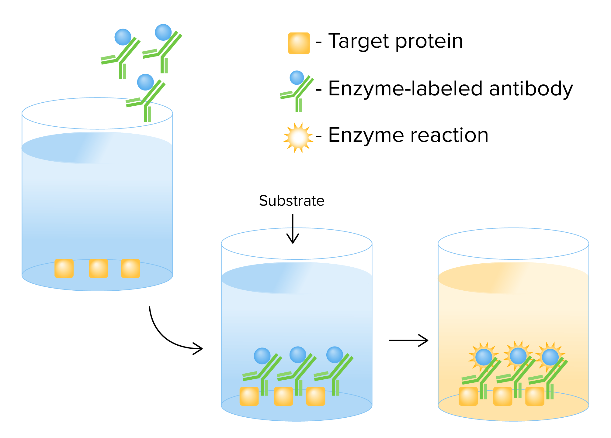
Mechanism of direct ELISA:
A target antigen is added to a plate along with enzyme-labeled antibodies specific to that antigen. After incubation, the excess unbound antibodies are removed by washing and a substrate is added. In the presence of the enzyme label, a reaction occurs that results in a color change.
Image by Lecturio.
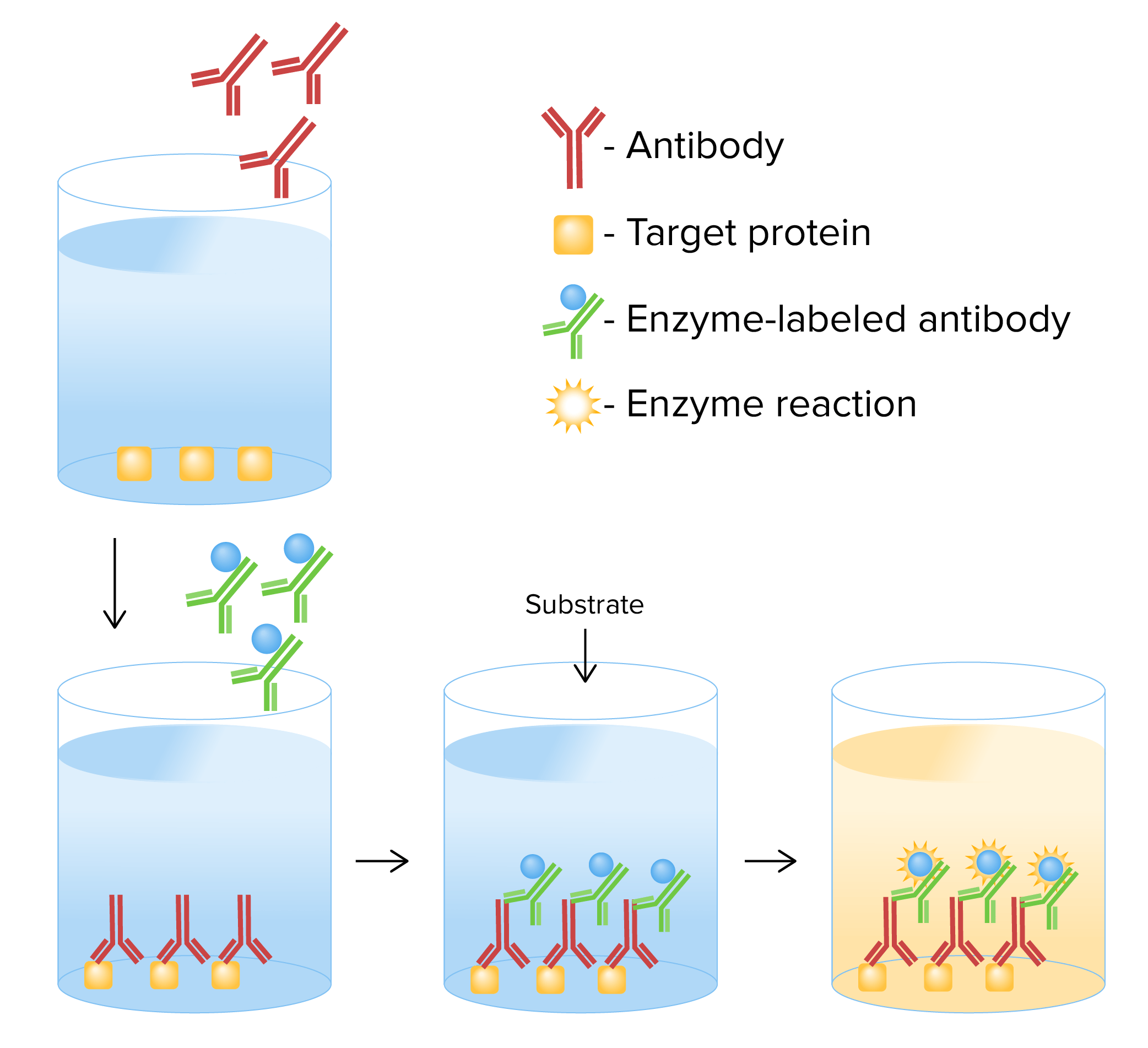
Mechanism of indirect ELISA:
A target antigen is added to a plate coated with primary antibodies. An antibody-antigen complex is formed after incubation. Excess antibody is removed by washing, and secondary enzyme-labeled antibodies are added, which bind to the antibody-antigen complex. Excess antibodies are washed away and the substrate is added. Presence of the enzyme label results in a color change.
Image by Lecturio.
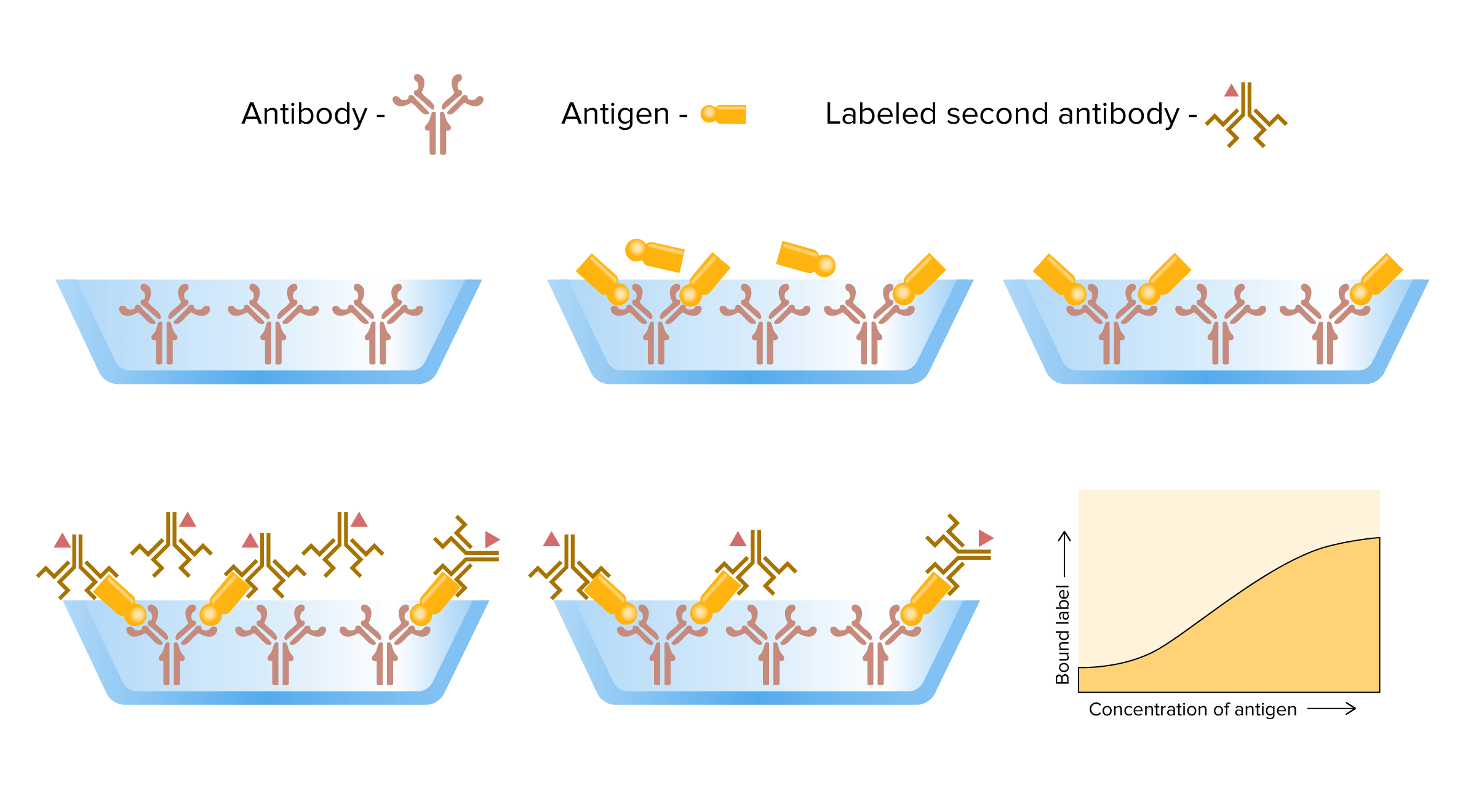
Mechanism of sandwich immunoassay: From left to right: Analyte antigen is added to an antibody-coated plate. Unbound antigens are removed by washing, and secondary enzyme-labeled antibodies are added, which bind to the antigens. The excess unbound, labeled antibodies are removed by washing. The substrate is added and the resulting color change is detected/measured.
Image by Lecturio.
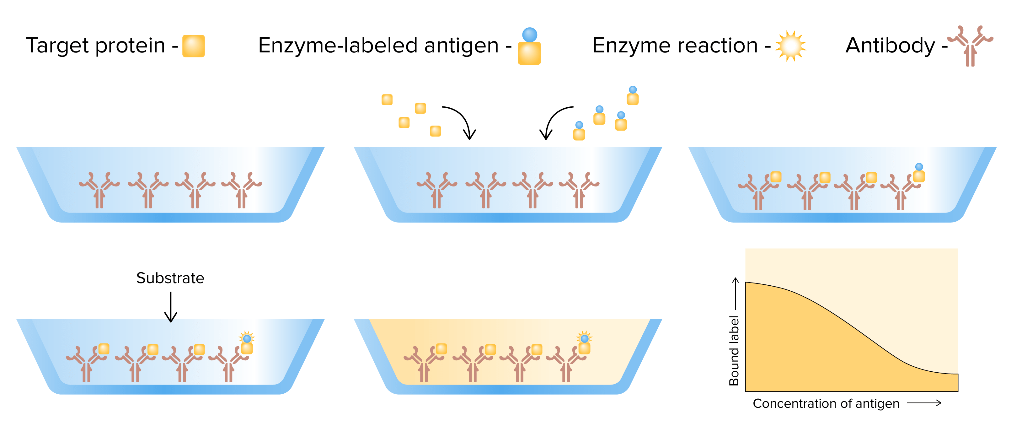
Mechanism of competitive ELISA:
From left to right: A target antigen and enzyme-labeled antigen are added to an antibody-coated plate. The antigens compete for binding to the antibodies. Next, a substrate is added and the subsequent color change is detected/measured. Unlike other forms of immunoassays, less color change indicates a higher concentration of the target antigen (as it does not contain the enzyme label).
Image by Lecturio.
Uses
Immunoassays have a wide range of clinical applications. The examples listed below are not exhaustive.
Infectious diseases
Immunoassays can be used to directly identify microorganisms (based on antigens or toxins) or indirectly assess for antibodiesAntibodiesImmunoglobulins (Igs), also known as antibodies, are glycoprotein molecules produced by plasma cells that act in immune responses by recognizing and binding particular antigens. The various Ig classes are IgG (the most abundant), IgM, IgE, IgD, and IgA, which differ in their biologic features, structure, target specificity, and distribution.Immunoglobulins: Types and Functions to the infectious agent. Some examples include:
Microorganism detection:
Legionella pneumophilaLegionella pneumophilaA species of gram-negative, aerobic bacteria that is the causative agent of legionnaires’ disease. It has been isolated from numerous environmental sites as well as from human lung tissue, respiratory secretions, and blood.Legionella/Legionellosis
Escherichia coliEscherichia coliThe gram-negative bacterium Escherichia coli is a key component of the human gut microbiota. Most strains of E. coli are avirulent, but occasionally they escape the GI tract, infecting the urinary tract and other sites. Less common strains of E. coli are able to cause disease within the GI tract, most commonly presenting as abdominal pain and diarrhea. Escherichia coli(toxin)
Clostridioides difficile (toxin)
Hepatitis BHepatitis BHepatitis B virus (HBV) is a partially double-stranded DNA virus, which belongs to the Orthohepadnavirus genus and the Hepadnaviridae family. Most individuals with acute HBV infection are asymptomatic or have mild, self-limiting symptoms. Chronic infection can be asymptomatic or create hepatic inflammation, leading to liver cirrhosis and hepatocellular carcinoma (HCC). Hepatitis B Virus
West Nile virusWest Nile VirusWest Nile virus is an enveloped, positive-sense, single-stranded RNA virus of the genus Flavivirus. Birds are the primary hosts and the disease is most often transmitted by Culex mosquitoes. Most people infected with West Nile virus are asymptomatic. Some patients develop West Nile fever (a self-limited, febrile illness) and a very small proportion of patients develop West Nile neuroinvasive disease. West Nile Virus
Hepatitis CHepatitis CHepatitis C is an infection of the liver caused by the hepatitis C virus (HCV). The infection can be transmitted through infectious blood or body fluids and may be transmitted during childbirth or through IV drug use or sexual intercourse. Hepatitis C virus can cause both acute and chronic hepatitis, ranging from a mild to a serious, lifelong illness including liver cirrhosis and hepatocellular carcinoma (HCC).Hepatitis C Virus
Borrelia burgdorferiBorrelia burgdorferiA specific species of bacteria, part of the borrelia burgdorferi group, whose common name is lyme disease spirochete.Borrelia
Treponema pallidumTreponema pallidumThe causative agent of venereal and non-venereal syphilis as well as yaws.Treponema
Cancer detection and monitoring
Immunoassays can be utilized to detect tumorTumorInflammation markers. Examples include:
PSAPSAA glycoprotein that is a kallikrein-like serine proteinase and an esterase, produced by epithelial cells of both normal and malignant prostate tissue. It is an important marker for the diagnosis of prostate cancer.Prostate Cancer
Carcinoembryonic antigenCarcinoembryonic antigenA glycoprotein that is secreted into the luminal surface of the epithelia in the gastrointestinal tract. It is found in the feces and pancreaticobiliary secretions and is used to monitor the response to colon cancer treatment.Serum Tumor Markers (CEACEAA glycoprotein that is secreted into the luminal surface of the epithelia in the gastrointestinal tract. It is found in the feces and pancreaticobiliary secretions and is used to monitor the response to colon cancer treatment.Serum Tumor Markers)
Immunoassays can be used to detect IgE antibodiesIgE antibodiesAn immunoglobulin associated with mast cells. Overexpression has been associated with allergic hypersensitivity.Type I Hypersensitivity Reaction to specific allergens:
Positive result:
Indicates sensitization to that allergen
Does not necessarily indicate whether that individual will have a clinical response upon exposure to that antigenAntigenSubstances that are recognized by the immune system and induce an immune reaction.Vaccination
Negative result:
Suggests no allergyAllergyAn abnormal adaptive immune response that may or may not involve antigen-specific IgEType I Hypersensitivity Reaction to the antigenAntigenSubstances that are recognized by the immune system and induce an immune reaction.Vaccination
Does not completely exclude allergyAllergyAn abnormal adaptive immune response that may or may not involve antigen-specific IgEType I Hypersensitivity Reaction to the antigenAntigenSubstances that are recognized by the immune system and induce an immune reaction.Vaccination (particularly, if there is a suggestive clinical history)
Medications
Therapeutic drug monitoring is an important application of immunoassays. Examples include monitoring the drug levels of:
DigoxinDigoxinA cardiotonic glycoside obtained mainly from digitalis lanata; it consists of three sugars and the aglycone digoxigenin. Digoxin has positive inotropic and negative chronotropic activity. It is used to control ventricular rate in atrial fibrillation and in the management of congestive heart failure with atrial fibrillation. Its use in congestive heart failure and sinus rhythm is less certain. The margin between toxic and therapeutic doses is small.Cardiac Glycosides
TheophyllineTheophyllineA methyl xanthine derivative from tea with diuretic, smooth muscle relaxant, bronchial dilation, cardiac and central nervous system stimulant activities. Theophylline inhibits the 3.Asthma Drugs
ImmunosuppressantsImmunosuppressantsImmunosuppressants are a class of drugs widely used in the management of autoimmune conditions and organ transplant rejection. The general effect is dampening of the immune response.Immunosuppressants (e.g., mycophenolic acid, cyclosporineCyclosporineA cyclic undecapeptide from an extract of soil fungi. It is a powerful immunosupressant with a specific action on T-lymphocytes. It is used for the prophylaxis of graft rejection in organ and tissue transplantation.Immunosuppressants)
Antibiotics (e.g., amikacin, vancomycinVancomycinAntibacterial obtained from streptomyces orientalis. It is a glycopeptide related to ristocetin that inhibits bacterial cell wall assembly and is toxic to kidneys and the inner ear.Glycopeptides, gentamicinGentamicinAminoglycosides)
Other diagnostic uses
Additional laboratory tests where immunoassays may be utilized include:
High-sensitivity troponin ITroponin IA troponin complex subunit that inhibits actomyosin ATPase activity thereby disrupting actin and myosin interaction. There are three troponin I subtypes: troponin i1, i2 and i3. Troponin i3 is cardiac-specific whereas troponin i1 and i2 are skeletal subtypes. Troponin i3 is a biomarker for damaged or injured cardiac myocytes and mutations in troponin i3 gene are associated with familial hypertrophic cardiomyopathy.Myocardial Infarction
ThyroidThyroidThe thyroid gland is one of the largest endocrine glands in the human body. The thyroid gland is a highly vascular, brownish-red gland located in the visceral compartment of the anterior region of the neck.Thyroid Gland: Anatomy function:
Thyroid-stimulating hormoneThyroid-stimulating hormoneA glycoprotein hormone secreted by the adenohypophysis. Thyrotropin stimulates thyroid gland by increasing the iodide transport, synthesis and release of thyroid hormones (thyroxine and triiodothyronine).Thyroid Hormones (TSH)
Free thyroxineThyroxineThe major hormone derived from the thyroid gland. Thyroxine is synthesized via the iodination of tyrosines (monoiodotyrosine) and the coupling of iodotyrosines (diiodotyrosine) in the thyroglobulin. Thyroxine is released from thyroglobulin by proteolysis and secreted into the blood.Thyroid Hormones (T4T4The major hormone derived from the thyroid gland. Thyroxine is synthesized via the iodination of tyrosines (monoiodotyrosine) and the coupling of iodotyrosines (diiodotyrosine) in the thyroglobulin. Thyroxine is released from thyroglobulin by proteolysis and secreted into the blood. Thyroxine is peripherally deiodinated to form triiodothyronine which exerts a broad spectrum of stimulatory effects on cell metabolism.Thyroid Hormones) and triiodothyroonine (T3T3A T3 thyroid hormone normally synthesized and secreted by the thyroid gland in much smaller quantities than thyroxine (T4). Most T3 is derived from peripheral monodeiodination of T4 at the 5′ position of the outer ring of the iodothyronine nucleus. The hormone finally delivered and used by the tissues is mainly t3.Thyroid Hormones)
Nutritional:
FolateFolateFolate and vitamin B12 are 2 of the most clinically important water-soluble vitamins. Deficiencies can present with megaloblastic anemia, GI symptoms, neuropsychiatric symptoms, and adverse pregnancy complications, including neural tube defects. Folate and Vitamin B12
Vitamin B12Vitamin B12A cobalt-containing coordination compound produced by intestinal microorganisms and found also in soil and water. Higher plants do not concentrate vitamin B 12 from the soil and so are a poor source of the substance as compared with animal tissues. Intrinsic factor is important for the assimilation of vitamin B 12.Folate and Vitamin B12
FerritinFerritinIron-containing proteins that are widely distributed in animals, plants, and microorganisms. Their major function is to store iron in a nontoxic bioavailable form. Each ferritin molecule consists of ferric iron in a hollow protein shell (apoferritins) made of 24 subunits of various sequences depending on the species and tissue types.Hereditary Hemochromatosis
Vitamin DVitamin DA vitamin that includes both cholecalciferols and ergocalciferols, which have the common effect of preventing or curing rickets in animals. It can also be viewed as a hormone since it can be formed in skin by action of ultraviolet rays upon the precursors, 7-dehydrocholesterol and ergosterol, and acts on vitamin D receptors to regulate calcium in opposition to parathyroid hormone.Fat-soluble Vitamins and their Deficiencies
Advantages and Errors
Advantages
Advantages depend on the type of immunoassay used; however, generally, they are:
Inexpensive
Sensitive
Specific
Relatively rapid and convenient
Sources of errorErrorRefers to any act of commission (doing something wrong) or omission (failing to do something right) that exposes patients to potentially hazardous situations.Disclosure of Information
Cross-reactivity (reagent antibodiesAntibodiesImmunoglobulins (Igs), also known as antibodies, are glycoprotein molecules produced by plasma cells that act in immune responses by recognizing and binding particular antigens. The various Ig classes are IgG (the most abundant), IgM, IgE, IgD, and IgA, which differ in their biologic features, structure, target specificity, and distribution.Immunoglobulins: Types and FunctionsbindBINDHyperbilirubinemia of the Newborn to molecules that are similar to the analyte → false positiveFalse positiveAn FP test result indicates that a person has the disease when they do not.Epidemiological Values of Diagnostic Tests/falsely elevated results)
AutoantibodiesAutoantibodiesAntibodies that react with self-antigens (autoantigens) of the organism that produced them.Blotting Techniques (endogenous antibodiesAntibodiesImmunoglobulins (Igs), also known as antibodies, are glycoprotein molecules produced by plasma cells that act in immune responses by recognizing and binding particular antigens. The various Ig classes are IgG (the most abundant), IgM, IgE, IgD, and IgA, which differ in their biologic features, structure, target specificity, and distribution.Immunoglobulins: Types and Functions, instead of reagent antibodiesAntibodiesImmunoglobulins (Igs), also known as antibodies, are glycoprotein molecules produced by plasma cells that act in immune responses by recognizing and binding particular antigens. The various Ig classes are IgG (the most abundant), IgM, IgE, IgD, and IgA, which differ in their biologic features, structure, target specificity, and distribution.Immunoglobulins: Types and Functions) bindBINDHyperbilirubinemia of the Newborn to the analyte → false negativeFalse negativeAn FN test result indicates a person does not have the disease when, in fact, they do.Epidemiological Values of Diagnostic Tests/falsely low results)
Antireagent antibodiesAntibodiesImmunoglobulins (Igs), also known as antibodies, are glycoprotein molecules produced by plasma cells that act in immune responses by recognizing and binding particular antigens. The various Ig classes are IgG (the most abundant), IgM, IgE, IgD, and IgA, which differ in their biologic features, structure, target specificity, and distribution.Immunoglobulins: Types and Functions (endogenous antibodiesAntibodiesImmunoglobulins (Igs), also known as antibodies, are glycoprotein molecules produced by plasma cells that act in immune responses by recognizing and binding particular antigens. The various Ig classes are IgG (the most abundant), IgM, IgE, IgD, and IgA, which differ in their biologic features, structure, target specificity, and distribution.Immunoglobulins: Types and FunctionsbindBINDHyperbilirubinemia of the Newborn to reagent antibodiesAntibodiesImmunoglobulins (Igs), also known as antibodies, are glycoprotein molecules produced by plasma cells that act in immune responses by recognizing and binding particular antigens. The various Ig classes are IgG (the most abundant), IgM, IgE, IgD, and IgA, which differ in their biologic features, structure, target specificity, and distribution.Immunoglobulins: Types and Functions → false positiveFalse positiveAn FP test result indicates that a person has the disease when they do not.Epidemiological Values of Diagnostic Tests/falsely elevated results)
References
Aydin, S. (2015). A short history, principles, and types of ELISA, and our laboratory experience with peptide/protein analyses using ELISA. Peptides, 72, 4–15. https://doi.org/10.1016/j.peptides.2015.04.012
Shah, K., & Maghsoudlou, P. (2016). Enzyme-linked immunosorbent assay (ELISA): The basics. British Journal of Hospital Medicine, 77(7), C98–C101. https://doi.org/10.12968/hmed.2016.77.7.C98
Konstantinou, G. N. (2017). Enzyme-linked immunosorbent assay (ELISA). In G. K. Makowski (Ed.), Methods in molecular biology (Vol. 1592, pp. 79–94). Humana Press. https://doi.org/10.1007/978-1-4939-6925-8_7
Kohl, T. O., & Ascoli, C. A. (2017). Direct competitive enzyme-linked immunosorbent assay (ELISA). Cold Spring Harbor Protocols, 2017(7), pdb.prot093740. https://doi.org/10.1101/pdb.prot093740
Kohl, T. O., & Ascoli, C. A. (2017). Indirect immunometric ELISA. Cold Spring Harbor Protocols, 2017(5), pdb.prot093708. https://doi.org/10.1101/pdb.prot093708
Kohl, T. O., & Ascoli, C. A. (2017). Immunometric double-antibody sandwich enzyme-linked immunosorbent assay. Cold Spring Harbor Protocols, 2017(6), pdb.prot093724. https://doi.org/10.1101/pdb.prot093724
Yin, Y., Cao, Y., Xu, Y., & Li, G. (2010). Colorimetric immunoassay for detection of tumor markers. International Journal of Molecular Sciences, 11(12), 5077–5094. https://doi.org/10.3390/ijms11125077
Hoofnagle, A. N., & Wener, M. H. (2009). The fundamental flaws of immunoassays and potential solutions using tandem mass spectrometry. Journal of Immunological Methods, 347(1–2), 3–11. https://doi.org/10.1016/j.jim.2009.06.003
Darwish, I. A. (2006). Immunoassay methods and their applications in pharmaceutical analysis: Basic methodology and recent advances. International Journal of Biomedical Science, 2(3), 217–235. PMID: 23674985.
Create your free account or log in to continue reading!