Elbow dislocation is the displacement of the radius and the ulna relative to the humerus. The most common mechanism of injury is falling on an outstretched hand Hand The hand constitutes the distal part of the upper limb and provides the fine, precise movements needed in activities of daily living. It consists of 5 metacarpal bones and 14 phalanges, as well as numerous muscles innervated by the median and ulnar nerves. Hand: Anatomy. Elbow dislocation presents with joint swelling Swelling Inflammation, pain Pain An unpleasant sensation induced by noxious stimuli which are detected by nerve endings of nociceptive neurons. Pain: Types and Pathways, and restricted range of motion Range of motion The distance and direction to which a bone joint can be extended. Range of motion is a function of the condition of the joints, muscles, and connective tissues involved. Joint flexibility can be improved through appropriate muscle strength exercises. Examination of the Upper Limbs. Dislocation of the elbow can be classified into simple or complex depending on the absence or presence, respectively, of a concomitant fracture Fracture A fracture is a disruption of the cortex of any bone and periosteum and is commonly due to mechanical stress after an injury or accident. Open fractures due to trauma can be a medical emergency. Fractures are frequently associated with automobile accidents, workplace injuries, and trauma. Overview of Bone Fractures. Dislocations are diagnosed with an X-ray X-ray Penetrating electromagnetic radiation emitted when the inner orbital electrons of an atom are excited and release radiant energy. X-ray wavelengths range from 1 pm to 10 nm. Hard x-rays are the higher energy, shorter wavelength x-rays. Soft x-rays or grenz rays are less energetic and longer in wavelength. The short wavelength end of the x-ray spectrum overlaps the gamma rays wavelength range. The distinction between gamma rays and x-rays is based on their radiation source. Pulmonary Function Tests. Management depends on whether the dislocation is simple or complex. Simple dislocations are managed with joint immobilization Immobilization Delirium and casting, while complex dislocations require open reduction and internal fixation. Elbow dislocations can be complicated by joint contractures Contractures Prolonged shortening of the muscle or other soft tissue around a joint, preventing movement of the joint. Wound Healing, which are due to fibrotic changes in the joint capsule Capsule An envelope of loose gel surrounding a bacterial cell which is associated with the virulence of pathogenic bacteria. Some capsules have a well-defined border, whereas others form a slime layer that trails off into the medium. Most capsules consist of relatively simple polysaccharides but there are some bacteria whose capsules are made of polypeptides. Bacteroides, or permanent joint instability, which are due to loose ligaments.
Last updated: Dec 26, 2025
Elbow dislocation is the displacement Displacement The process by which an emotional or behavioral response that is appropriate for one situation appears in another situation for which it is inappropriate. Defense Mechanisms of the radius Radius The outer shorter of the two bones of the forearm, lying parallel to the ulna and partially revolving around it. Forearm: Anatomy and the ulna Ulna The inner and longer bone of the forearm. Forearm: Anatomy relative to the humerus Humerus Bone in humans and primates extending from the shoulder joint to the elbow joint. Arm: Anatomy.
The elbow is a synovial hinge joint between the upper arm Upper Arm The arm, or “upper arm” in common usage, is the region of the upper limb that extends from the shoulder to the elbow joint and connects inferiorly to the forearm through the cubital fossa. It is divided into 2 fascial compartments (anterior and posterior). Arm: Anatomy and the forearm Forearm The forearm is the region of the upper limb between the elbow and the wrist. The term “forearm” is used in anatomy to distinguish this area from the arm, a term that is commonly used to describe the entire upper limb. The forearm consists of 2 long bones (the radius and the ulna), the interosseous membrane, and multiple arteries, nerves, and muscles. Forearm: Anatomy.
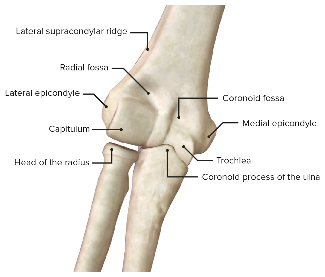
Anterior view of the elbow (humeroulnar joint), featuring the articular surfaces
Image by BioDigital, edited by Lecturio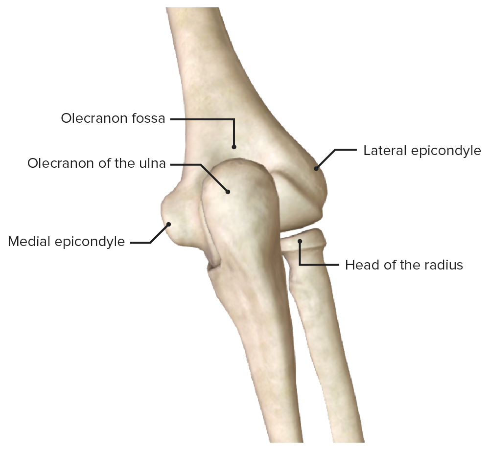
Posterior view of the elbow (humeroulnar joint), featuring the olecranon:
Trochlea and olecranon fossa maintain anterior-posterior stability in extension while coronoid fossa, radiocapitellar joint and biceps-triceps-brachialis maintain anterior-posterior stability in flexion.
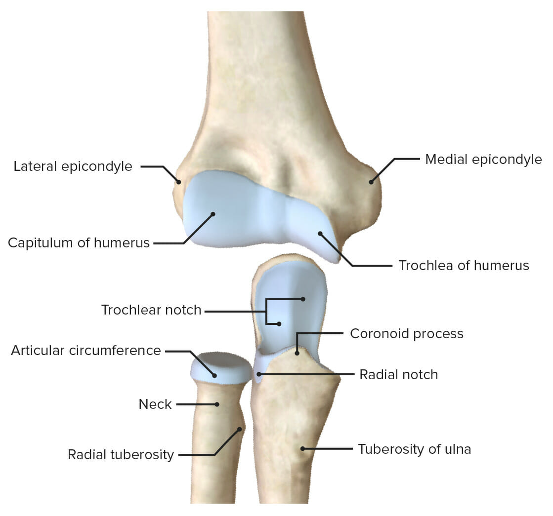
Articular surfaces of the elbow joint
Image by BioDigital, edited by Lecturio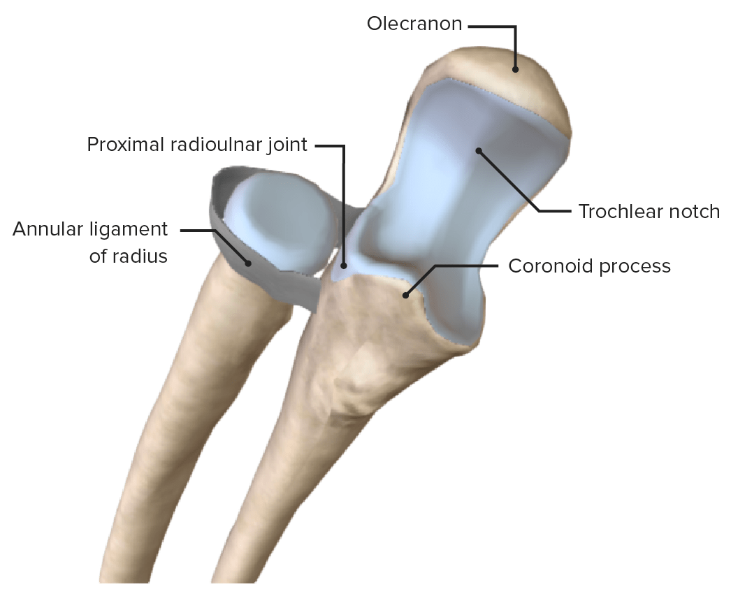
Proximal radioulnar joint, featuring its main supporting ligament, the annular ligament of the radius
Image by BioDigital, edited by Lecturio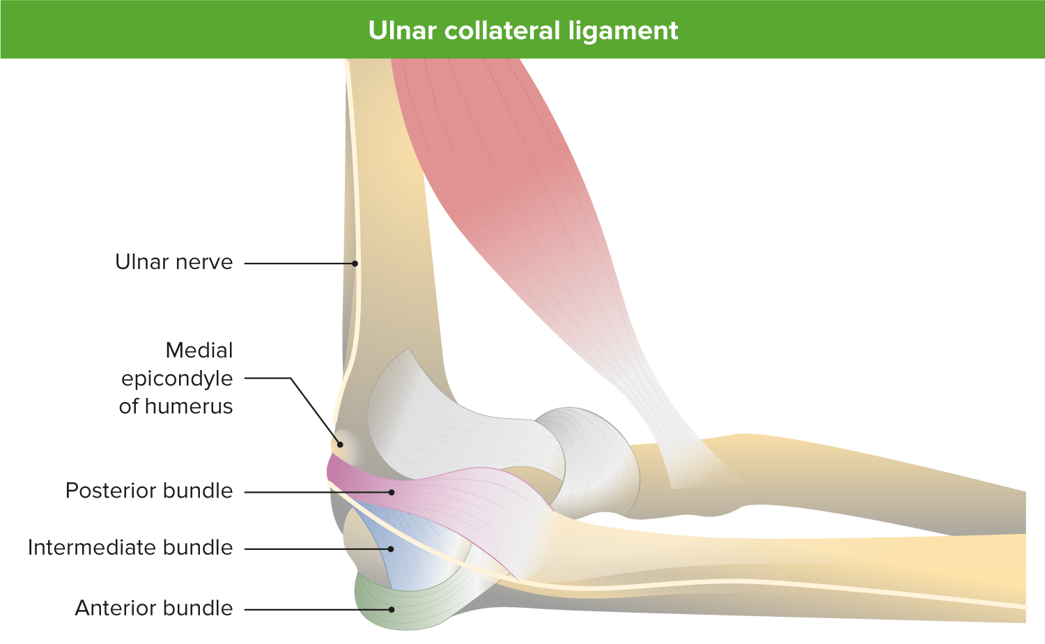
Medial view of the elbow illustrating the 3 bands of the medial ulnar collateral ligament: The anterior bundle is the primary static stabilizer against valgus stress in flexion and extension.
Image by Lecturio.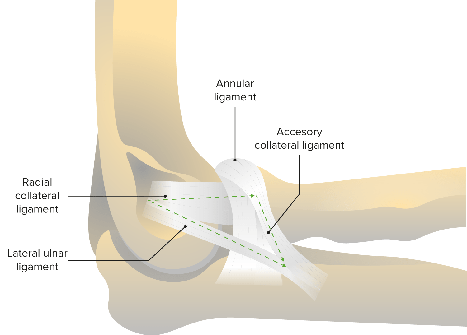
Lateral view of the elbow joint, featuring the annular ligament and radial collateral ligament: The radial collateral ligament is the primary static stabilizer against valgus stress.
Image by Lecturio.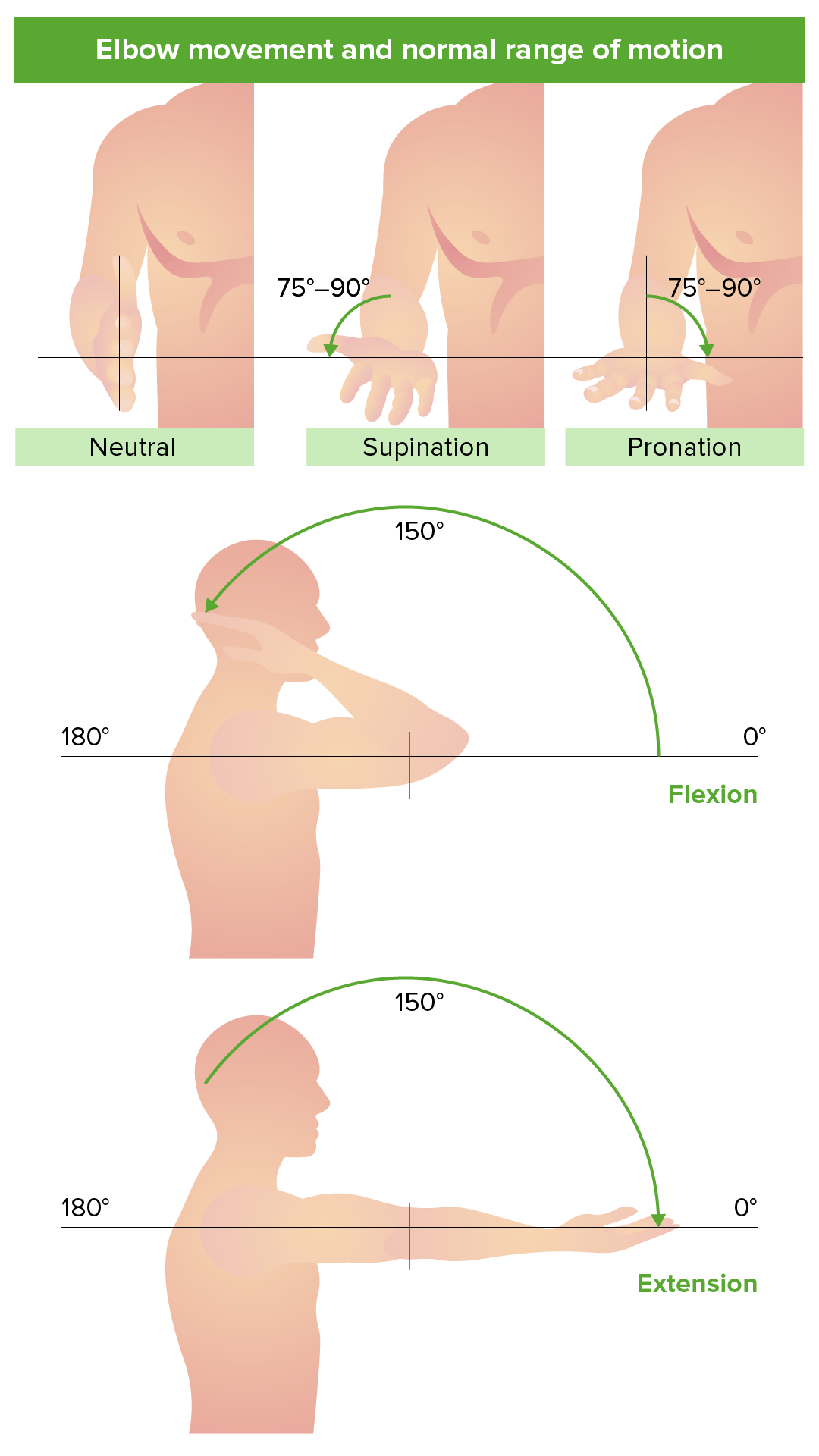
ange of motion and movements of the elbow joint
Image by Lecturio.Trauma is the most common cause, and the direction of the force affecting the elbow joint Elbow joint The elbow is the synovial hinge joint between the humerus in the upper arm and the radius and ulna in the forearm. The elbow consists of 3 joints, which form a functional unit enclosed within a single articular capsule. The elbow is the link between the powerful motions of the shoulder and the intricate fine-motor function of the hand. Elbow Joint: Anatomy determines the direction of the dislocation.
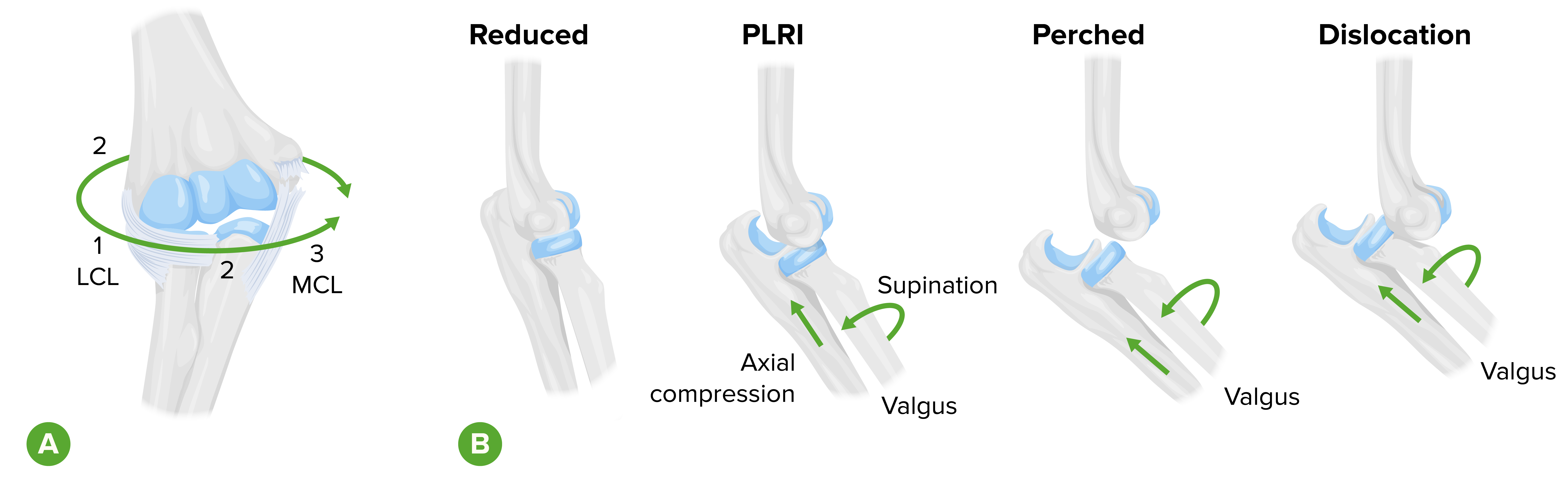
Soft tissue injury during elbow dislocation progresses from lateral to medial, with the anterior band of the medial collateral ligament the last structure to rupture. After a series of posterolateral rotatory instability, the final stage results in complete dislocation.
LCL: lateral collateral ligament
MCL: medical collateral ligament
PLRI: posterolateral rotatory instability
The classification of elbow dislocations can be anatomical or clinical.
Clinical classification:
Anatomical classification:
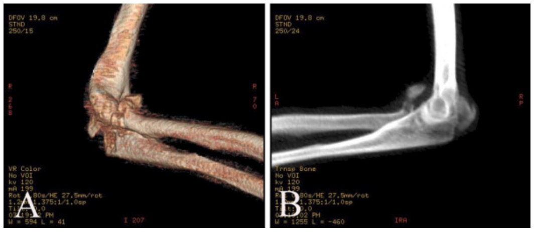
3-Dimensional CT of posterior elbow dislocation with fracture of the olecranon and radial head, without coronoid fracture
Image: “CT of posterior elbow dislocation with fracture” by Department of Orthopaedics and Trauma Surgery, PLA General Hospital, Beijing, PR China. License: CC BY 2.0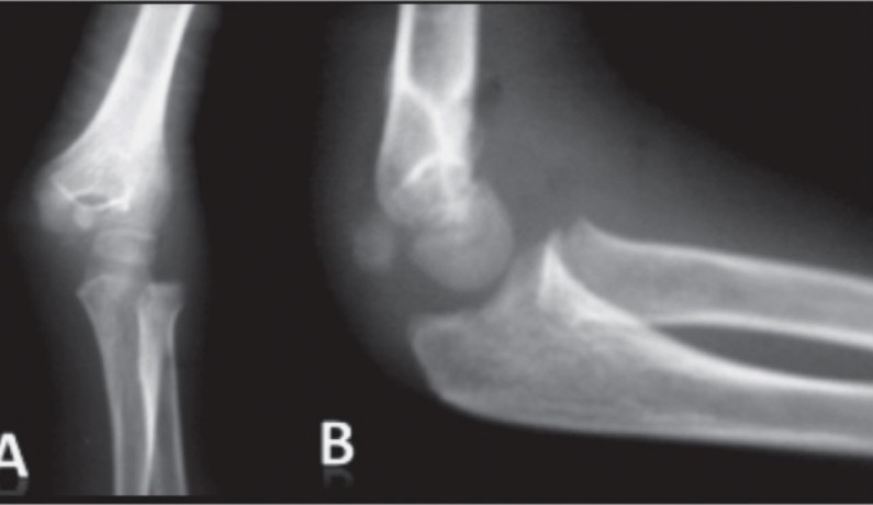
Anteroposterior (A) and lateral (B) views of a dislocated elbow showing a lateral displacement of the radioulnar joint from the distal humerus with a fracture entrapment of the medial epicondyle
Image: “Anteroposterior (A) and lateral (B) views of the sustained elbow” by H Kaziz, MD et al. License: CC BY 3.0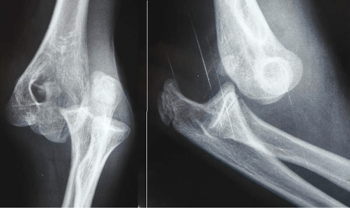
Radiographs showing a posterolateral dislocation of the elbow
Image on left: anteroposterior view
Image on right: lateral view
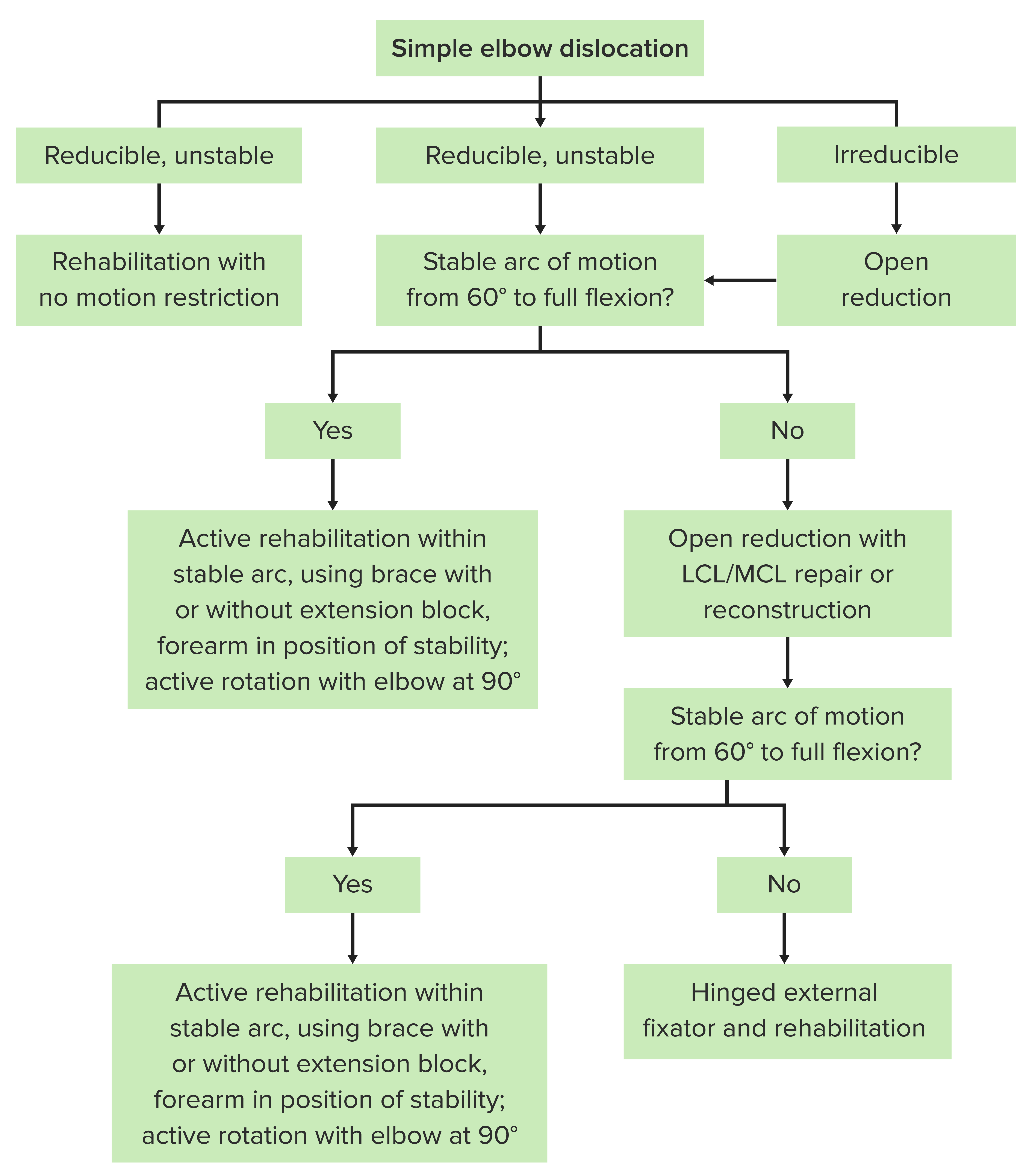
Algorithm for nonsurgical and surgical treatment of simple elbow dislocation
LCL: lateral collateral ligament
MCL: medial collateral ligament
Elbow dislocation can commonly be observed in the following underlying conditions: