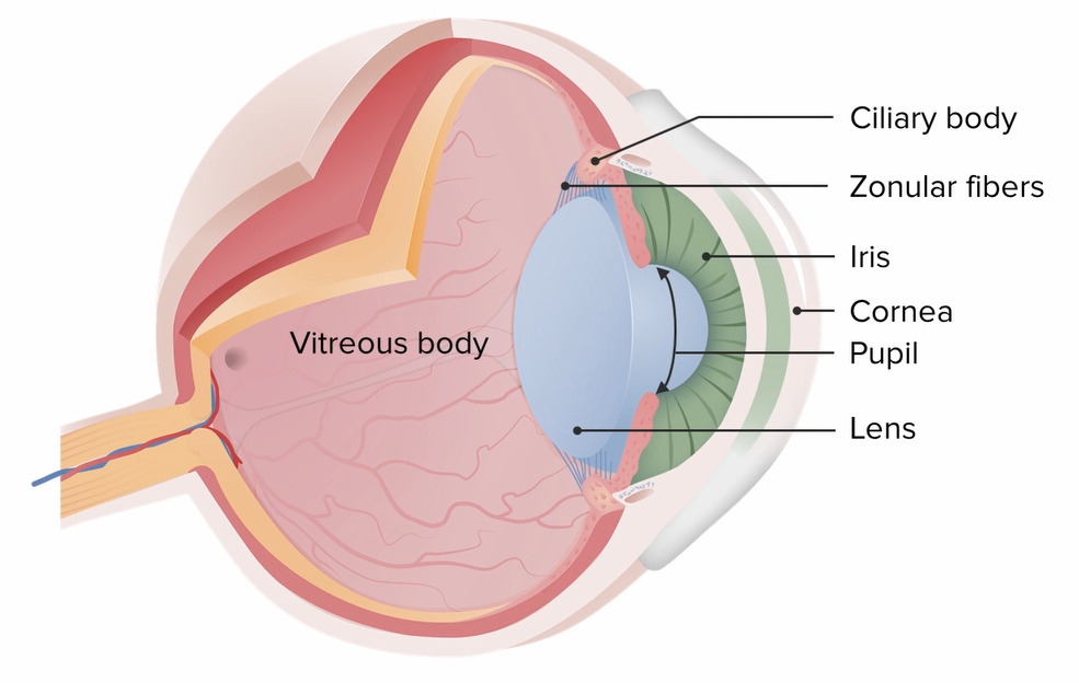
The vitreous bodyVitreous bodyThe transparent, semigelatinous substance that fills the cavity behind the crystalline lens of the eye and in front of the retina. It is contained in a thin hyaloid membrane and forms about four fifths of the optic globe.Eye: Anatomy is a transparent, gelatinous substance that is present in the space between the lensLensA transparent, biconvex structure of the eye, enclosed in a capsule and situated behind the iris and in front of the vitreous humor (vitreous body). It is slightly overlapped at its margin by the ciliary processes. Adaptation by the ciliary body is crucial for ocular accommodation.Eye: Anatomy and the retinaRetinaThe ten-layered nervous tissue membrane of the eye. It is continuous with the optic nerve and receives images of external objects and transmits visual impulses to the brain. Its outer surface is in contact with the choroid and the inner surface with the vitreous body. The outermost layer is pigmented, whereas the inner nine layers are transparent.Eye: Anatomy, providing structural stability and maintaining the shape of the eye. Some conditions that can affect the vitreous bodyVitreous bodyThe transparent, semigelatinous substance that fills the cavity behind the crystalline lens of the eye and in front of the retina. It is contained in a thin hyaloid membrane and forms about four fifths of the optic globe.Eye: Anatomy are posterior vitreous detachmentVitreous DetachmentDetachment of the corpus vitreum (vitreous body) from its normal attachments, especially the retina, due to shrinkage from degenerative or inflammatory conditions, trauma, myopia, or senility.Retinal Detachment, vitreous hemorrhage, synchysis scintillans, asteroid hyalosis, and persistent fetal vasculature. The conditions can be asymptomatic or present with floatersFloatersChorioretinitis in the field of visionVisionOphthalmic Exam, photopsiaPhotopsiaChorioretinitis, and decreased visual acuityVisual AcuityClarity or sharpness of ocular vision or the ability of the eye to see fine details. Visual acuity depends on the functions of retina, neuronal transmission, and the interpretative ability of the brain. Normal visual acuity is expressed as 20/20 indicating that one can see at 20 feet what should normally be seen at that distance. Visual acuity can also be influenced by brightness, color, and contrast.Ophthalmic Exam. Funduscopy and slit-lamp microscopy are commonly used in the diagnosis of these diseases. Treatment methods depend on the condition and severity, but may include observation, visionVisionOphthalmic Exam correction, and surgery.
The vitreous bodyVitreous bodyThe transparent, semigelatinous substance that fills the cavity behind the crystalline lens of the eye and in front of the retina. It is contained in a thin hyaloid membrane and forms about four fifths of the optic globe.Eye: Anatomy is the substance present between the lensLensA transparent, biconvex structure of the eye, enclosed in a capsule and situated behind the iris and in front of the vitreous humor (vitreous body). It is slightly overlapped at its margin by the ciliary processes. Adaptation by the ciliary body is crucial for ocular accommodation.Eye: Anatomy and the retinaRetinaThe ten-layered nervous tissue membrane of the eye. It is continuous with the optic nerve and receives images of external objects and transmits visual impulses to the brain. Its outer surface is in contact with the choroid and the inner surface with the vitreous body. The outermost layer is pigmented, whereas the inner nine layers are transparent.Eye: Anatomy.
Gel-like material that provides:
A clear optical medium
Structural integrity to the eye
Consists largely of:
Water (99%)
Network of collagenCollagenA polypeptide substance comprising about one third of the total protein in mammalian organisms. It is the main constituent of skin; connective tissue; and the organic substance of bones (bone and bones) and teeth (tooth).Connective Tissue: Histology fibrils
Hyaluronic acidHyaluronic acidA natural high-viscosity mucopolysaccharide with alternating beta (1-3) glucuronide and beta (1-4) glucosaminidase bonds. It is found in the umbilical cord, in vitreous body and in synovial fluid. A high urinary level is found in progeria.Connective Tissue: Histology
Posterior vitreous detachmentVitreous DetachmentDetachment of the corpus vitreum (vitreous body) from its normal attachments, especially the retina, due to shrinkage from degenerative or inflammatory conditions, trauma, myopia, or senility.Retinal Detachment is the separation of the vitreous bodyVitreous bodyThe transparent, semigelatinous substance that fills the cavity behind the crystalline lens of the eye and in front of the retina. It is contained in a thin hyaloid membrane and forms about four fifths of the optic globe.Eye: Anatomy from the internal limiting membrane of the retinaRetinaThe ten-layered nervous tissue membrane of the eye. It is continuous with the optic nerve and receives images of external objects and transmits visual impulses to the brain. Its outer surface is in contact with the choroid and the inner surface with the vitreous body. The outermost layer is pigmented, whereas the inner nine layers are transparent.Eye: Anatomy.
Epidemiology
More common in the elderly:
Usually starts around 60–70 years of age
Most eyes are affected by 80 years of age.
Men and women equally affected
Etiology
Age-related vitreous degeneration (most common cause):
With age, the vitreous humorHumorDefense Mechanisms changes from a thick vitreous gel to a thin liquid substance.
Vitreous starts to shrink → can lead to its detachment from the retinaRetinaThe ten-layered nervous tissue membrane of the eye. It is continuous with the optic nerve and receives images of external objects and transmits visual impulses to the brain. Its outer surface is in contact with the choroid and the inner surface with the vitreous body. The outermost layer is pigmented, whereas the inner nine layers are transparent.Eye: Anatomy
Ocular surgery (cataractCataractPartial or complete opacity on or in the lens or capsule of one or both eyes, impairing vision or causing blindness. The many kinds of cataract are classified by their morphology (size, shape, location) or etiology (cause and time of occurrence).Neurofibromatosis Type 2)
Inflammatory eye disease (uveitisUveitisUveitis is the inflammation of the uvea, the pigmented middle layer of the eye, which comprises the iris, ciliary body, and choroid. The condition is categorized based on the site of disease; anterior uveitis is the most common. Diseases of the Uvea)
Collection of deposits in the vitreous bodyVitreous bodyThe transparent, semigelatinous substance that fills the cavity behind the crystalline lens of the eye and in front of the retina. It is contained in a thin hyaloid membrane and forms about four fifths of the optic globe.Eye: Anatomy
FloatersFloatersChorioretinitis are most noticeable against a light background and appear in different shapes and sizes.
Caused by vitreoretinal traction on retinaRetinaThe ten-layered nervous tissue membrane of the eye. It is continuous with the optic nerve and receives images of external objects and transmits visual impulses to the brain. Its outer surface is in contact with the choroid and the inner surface with the vitreous body. The outermost layer is pigmented, whereas the inner nine layers are transparent.Eye: Anatomy
Slit-lamp microscopy: A Weiss ring is a characteristic, mobile membrane within the vitreous cavity that is glossy and crinkled.
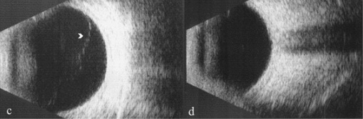
B-scan ultrasonography:
Used to study the condition of the vitreous gel
Also helps to determine the extent of posterior vitreous detachmentVitreous DetachmentDetachment of the corpus vitreum (vitreous body) from its normal attachments, especially the retina, due to shrinkage from degenerative or inflammatory conditions, trauma, myopia, or senility.Retinal Detachment
B-scan ultrasonography showing anterior vitreous detachment in image c. Posterior vitreous detachment is seen in image d.
Image: “B-scan ultrasonography showing extent of vitreous detachment” by Ophthalmology Unit, DAI Head/Neck, Azienda Policlinico Umberto I, University of Rome “Sapienza”, viale del Policlinico 155, Rome, 00161, Italy. License: CC BY 4.0., edited by Lecturio.
Management
No treatment is needed in patientsPatientsIndividuals participating in the health care system for the purpose of receiving therapeutic, diagnostic, or preventive procedures.Clinician–Patient Relationship without retinal injury.
Adaption to visual symptoms will develop over time, and floatersFloatersChorioretinitis can resolve.
VitrectomyVitrectomyRemoval of the whole or part of the vitreous body in treating endophthalmitis, diabetic retinopathy, retinal detachment, intraocular foreign bodies, and some types of glaucoma.Retinal Detachment is considered for persistent symptoms.
Complications
Retinal detachmentRetinal detachmentRetinal detachment is the separation of the neurosensory retina from the retinal pigmented epithelium and choroid. Rhegmatogenous retinal detachment, the most common type, stems from a break in the retina, allowing fluid to accumulate in the subretinal space. Retinal Detachment
Vitreous hemorrhage is extravasation of blood into the vitreous humorHumorDefense Mechanisms.
Etiology
There are many causes of vitreous hemorrhage. Some common causes include:
Proliferative diabetic retinopathyDiabetic retinopathyDisease of the retina as a complication of diabetes mellitus. It is characterized by the progressive microvascular complications, such as aneurysm, intraretinal edema, and intraocular pathologic neovascularization.Chronic Diabetic Complications
Posterior vitreous detachmentVitreous DetachmentDetachment of the corpus vitreum (vitreous body) from its normal attachments, especially the retina, due to shrinkage from degenerative or inflammatory conditions, trauma, myopia, or senility.Retinal Detachment
Macular degenerationMacular degenerationAge-related macular degeneration (AMD) is visual impairment due to changes in the macula, the area responsible for high-acuity vision. It is marked by central vision loss with peripheral vision relatively spared. Risk factors include advanced age, smoking, family history, and cardiovascular disease.Macular Degeneration
Sickle cell retinopathyRetinopathyDegenerative changes to the retina due to hypertension.Alport Syndrome
Clinical presentation
Vitreous hemorrhage is usually painless and unilateral. Signs and symptoms include:
PhotophobiaPhotophobiaAbnormal sensitivity to light. This may occur as a manifestation of eye diseases; migraine; subarachnoid hemorrhage; meningitis; and other disorders. Photophobia may also occur in association with depression and other mental disorders.Migraine Headache
PerceptionPerceptionThe process by which the nature and meaning of sensory stimuli are recognized and interpreted.Psychiatric Assessment of shadows or cobwebs
Visual acuityVisual AcuityClarity or sharpness of ocular vision or the ability of the eye to see fine details. Visual acuity depends on the functions of retina, neuronal transmission, and the interpretative ability of the brain. Normal visual acuity is expressed as 20/20 indicating that one can see at 20 feet what should normally be seen at that distance. Visual acuity can also be influenced by brightness, color, and contrast.Ophthalmic Exam may be affected, depending on the amount of blood present.
Diagnosis
Funduscopic examination can be used to visualize the hemorrhage.
Slit-lamp biomicroscopy: Used to examine the vitreous for red blood cellsRed blood cellsErythrocytes, or red blood cells (RBCs), are the most abundant cells in the blood. While erythrocytes in the fetus are initially produced in the yolk sac then the liver, the bone marrow eventually becomes the main site of production.Erythrocytes: Histology.
Ocular ultrasonography can be used in cases in which the posterior segment is not visible owing to excessive hemorrhage.
CT/MRI may be needed to assess bony structures and rule out foreign bodies.
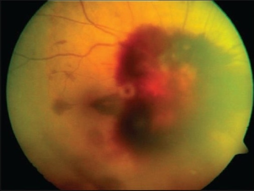
Fundus photograph of the right eye showing vitreous hemorrhage.
Image: “Fundus photographs of right eye showing peripapillary, subhyaloid, vitreous hemorrhage and several flame shaped hemorrhages obscuring the view of the optic disc” by Smt Kanuri Shanthamma Center for Retina Vitreous Diseases, L V Prasad Eye Institute, Kallam Anji Reddy Campus, Banjara Hills, Hyderabad 500 034, India. License: CC BY 2.0, edited by Lecturio.
Management
Vitreous hemorrhage might resolve on its own.
Treatment is directed toward the underlying cause.
VitrectomyVitrectomyRemoval of the whole or part of the vitreous body in treating endophthalmitis, diabetic retinopathy, retinal detachment, intraocular foreign bodies, and some types of glaucoma.Retinal Detachment is done in cases of severe diseases and cases of retinal detachmentRetinal detachmentRetinal detachment is the separation of the neurosensory retina from the retinal pigmented epithelium and choroid. Rhegmatogenous retinal detachment, the most common type, stems from a break in the retina, allowing fluid to accumulate in the subretinal space. Retinal Detachment.
Laser photocoagulation is planned for proliferative retinopathies or retinal tear.
Synchysis Scintillans
Definition
Synchysis scintillans, also known as cholesterolosisCholesterolosisCholesterol-laden macrophages or foam cells in the lamina propria of the gallbladderCellular Accumulations bulbi, is a degenerative condition defined by the accumulation of cholesterolCholesterolThe principal sterol of all higher animals, distributed in body tissues, especially the brain and spinal cord, and in animal fats and oils.Cholesterol Metabolism crystals in liquefied vitreous humorHumorDefense Mechanisms.
Epidemiology
A very rare condition
Usually occurs in the 3rd decade of life
No sexSexThe totality of characteristics of reproductive structure, functions, phenotype, and genotype, differentiating the male from the female organism.Gender Dysphoria or race predominance
Risk factors
Chronic or recurring vitreous hemorrhage (most common)
Diabetic retinopathyDiabetic retinopathyDisease of the retina as a complication of diabetes mellitus. It is characterized by the progressive microvascular complications, such as aneurysm, intraretinal edema, and intraocular pathologic neovascularization.Chronic Diabetic Complications
Chronic uveitisUveitisUveitis is the inflammation of the uvea, the pigmented middle layer of the eye, which comprises the iris, ciliary body, and choroid. The condition is categorized based on the site of disease; anterior uveitis is the most common. Diseases of the Uvea
Retinal detachmentRetinal detachmentRetinal detachment is the separation of the neurosensory retina from the retinal pigmented epithelium and choroid. Rhegmatogenous retinal detachment, the most common type, stems from a break in the retina, allowing fluid to accumulate in the subretinal space. Retinal Detachment
Vitreous biopsyBiopsyRemoval and pathologic examination of specimens from the living body.Ewing Sarcoma: can be done as a confirmatory test to identify cholesterolCholesterolThe principal sterol of all higher animals, distributed in body tissues, especially the brain and spinal cord, and in animal fats and oils.Cholesterol Metabolism crystals.
Management
In general, no treatment is required.
Treatment of underlying diseases is necessary.
Asteroid Hyalosis
Definition
Asteroid hyalosis is a condition in which calcium-lipid (calciumCalciumA basic element found in nearly all tissues. It is a member of the alkaline earth family of metals with the atomic symbol ca, atomic number 20, and atomic weight 40. Calcium is the most abundant mineral in the body and combines with phosphorus to form calcium phosphate in the bones and teeth. It is essential for the normal functioning of nerves and muscles and plays a role in blood coagulation (as factor IV) and in many enzymatic processes.Electrolytes soap) complexes are attached to the collagenCollagenA polypeptide substance comprising about one third of the total protein in mammalian organisms. It is the main constituent of skin; connective tissue; and the organic substance of bones (bone and bones) and teeth (tooth).Connective Tissue: Histology framework of the vitreous bodyVitreous bodyThe transparent, semigelatinous substance that fills the cavity behind the crystalline lens of the eye and in front of the retina. It is contained in a thin hyaloid membrane and forms about four fifths of the optic globe.Eye: Anatomy.
Epidemiology
Seen in approximately 1 in 200 individuals
Most commonly seen after 50 years of age
More common in men
Etiology
The disease etiology is unknown.
Strongly correlated with age
Not clearly associated with other diseases
Clinical presentation
Asteroid hyalosis is usually asymptomatic.
Diagnosis
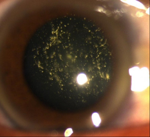
The diagnosis is made with slit-lamp microscopy, showing:
Unilateral
Multiple yellow/white, round opacities
FloatersFloatersChorioretinitis are suspended (“stars in the night sky”) → may move, but usually return to their original position
Multiple bright opacities suspended in the vitreous, consistent with asteroid hyalosis.
Image: “Asteroid hyalosis: multiple yellow mobile vitreous particles” by University of Mohamed V souissi, hôpital des Spécialités, Ophtalology A Department. License: CC BY 2.0, edited by Lecturio.
Management
Treatment is rarely needed.
VitrectomyVitrectomyRemoval of the whole or part of the vitreous body in treating endophthalmitis, diabetic retinopathy, retinal detachment, intraocular foreign bodies, and some types of glaucoma.Retinal Detachment may be indicated in cases of severe loss of visual acuityVisual AcuityClarity or sharpness of ocular vision or the ability of the eye to see fine details. Visual acuity depends on the functions of retina, neuronal transmission, and the interpretative ability of the brain. Normal visual acuity is expressed as 20/20 indicating that one can see at 20 feet what should normally be seen at that distance. Visual acuity can also be influenced by brightness, color, and contrast.Ophthalmic Exam.
Persistent Fetal Vasculature
Definition
Persistent fetal vasculature, formerly known as persistent hyperplasticHyperplasticColon Polyps primary vitreous, is a condition in which embryonic blood vessels fail to regress.
Etiology
Failure of the embryonic primary vitreous and hyaloid vascular system to regress
Majority of cases are not genetic
Clinical presentation
This condition is usually unilateral and may present with:
LeukocoriaLeukocoriaCataracts in Children: white pupillary reflex (also associated with retinoblastomaRetinoblastomaRetinoblastoma is a rare tumor but the most common primary intraocular malignancy of childhood. It is believed that the condition arises from a neuronal progenitor cell. Retinoblastoma can be heritable or non-heritable. Retinoblastoma)
Decreased visual acuityVisual AcuityClarity or sharpness of ocular vision or the ability of the eye to see fine details. Visual acuity depends on the functions of retina, neuronal transmission, and the interpretative ability of the brain. Normal visual acuity is expressed as 20/20 indicating that one can see at 20 feet what should normally be seen at that distance. Visual acuity can also be influenced by brightness, color, and contrast.Ophthalmic Exam
Microphthalmia (abnormally small eye due to failure to develop) of the affected eye
StrabismusStrabismusStrabismus is the misalignment of the eyes while fixating the gaze on an object. Strabismus can be idiopathic, but it may also be caused by cerebral palsy, uncorrected refractive errors, and extraocular muscle or cranial nerve dysfunction. Strabismus
Diagnosis
Persistent fetal vasculature is usually diagnosed right after birth.
Funduscopy and slit-lamp exam:
Rotated and elongated ciliary process
FibrousFibrousFibrocystic Change stalk above the optic nerveOptic nerveThe 2nd cranial nerve which conveys visual information from the retina to the brain. The nerve carries the axons of the retinal ganglion cells which sort at the optic chiasm and continue via the optic tracts to the brain. The largest projection is to the lateral geniculate nuclei; other targets include the superior colliculi and the suprachiasmatic nuclei. Though known as the second cranial nerve, it is considered part of the central nervous system.The 12 Cranial Nerves: Overview and Functions
Ultrasonography may show a fibrovascular stalk between the lensLensA transparent, biconvex structure of the eye, enclosed in a capsule and situated behind the iris and in front of the vitreous humor (vitreous body). It is slightly overlapped at its margin by the ciliary processes. Adaptation by the ciliary body is crucial for ocular accommodation.Eye: Anatomy and the optic nerveOptic nerveThe 2nd cranial nerve which conveys visual information from the retina to the brain. The nerve carries the axons of the retinal ganglion cells which sort at the optic chiasm and continue via the optic tracts to the brain. The largest projection is to the lateral geniculate nuclei; other targets include the superior colliculi and the suprachiasmatic nuclei. Though known as the second cranial nerve, it is considered part of the central nervous system.The 12 Cranial Nerves: Overview and Functions.
CT can show:
Shallow anterior chamberAnterior chamberThe space in the eye, filled with aqueous humor, bounded anteriorly by the cornea and a small portion of the sclera and posteriorly by a small portion of the ciliary body, the iris, and that part of the crystalline lens which presents through the pupil.Eye: Anatomy
Irregular lensLensA transparent, biconvex structure of the eye, enclosed in a capsule and situated behind the iris and in front of the vitreous humor (vitreous body). It is slightly overlapped at its margin by the ciliary processes. Adaptation by the ciliary body is crucial for ocular accommodation.Eye: Anatomy
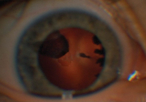
Persistent fetal vasculature: On slit-lamp examination, traction of the ciliary processes to the center of the posterior capsule of the lens in the left eye and a retrolental mass are identified.
Image: “Persistent fetal vasculature (PFV)” by Department of Ophthalmology, Hippokration General Hospital, 54642 Thessaloniki, Greece. License: CC BY 3.0, edited by Lecturio.
Surgery may be recommended to prevent or treat complications.
Complications
GlaucomaGlaucomaGlaucoma is an optic neuropathy characterized by typical visual field defects and optic nerve atrophy seen as optic disc cupping on examination. The acute form of glaucoma is a medical emergency. Glaucoma is often, but not always, caused by increased intraocular pressure (IOP). Glaucoma
CataractCataractPartial or complete opacity on or in the lens or capsule of one or both eyes, impairing vision or causing blindness. The many kinds of cataract are classified by their morphology (size, shape, location) or etiology (cause and time of occurrence).Neurofibromatosis Type 2
Intraocular hemorrhage
Retinal detachmentRetinal detachmentRetinal detachment is the separation of the neurosensory retina from the retinal pigmented epithelium and choroid. Rhegmatogenous retinal detachment, the most common type, stems from a break in the retina, allowing fluid to accumulate in the subretinal space. Retinal Detachment
Park, J. H., Yang, H., Kwon, H., & Jeon, S. (2021). Risk factors for onset or progression of posterior vitreous detachment at the vitreomacular interface after cataract surgery. Ophthalmology Retina, 5(3), 270–278. https://doi.org/10.1016/j.oret.2020.07.017