La miocarditis es una enfermedad inflamatoria del miocardio, que puede aparecer de forma aislada o asociada a un proceso sistémico. Existen numerosas etiologías de la miocarditis, pero todas conducen a la inflamación y a la lesión de losLOSNeisseria miocitos, lo que suele provocar signos y síntomas de insuficiencia cardíaca. La evolución de la miocarditis puede variar enENErythema nodosum is an immune-mediated panniculitis (inflammation of the subcutaneous fat) caused by a type IV (delayed-type) hypersensitivity reaction. It commonly manifests in young women as tender, erythematous nodules on the shins.Erythema Nodosum función de la etiología y del tiempo de progresión de losLOSNeisseria síntomas. El diagnóstico se apoya enENErythema nodosum is an immune-mediated panniculitis (inflammation of the subcutaneous fat) caused by a type IV (delayed-type) hypersensitivity reaction. It commonly manifests in young women as tender, erythematous nodules on the shins.Erythema NodosumlosLOSNeisseria hallazgos clínicos, las pruebas de laboratorio y la imagenología cardíaca. Raramente, se requiere un diagnóstico de confirmación mediante biopsia endomiocárdica. El tratamiento es de soporte y está dirigido a tratar las complicaciones.
La miocarditis es una enfermedad inflamatoria del miocardio.
Epidemiología
Incidencia: aproximadamente 10–22 casos por cada 100 000 personas
Alrededor del 1%–5% de las infecciones virales afectan alALAmyloidosis miocardio.
Afecta más a losLOSNeisseria hombres que a las mujeres
Más común enENErythema nodosum is an immune-mediated panniculitis (inflammation of the subcutaneous fat) caused by a type IV (delayed-type) hypersensitivity reaction. It commonly manifests in young women as tender, erythematous nodules on the shins.Erythema Nodosum adultos jóvenes
VirusVirusViruses are infectious, obligate intracellular parasites composed of a nucleic acid core surrounded by a protein capsid. Viruses can be either naked (non-enveloped) or enveloped. The classification of viruses is complex and based on many factors, including type and structure of the nucleoid and capsid, the presence of an envelope, the replication cycle, and the host range. Virology (más común enENErythema nodosum is an immune-mediated panniculitis (inflammation of the subcutaneous fat) caused by a type IV (delayed-type) hypersensitivity reaction. It commonly manifests in young women as tender, erythematous nodules on the shins.Erythema Nodosum Norteamérica y Europa)
Bacterias
Protozoarios (más comunes enENErythema nodosum is an immune-mediated panniculitis (inflammation of the subcutaneous fat) caused by a type IV (delayed-type) hypersensitivity reaction. It commonly manifests in young women as tender, erythematous nodules on the shins.Erythema Nodosum África, AsiaASIASpinal Cord Injuries y Sudamérica)
Hongos
Trastornos mediados por el sistema inmunitario:
Fiebre reumática
Reacciones alérgicas
Rechazo de trasplante
Enfermedad de Kawasaki
SarcoidosisSarcoidosisSarcoidosis is a multisystem inflammatory disease that causes noncaseating granulomas. The exact etiology is unknown. Sarcoidosis usually affects the lungs and thoracic lymph nodes, but it can also affect almost every system in the body, including the skin, heart, and eyes, most commonly. Sarcoidosis
Lupus eritematoso sistémico
Polimiositis
Dermatomiositis
Esclerodermia
Artritis idiopática juvenil
VasculitisVasculitisInflammation of any one of the blood vessels, including the arteries; veins; and rest of the vasculature system in the body.Systemic Lupus Erythematosus
Agentes exógenos
Predisposición genética:
Defectos genéticos enENErythema nodosum is an immune-mediated panniculitis (inflammation of the subcutaneous fat) caused by a type IV (delayed-type) hypersensitivity reaction. It commonly manifests in young women as tender, erythematous nodules on the shins.Erythema Nodosum las proteínas estructurales de losLOSNeisseria miocitos
La siguiente tabla resume las causas infecciosas de la miocarditis. Tenga enENErythema nodosum is an immune-mediated panniculitis (inflammation of the subcutaneous fat) caused by a type IV (delayed-type) hypersensitivity reaction. It commonly manifests in young women as tender, erythematous nodules on the shins.Erythema Nodosum cuenta que esta lista no abarca la totalidad.
Tabla: Causas de la miocarditis infecciosa
Viral
Bacteriana
Parasitaria
Fúngica
VirusVirusViruses are infectious, obligate intracellular parasites composed of a nucleic acid core surrounded by a protein capsid. Viruses can be either naked (non-enveloped) or enveloped. The classification of viruses is complex and based on many factors, including type and structure of the nucleoid and capsid, the presence of an envelope, the replication cycle, and the host range. Virology Coxsackie B
AdenovirusAdenovirusAdenovirus (member of the family Adenoviridae) is a nonenveloped, double-stranded DNA virus. Adenovirus is transmitted in a variety of ways, and it can have various presentations based on the site of entry. Presentation can include febrile pharyngitis, conjunctivitis, acute respiratory disease, atypical pneumonia, and gastroenteritis. Adenovirus
Parvovirus B19Parvovirus B19Primate erythroparvovirus 1 (generally referred to as parvovirus B19, B19 virus, or sometimes erythrovirus B19) ranks among the smallest DNA viruses. Parvovirus B19 is of the family Parvoviridae and genus Erythrovirus. In immunocompetent humans, parvovirus B19 classically results in erythema infectiosum (5th disease) or “slapped cheek syndrome.”Parvovirus B19
Herpesvirus humano 6
VirusVirusViruses are infectious, obligate intracellular parasites composed of a nucleic acid core surrounded by a protein capsid. Viruses can be either naked (non-enveloped) or enveloped. The classification of viruses is complex and based on many factors, including type and structure of the nucleoid and capsid, the presence of an envelope, the replication cycle, and the host range. Virology de Epstein-Barr
Citomegalovirus
Hepatitis CHepatitis CHepatitis C is an infection of the liver caused by the hepatitis C virus (HCV). The infection can be transmitted through infectious blood or body fluids and may be transmitted during childbirth or through IV drug use or sexual intercourse. Hepatitis C virus can cause both acute and chronic hepatitis, ranging from a mild to a serious, lifelong illness including liver cirrhosis and hepatocellular carcinoma (HCC).Hepatitis C Virus
InfluenzaInfluenzaInfluenza viruses are members of the Orthomyxoviridae family and the causative organisms of influenza, a highly contagious febrile respiratory disease. There are 3 primary influenza viruses (A, B, and C) and various subtypes, which are classified based on their virulent surface antigens, hemagglutinin (HA) and neuraminidase (NA). Influenza typically presents with a fever, myalgia, headache, and symptoms of an upper respiratory infection. Influenza Viruses/Influenza
PoliovirusPoliovirusPoliomyelitis is an infectious disease caused by the poliovirus. This virus is a member of the Picornaviridae family. It is a small, single-stranded, positive-sense RNA virus without a lipid envelope. Transmission occurs through the fecal-oral route and, occasionally, through respiratory aerosols. Poliovirus/Poliomyelitis
VIH
Borrelia burgdorferiBorrelia burgdorferiA specific species of bacteria, part of the borrelia burgdorferi group, whose common name is lyme disease spirochete.Borrelia
Mycoplasma pneumoniaeMycoplasma pneumoniaeShort filamentous organism of the genus mycoplasma, which binds firmly to the cells of the respiratory epithelium. It is one of the etiologic agents of non-viral primary atypical pneumonia in man.Mycoplasma
Mycobacterium tuberculosisMycobacterium tuberculosisTuberculosis (TB) is an infectious disease caused by Mycobacterium tuberculosis complex bacteria. The bacteria usually attack the lungs but can also damage other parts of the body. Approximately 30% of people around the world are infected with this pathogen, with the majority harboring a latent infection. Tuberculosis spreads through the air when a person with active pulmonary infection coughs or sneezes.Tuberculosis
Corynebacterium diphtheriaeCorynebacterium diphtheriaeDiphtheria is an infectious disease caused by corynebacterium diphtheriae that most often results in respiratory disease with membranous inflammation of the pharynx, sore throat, fever, swollen glands, and weakness. The hallmark sign is a sheet of thick, gray material covering the back of the throat.Diphtheria
StaphylococcusStaphylococcusStaphylococcus is a medically important genera of Gram-positive, aerobic cocci. These bacteria form clusters resembling grapes on culture plates. Staphylococci are ubiquitous for humans, and many strains compose the normal skin flora.Staphylococcus
Neisseria gonorrhoeaeNeisseria gonorrhoeaeA species of gram-negative, aerobic bacteria primarily found in purulent venereal discharges. It is the causative agent of gonorrhea.Neisseria
StreptococcusStreptococcusStreptococcus is one of the two medically important genera of gram-positive cocci, the other being Staphylococcus. Streptococci are identified as different species on blood agar on the basis of their hemolytic pattern and sensitivity to optochin and bacitracin. There are many pathogenic species of streptococci, including S. pyogenes, S. agalactiae, S. pneumoniae, and the viridans streptococci.Streptococcus
BrucellaBrucellaBrucellosis (also known as undulant fever, Mediterranean fever, or Malta fever) is a zoonotic infection that spreads predominantly through ingestion of unpasteurized dairy products or direct contact with infected animal products. Clinical manifestations include fever, arthralgias, malaise, lymphadenopathy, and hepatosplenomegaly. Brucella/Brucellosis
Haemophilus influenzaeHaemophilus InfluenzaeA species of Haemophilus found on the mucous membranes of humans and a variety of animals. The species is further divided into biotypes I through viii.Haemophilus
Treponema pallidumTreponema pallidumThe causative agent of venereal and non-venereal syphilis as well as yaws.Treponema
Coxiella burnetti
Rickettsia rickettsiiRickettsia rickettsiiA species of gram-negative, aerobic bacteria that is the etiologic agent of rocky mountain spotted fever. Its cells are slightly smaller and more uniform in size than those of rickettsia prowazekii.Rickettsia
Trypanosoma cruziTrypanosoma cruziChagas disease is an infection caused by the American trypanosome Trypanosoma cruzi. This parasitic protozoan is transmitted in the feces of reduviid bugs in South and Central America. Acute infection may present with inflammation at the inoculation site (chagoma), fever, and lymphadenopathy. Untreated, chronic infection can progress to severe complications.Trypanosoma cruzi/Chagas disease
ToxoplasmaToxoplasmaToxoplasmosis is an infectious disease caused by Toxoplasma gondii, an obligate intracellular protozoan parasite. Felines are the definitive host, but transmission to humans can occur through contact with cat feces or the consumption of contaminated foods. The clinical presentation and complications depend on the host’s immune status. Toxoplasma/Toxoplasmosis gondii
Entamoeba histolyticaEntamoeba HistolyticaA species of parasitic protozoa causing entamoebiasis and amebic dysentery (dysentery, amebic). Characteristics include a single nucleus containing a small central karyosome and peripheral chromatin that is finely and regularly beaded.Amebicides
LeishmaniaLeishmaniaLeishmania species are obligate intracellular parasites that are transmitted by an infected sandfly. The disease is endemic to Asia, the Middle East, Africa, the Mediterranean, and South and Central America. Clinical presentation varies, dependent on the pathogenicity of the species and the host’s immune response. Leishmania/Leishmaniasis
AspergillusAspergillusA genus of mitosporic fungi containing about 100 species and eleven different teleomorphs in the family trichocomaceae.Echinocandins
CandidaCandidaCandida is a genus of dimorphic, opportunistic fungi. Candida albicans is part of the normal human flora and is the most common cause of candidiasis. The clinical presentation varies and can include localized mucocutaneous infections (e.g., oropharyngeal, esophageal, intertriginous, and vulvovaginal candidiasis) and invasive disease (e.g., candidemia, intraabdominal abscess, pericarditis, and meningitis). Candida/Candidiasis
ActinomycesActinomycesActinomyces is an anaerobic, gram-positive, branching, filamentous rod. Actinomyces israelii is the most common species involved in human disease. The organism is commonly found as part of the normal flora in the oral cavity, gastrointestinal tract, and reproductive tract. Actinomyces/Actinomycosis
BlastomycesBlastomycesBlastomycosis is an infection caused by inhalation of the spores of the fungus, Blastomyces. Blastomyces species thrive in moist soil and decaying material and are common in the Ohio and Mississippi River valleys and the Great Lakes regions of the United States and Canada. Although most patients are asymptomatic, some can develop pneumonia.Blastomyces/Blastomycosis
CoccidioidesCoccidioidesCoccidioidomycosis, commonly known as San Joaquin Valley fever, is a fungal disease caused by Coccidioides immitis or Coccidioides posadasii. When Coccidioides spores are inhaled, they transform into spherules that result in infection. Coccidioidomycosis is also a common cause of community-acquired pneumonia and can cause severe disease in the immunocompromised.Coccidioides/Coccidioidomycosis
HistoplasmaHistoplasmaHistoplasmosis is an infection caused by Histoplasma capsulatum, a dimorphic fungus. The fungus exists as a mold at low temperatures and as yeast at high temperatures. H. capsulatum is the most common endemic fungal infection in the US and is most prevalent in the midwestern and central states along the Ohio and Mississippi River valleys.Histoplasma/Histoplasmosis
CryptococcusCryptococcusCryptococcosis is an opportunistic, fungal infection caused by the Cryptococcus species. The principal pathogens in humans are C. neoformans (primary) and C. gattii. Cryptococcus neoformans is typically found in pigeon droppings and acquired by inhaling dust from contaminated soil. The majority of affected patients are immunocompromised. Cryptococcus/Cryptococcosis
Mucormyocises
NocardiaNocardiaNocardia is a branching, filamentous, gram-positive bacilli. It is partially acid fast due to the presence of mycolic acids in the cell wall. Nocardia is a ubiquitous soil organism that most commonly affects immunocompromised patients. Nocardia is transmitted via inhalation of aerosolized bacteria or less commonly, via direct contact with wounds. Nocardia/Nocardiosis
Inflamación y lesión miocárdica → necrosisNecrosisThe death of cells in an organ or tissue due to disease, injury or failure of the blood supply.Ischemic Cell Damage miocárdica
Daño grave y prolongado → fibrosisFibrosisAny pathological condition where fibrous connective tissue invades any organ, usually as a consequence of inflammation or other injury.Bronchiolitis Obliterans → remodelación de la cavidad cardíaca → miocardiopatía dilatada
Consecuencias:
Insuficiencia cardíaca
Arritmia cardíaca
Extensión alALAmyloidosis pericardio → pericarditisPericarditisPericarditis is an inflammation of the pericardium, often with fluid accumulation. It can be caused by infection (often viral), myocardial infarction, drugs, malignancies, metabolic disorders, autoimmune disorders, or trauma. Acute, subacute, and chronic forms exist. Pericarditis
La presentación clínica puede variar enENErythema nodosum is an immune-mediated panniculitis (inflammation of the subcutaneous fat) caused by a type IV (delayed-type) hypersensitivity reaction. It commonly manifests in young women as tender, erythematous nodules on the shins.Erythema Nodosum función de la gravedad y la progresión de losLOSNeisseria síntomas. La mayoría de losLOSNeisseria signos y síntomas están relacionados con la insuficiencia cardíaca.
Clasificación
La miocarditis se clasifica enENErythema nodosum is an immune-mediated panniculitis (inflammation of the subcutaneous fat) caused by a type IV (delayed-type) hypersensitivity reaction. It commonly manifests in young women as tender, erythematous nodules on the shins.Erythema Nodosum función de su progresión temporal.
Subclínica: ausencia de síntomas o síntomas leves
Aguda: la insuficiencia cardíaca se desarrolla enENErythema nodosum is an immune-mediated panniculitis (inflammation of the subcutaneous fat) caused by a type IV (delayed-type) hypersensitivity reaction. It commonly manifests in young women as tender, erythematous nodules on the shins.Erythema Nodosum < 3 meses.
Crónica: la insuficiencia cardíaca se desarrolla enENErythema nodosum is an immune-mediated panniculitis (inflammation of the subcutaneous fat) caused by a type IV (delayed-type) hypersensitivity reaction. It commonly manifests in young women as tender, erythematous nodules on the shins.Erythema Nodosum > 3 meses.
Disminución de la tolerancia alALAmyloidosis ejercicio
Examen físico
Taquicardia
Arritmia
EdemaEdemaEdema is a condition in which excess serous fluid accumulates in the body cavity or interstitial space of connective tissues. Edema is a symptom observed in several medical conditions. It can be categorized into 2 types, namely, peripheral (in the extremities) and internal (in an organ or body cavity). Edema periférico
Hepatomegalia
Estertores pulmonares
Distensión venosa yugular
Un soplo → puede indicar una agrandamiento de la cavidad cardíaca:
Roce pericárdico (pericarditisPericarditisPericarditis is an inflammation of the pericardium, often with fluid accumulation. It can be caused by infection (often viral), myocardial infarction, drugs, malignancies, metabolic disorders, autoimmune disorders, or trauma. Acute, subacute, and chronic forms exist. Pericarditis)
Complicaciones
ShockShockShock is a life-threatening condition associated with impaired circulation that results in tissue hypoxia. The different types of shock are based on the underlying cause: distributive (↑ cardiac output (CO), ↓ systemic vascular resistance (SVR)), cardiogenic (↓ CO, ↑ SVR), hypovolemic (↓ CO, ↑ SVR), obstructive (↓ CO), and mixed. Types of Shock cardiogénico
LosLOSNeisseria estudios virales pueden ayudar a identificar el agente causante.
Tamizaje reumatológico de causas autoinmunes
ECGECGAn electrocardiogram (ECG) is a graphic representation of the electrical activity of the heart plotted against time. Adhesive electrodes are affixed to the skin surface allowing measurement of cardiac impulses from many angles. The ECG provides 3-dimensional information about the conduction system of the heart, the myocardium, and other cardiac structures. Electrocardiogram (ECG):
Cambios enENErythema nodosum is an immune-mediated panniculitis (inflammation of the subcutaneous fat) caused by a type IV (delayed-type) hypersensitivity reaction. It commonly manifests in young women as tender, erythematous nodules on the shins.Erythema Nodosum el ST:
A menudo inespecíficos
Elevación difusa del ST observadas enENErythema nodosum is an immune-mediated panniculitis (inflammation of the subcutaneous fat) caused by a type IV (delayed-type) hypersensitivity reaction. It commonly manifests in young women as tender, erythematous nodules on the shins.Erythema Nodosum la pericarditisPericarditisPericarditis is an inflammation of the pericardium, often with fluid accumulation. It can be caused by infection (often viral), myocardial infarction, drugs, malignancies, metabolic disorders, autoimmune disorders, or trauma. Acute, subacute, and chronic forms exist. Pericarditis
Arritmia
Retrasos enENErythema nodosum is an immune-mediated panniculitis (inflammation of the subcutaneous fat) caused by a type IV (delayed-type) hypersensitivity reaction. It commonly manifests in young women as tender, erythematous nodules on the shins.Erythema Nodosum la conducción
Radiografía de tórax:
Corazón normal-agrandado
Indicaciones de insuficiencia cardíaca:
Congestión venosa pulmonar
Derrame pleural
Imagenología cardíaca
Ultrasonido cardiaco:
Debe realizarse enENErythema nodosum is an immune-mediated panniculitis (inflammation of the subcutaneous fat) caused by a type IV (delayed-type) hypersensitivity reaction. It commonly manifests in young women as tender, erythematous nodules on the shins.Erythema Nodosum todos losLOSNeisseria pacientes con sospecha de miocarditis
Puede ser normal enENErythema nodosum is an immune-mediated panniculitis (inflammation of the subcutaneous fat) caused by a type IV (delayed-type) hypersensitivity reaction. It commonly manifests in young women as tender, erythematous nodules on the shins.Erythema Nodosum estadios precoces o enENErythema nodosum is an immune-mediated panniculitis (inflammation of the subcutaneous fat) caused by a type IV (delayed-type) hypersensitivity reaction. It commonly manifests in young women as tender, erythematous nodules on the shins.Erythema Nodosum casos leves
Posibles hallazgos:
Dilatación del ventrículo izquierdo
Disfunción sistólica ventricular
Regurgitación mitral o tricúspide
Derrame pericárdico
Trombos intracardíacos
Excluye otras causas de insuficiencia cardíaca
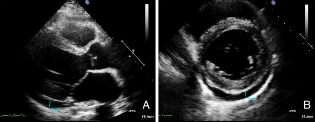
Imágenes del ecocardiograma transtorácico de un paciente con miocarditis: (A) Vista del eje largo y (B) vista de eje corto que muestra un derrame pericárdico (marcado en azul)
EdemaEdemaEdema is a condition in which excess serous fluid accumulates in the body cavity or interstitial space of connective tissues. Edema is a symptom observed in several medical conditions. It can be categorized into 2 types, namely, peripheral (in the extremities) and internal (in an organ or body cavity). Edema miocárdico
Hiperemia miocárdica
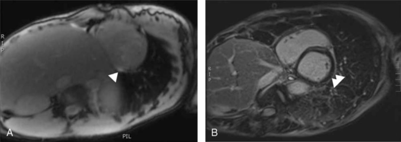
RM cardíaca sugestiva de miocarditis. Las flechas blancas marcan el realce del miocardio, que es sugestivo de edema
Imagen: “Myocarditis in Patients With Antisynthetase Syndrome: Prevalence, Presentation, and Outcomes” por Dieval, C. et al. Licencia: CC BY 4.0
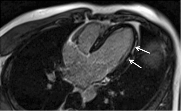
RM cardíaca muestra el realce de la pared lateral con gadolinio, que es un hallazgo característico
Imageen: “Myopericarditis complicated by pulmonary embolism in an immunocompetent patient with acute cytomegalovirus infection: A case report” por Vandamme, Y.M. et al. Licencia: CC BY 2.0
Biopsia endomiocárdica
La biopsia endomiocárdica es el estándar de oro para el diagnóstico; sin embargo, rara vez es necesaria.
Indicaciones:
Deterioro agudo de la función cardíaca sin etiología conocida
NecrosisNecrosisThe death of cells in an organ or tissue due to disease, injury or failure of the blood supply.Ischemic Cell Damage de miocitos
FibrosisFibrosisAny pathological condition where fibrous connective tissue invades any organ, usually as a consequence of inflammation or other injury.Bronchiolitis Obliterans intersticial
Hipertrofia de las miofibras
Cambios granulomatosos:
MycobacteriumMycobacteriumMycobacterium is a genus of the family Mycobacteriaceae in the phylum Actinobacteria. Mycobacteria comprise more than 150 species of facultative intracellular bacilli that are mostly obligate aerobes. Mycobacteria are responsible for multiple human infections including serious diseases, such as tuberculosis (M. tuberculosis), leprosy (M. leprae), and M. avium complex infections.Mycobacterium
Hongos
Parásitos
Miocarditis sarcoidea (no caseificante)
Células gigantes: miocarditis de células gigantes
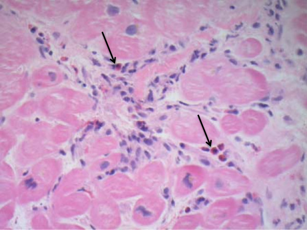
Biopsia endomiocárdica en la miocarditis eosinofílica: Hay una infiltración de eosinófilos (flechas) que se observa en la tinción con hematoxilina y eosina.
Imagen: “Eosinophilic myocarditis: Two case reports and review of the literature” por Rizkallah, J. et al. Licencia: CC BY 2.0
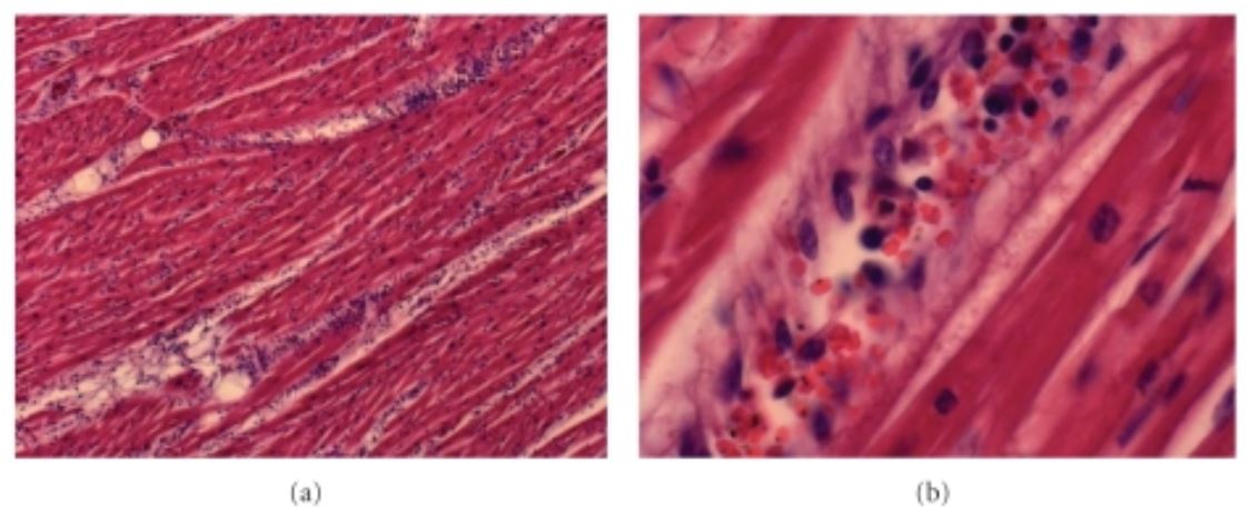
Miocarditis aguda por Plasmodium falciparum: Existe una amplia infiltración linfocítica.
Imagen: “Fatal Myocarditis in Course of Plasmodium falciparum Infection: Case Report and Review of Cardiac Complications in Malaria” por Costenaro P, Benedetti P, Facchin C, Mengoli C, Pellizzer G. Licencia: CC BY 3.0
Tratamiento
Principios generales
El tratamiento de la miocarditis es generalmente de soporte, pero a menudo incluye:
Tratamiento de la insuficiencia cardíaca
Tratamiento de arritmias
Tratamiento de la etiología subyacente (cuando sea posible)
Tratamiento de la insuficiencia cardíaca
Tratamiento médico de la insuficiencia cardíaca:
Diuréticos
Inhibidores de la enzima convertidora de angiotensina
Betabloqueadores
Antagonistas de losLOSNeisseria receptores de la angiotensina II
Antagonistas de la aldosterona
Para la insuficiencia cardíaca fulminante:
Soporte inotrópico
Balón intraaórtico de contrapulsación
Dispositivo de asistencia ventricular izquierda
Trasplante cardíaco
Tratamiento de arritmias
Terapia antiarrítmica
Cardioversión
Estimulación temporal o permanente
Otras consideraciones
LosLOSNeisseria anticoagulantes están indicados para:
Trombos intracardíacos
Evidencia de embolia sistémica
Fibrilación auricular
Evitar:
AINE
Consumo de alcohol
Ejercicio
Seguimiento a largo plazo
La mayoría de losLOSNeisseria pacientes tendrán una recuperación parcial o total. Sin embargo, se recomienda realizar un seguimiento y control por tiempo prolongado.
Reanudación gradual de las actividades físicas
Monitorización con ultrasonido cardiaco de forma seriada
Reevaluación y titulación de la medicación, según corresponda
PericarditisPericarditisPericarditis is an inflammation of the pericardium, often with fluid accumulation. It can be caused by infection (often viral), myocardial infarction, drugs, malignancies, metabolic disorders, autoimmune disorders, or trauma. Acute, subacute, and chronic forms exist. Pericarditis: inflamación del revestimiento exterior del corazón como consecuencia de una infección, enfermedad autoinmune, radiación, intervención quirúrgica o IAM. La pericarditisPericarditisPericarditis is an inflammation of the pericardium, often with fluid accumulation. It can be caused by infection (often viral), myocardial infarction, drugs, malignancies, metabolic disorders, autoimmune disorders, or trauma. Acute, subacute, and chronic forms exist. Pericarditis se presenta clínicamente con fiebre y dolorDolorInflammation torácico pleurítico que aumenta alALAmyloidosis acostarse enENErythema nodosum is an immune-mediated panniculitis (inflammation of the subcutaneous fat) caused by a type IV (delayed-type) hypersensitivity reaction. It commonly manifests in young women as tender, erythematous nodules on the shins.Erythema Nodosum posición supina, y roce pericárdico a la auscultación. Un ECGECGAn electrocardiogram (ECG) is a graphic representation of the electrical activity of the heart plotted against time. Adhesive electrodes are affixed to the skin surface allowing measurement of cardiac impulses from many angles. The ECG provides 3-dimensional information about the conduction system of the heart, the myocardium, and other cardiac structures. Electrocardiogram (ECG) con elevación difusa del segmento ST y un ecocardiograma que muestre un derrame pericárdico pueden confirmar el diagnóstico. El tratamiento es de soporte.
IAM: isquemia del miocardio debido a la obstrucción parcial o total de las arterias coronarias. LosLOSNeisseria pacientes se presentan con dolorDolorInflammation torácico de inicio agudo. Como enENErythema nodosum is an immune-mediated panniculitis (inflammation of the subcutaneous fat) caused by a type IV (delayed-type) hypersensitivity reaction. It commonly manifests in young women as tender, erythematous nodules on the shins.Erythema Nodosum el caso de la miocarditis, puede desarrollarse una insuficiencia cardíaca, que suele ir acompañada de una elevación de las troponinas y de losLOSNeisseria típicos cambios isquémicos enENErythema nodosum is an immune-mediated panniculitis (inflammation of the subcutaneous fat) caused by a type IV (delayed-type) hypersensitivity reaction. It commonly manifests in young women as tender, erythematous nodules on the shins.Erythema Nodosum el ECGECGAn electrocardiogram (ECG) is a graphic representation of the electrical activity of the heart plotted against time. Adhesive electrodes are affixed to the skin surface allowing measurement of cardiac impulses from many angles. The ECG provides 3-dimensional information about the conduction system of the heart, the myocardium, and other cardiac structures. Electrocardiogram (ECG) (elevaciones o depresiones del ST). Las anomalías de la contractilidad de la pared pueden diagnosticarse a partir de un ultrasonido transtorácico. El tratamiento es con medicamentos antiplaquetarios, nitratos, betabloqueadores y revascularización.
Derrame pericárdico y taponamiento: presencia de líquido enENErythema nodosum is an immune-mediated panniculitis (inflammation of the subcutaneous fat) caused by a type IV (delayed-type) hypersensitivity reaction. It commonly manifests in young women as tender, erythematous nodules on the shins.Erythema Nodosum el saco pericárdico, que puede provocar la compresión del corazón y dar lugar a la fisiología del taponamiento. La fisiología del taponamiento impide que el corazón se llene y provoca un colapso hemodinámico. La tríada de Beck: dilatación de las venas del cuello, hipotensión y sonidos cardíacos hipofonéticos o distantes se observa a menudo enENErythema nodosum is an immune-mediated panniculitis (inflammation of the subcutaneous fat) caused by a type IV (delayed-type) hypersensitivity reaction. It commonly manifests in young women as tender, erythematous nodules on the shins.Erythema Nodosum el examen físico. El diagnóstico se confirma mediante ultrasonido transtorácico y el tratamiento consiste enENErythema nodosum is an immune-mediated panniculitis (inflammation of the subcutaneous fat) caused by a type IV (delayed-type) hypersensitivity reaction. It commonly manifests in young women as tender, erythematous nodules on the shins.Erythema Nodosum el drenaje pericárdico.
Embolia pulmonar: obstrucción de las arterias pulmonares, que enENErythema nodosum is an immune-mediated panniculitis (inflammation of the subcutaneous fat) caused by a type IV (delayed-type) hypersensitivity reaction. It commonly manifests in young women as tender, erythematous nodules on the shins.Erythema Nodosum la mayoría de losLOSNeisseria casos se debe a la migración de trombos desde el sistema venoso profundo. LosLOSNeisseria signos y síntomas incluyen dolorDolorInflammation torácico pleurítico, disnea, taquipnea y taquicardia. LosLOSNeisseria casos graves de embolia pulmonar pueden provocar inestabilidad hemodinámica o un paro cardiopulmonar. Una TC de tórax con angiografía es el método principal de diagnóstico. El tratamiento enENErythema nodosum is an immune-mediated panniculitis (inflammation of the subcutaneous fat) caused by a type IV (delayed-type) hypersensitivity reaction. It commonly manifests in young women as tender, erythematous nodules on the shins.Erythema Nodosum pacientes inestables incluye la oxigenación y las terapias anticoagulantes y trombolíticas.
Referencias
Leone, O., Pieroni, M., Rapezzi, C., et al. (2019). The spectrum of myocarditis: From pathology to the clinics. Virchows Arch. 475, 279–301. https://doi.org/10.1007/s00428-019-02615-8
Obtenga Medical Premium para poner a prueba sus conocimientos
Lecturio Medical Premium le brinda acceso completo a todo el contenido y las funciones
Obtenga Premium para ver todos los vídeos
Verifica tu correo electrónico para obtener una prueba gratuita.
Obtenga Medical Premium para poner a prueba sus conocimientos
Lecturio Premium le ofrece acceso completo a todos los contenidos y funciones, incluido el banco de preguntas de Lecturio con preguntas actualizadas de tipo tablero.