The thigh is the region of the lower limb found between the hip and the knee joint Knee joint The knee joint is made up of the articulations between the femur, tibia, and patella bones, and is one of the largest and most complex joints of the human body. The knee is classified as a synovial hinge joint, which primarily allows for flexion and extension with a more limited degree of translation and rotation. Knee Joint: Anatomy. There is a single bone Bone Bone is a compact type of hardened connective tissue composed of bone cells, membranes, an extracellular mineralized matrix, and central bone marrow. The 2 primary types of bone are compact and spongy. Bones: Structure and Types in the thigh called the femur, which is surrounded by large muscles grouped into 3 fascial compartments. The thigh is supplied primarily by the femoral artery Femoral Artery The main artery of the thigh, a continuation of the external iliac artery. Femoral Region and Hernias: Anatomy and its branches, drained by deep and superficial venous networks, and innervated by branches of the lumbar and sacral plexuses.
Last updated: Dec 15, 2025
| Segment | Important landmarks |
|---|---|
| Proximal end |
|
| Shaft |
|
| Distal end |
|

Anterior view of the right femur
Image by BioDigital, edited by Lecturio.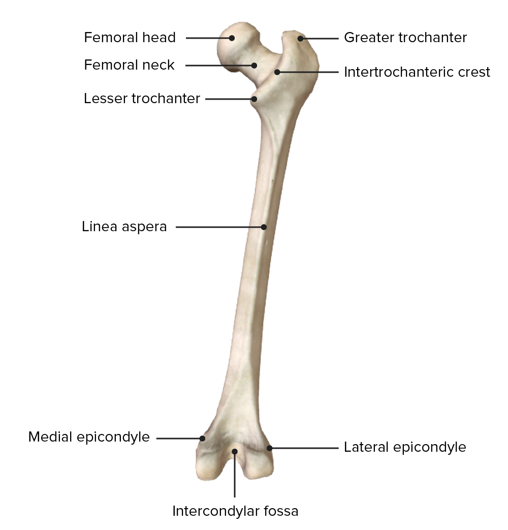
Posterior view of the right femur
Image by BioDigital, edited by Lecturio.The iliotibial tract or iliotibial band is a thickening of the fascia lata Fascia lata Femoral Region and Hernias: Anatomy located on the lateral surface of the thigh. The iliotibial tract is a stabilizer of the hip and knee.
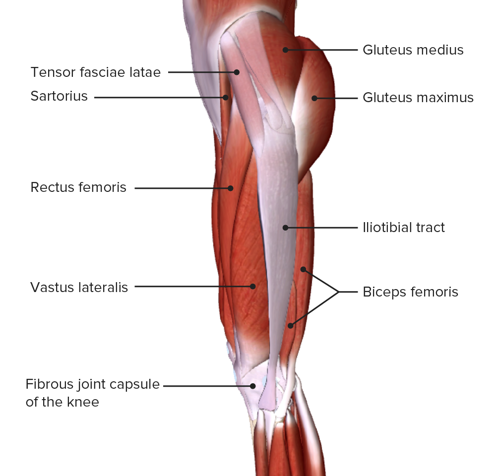
Lateral view of the thigh, featuring the iliotibial tract and tensor fasciae latae muscle
Image by BioDigital, edited by Lecturio.The 3 intermuscular septa arise from the fascia lata Fascia lata Femoral Region and Hernias: Anatomy and attach to the linea aspera of the femur. The lateral, medial, and posterior intermuscular septums divide the thigh into the following:
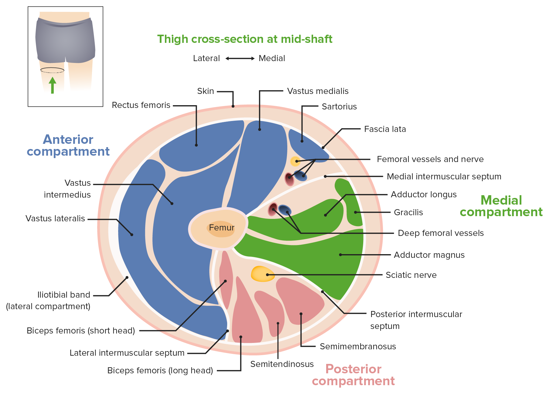
Thigh cross-section at mid-shaft
Image by Lecturio.| Muscle | Origin | Insertion | Innervation | Function |
|---|---|---|---|---|
| Rectus femoris | Anterior inferior iliac spine Spine The human spine, or vertebral column, is the most important anatomical and functional axis of the human body. It consists of 7 cervical vertebrae, 12 thoracic vertebrae, and 5 lumbar vertebrae and is limited cranially by the skull and caudally by the sacrum. Vertebral Column: Anatomy | Tibial tuberosity Tibial tuberosity Leg: Anatomy via common quadriceps and patellar ligament Patellar ligament A band of fibrous tissue that attaches the apex of the patella to the lower part of the tubercle of the tibia. The ligament is actually the caudal continuation of the common tendon of the quadriceps femoris. The patella is embedded in that tendon. As such, the patellar ligament can be thought of as connecting the quadriceps femoris tendon to the tibia, and therefore it is sometimes called the patellar tendon. Osgood-Schlatter Disease | Femoral nerve Femoral Nerve A nerve originating in the lumbar spinal cord (usually L2 to L4) and traveling through the lumbar plexus to provide motor innervation to extensors of the thigh and sensory innervation to parts of the thigh, lower leg, and foot, and to the hip and knee joints. Femoral Region and Hernias: Anatomy (L3, L4) |
|
| Vastus lateralis | Greater trochanter and the lateral lip of linea aspera | |||
| Vastus intermedius | Anterior shaft of the femur | |||
| Vastus medialis | Intertrochanteric line and medial lip of linea aspera | |||
| Sartorius | Anterior superior iliac spine Anterior Superior Iliac Spine Chronic Apophyseal Injury | Medial surface of the proximal tibia Tibia The second longest bone of the skeleton. It is located on the medial side of the lower leg, articulating with the fibula laterally, the talus distally, and the femur proximally. Knee Joint: Anatomy | Femoral nerve Femoral Nerve A nerve originating in the lumbar spinal cord (usually L2 to L4) and traveling through the lumbar plexus to provide motor innervation to extensors of the thigh and sensory innervation to parts of the thigh, lower leg, and foot, and to the hip and knee joints. Femoral Region and Hernias: Anatomy (L2) |
|
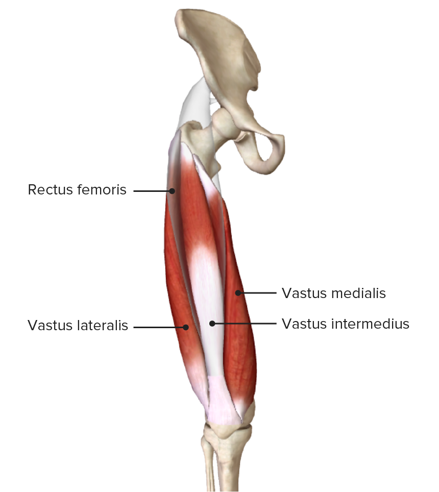
Anterior view of the thigh showing the origin and insertion of the quadriceps femoris
Image by BioDigital, edited by Lecturio.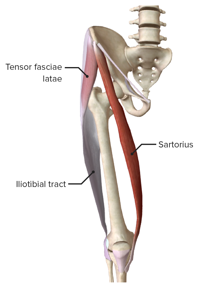
Tensor fasciae latae, sartorius muscles, and iliotibial tract
Image by BioDigital, edited by Lecturio.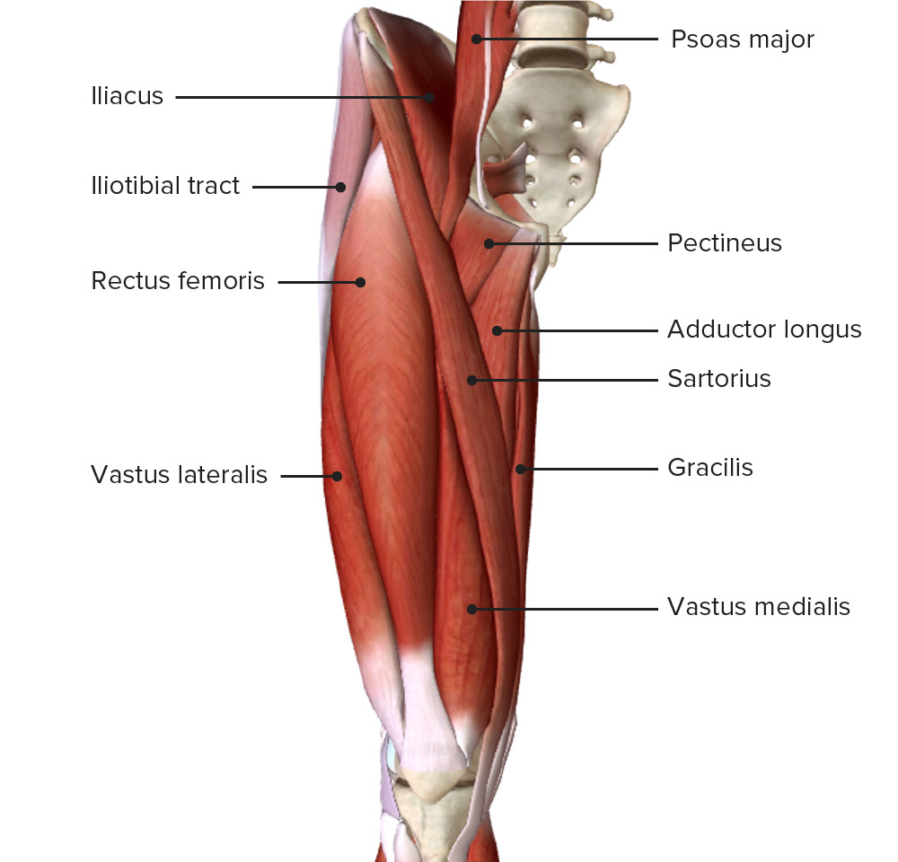
Anterior view of the thigh showing the muscles of the anterior compartment (quadriceps femoris and sartorius) and their relationship to neighboring muscles and each other
Image by BioDigital, edited by Lecturio.| Muscle | Origin | Insertion | Innervation | Function |
|---|---|---|---|---|
| Biceps Biceps Arm: Anatomy femoris |
|
Lateral surface of the fibula Fibula The bone of the lower leg lateral to and smaller than the tibia. In proportion to its length, it is the most slender of the long bones. Leg: Anatomy |
|
|
| Semitendinosus | Ischial tuberosity Ischial Tuberosity Chronic Apophyseal Injury | Medial surface of the proximal tibia Tibia The second longest bone of the skeleton. It is located on the medial side of the lower leg, articulating with the fibula laterally, the talus distally, and the femur proximally. Knee Joint: Anatomy | Tibial division of sciatic nerve Sciatic Nerve A nerve which originates in the lumbar and sacral spinal cord (l4 to s3) and supplies motor and sensory innervation to the lower extremity. The sciatic nerve, which is the main continuation of the sacral plexus, is the largest nerve in the body. It has two major branches, the tibial nerve and the peroneal nerve. Gluteal Region: Anatomy (L5, S1 S1 Heart Sounds) | Extends the hip, flexes the knee, and medially rotates the knee when flexed |
| Semimembranosus | Posterior surface of medial condyle of tibia Tibia The second longest bone of the skeleton. It is located on the medial side of the lower leg, articulating with the fibula laterally, the talus distally, and the femur proximally. Knee Joint: Anatomy |
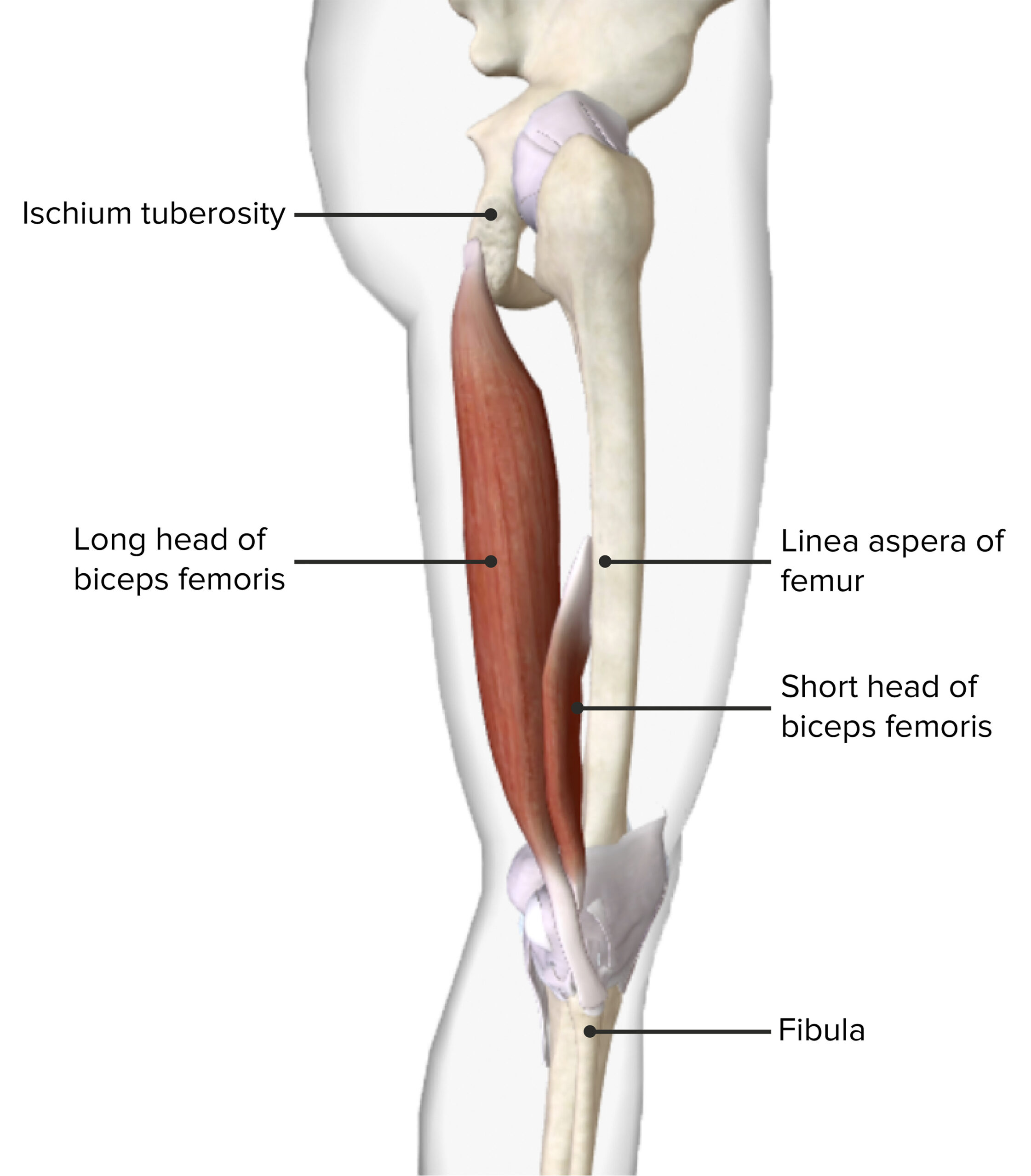
Lateral view of the thigh showing the origin and insertion of the biceps femoris muscle
Image by BioDigital, edited by Lecturio.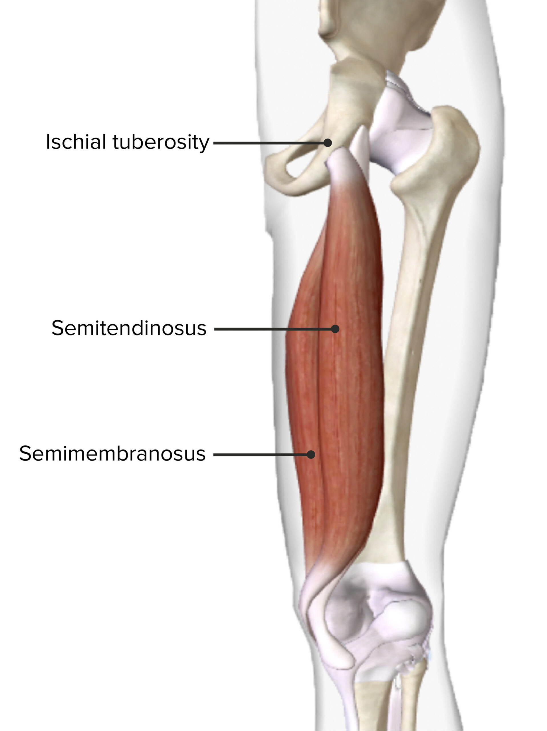
Posterior view of the right thigh showing the origin and insertion of the semitendinosus and semimembranosus muscles
Image by BioDigital, edited by Lecturio.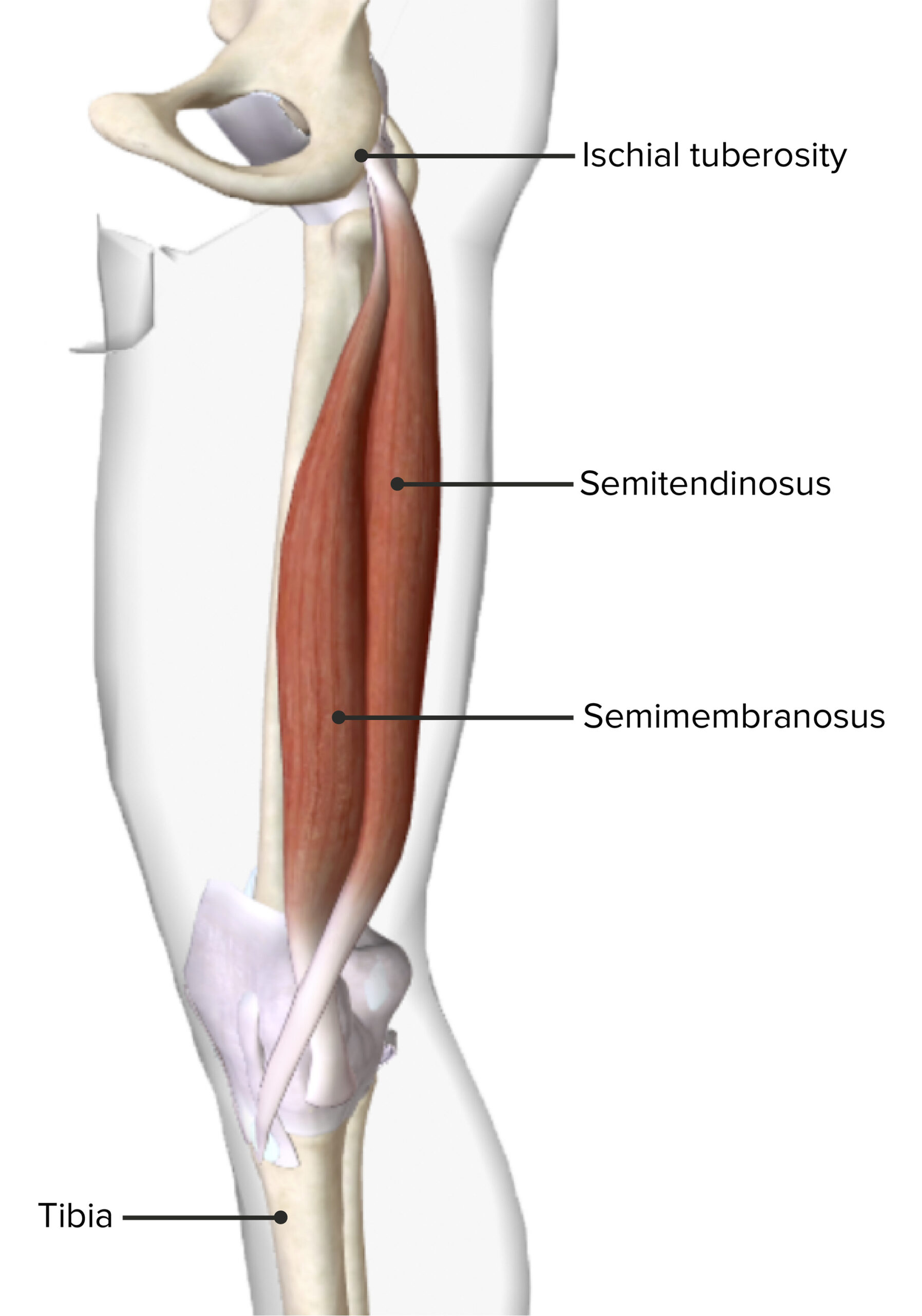
Medial view of the right thigh showing the origin and insertion of the semitendinosus and semimembranosus muscles
Image by BioDigital, edited by Lecturio.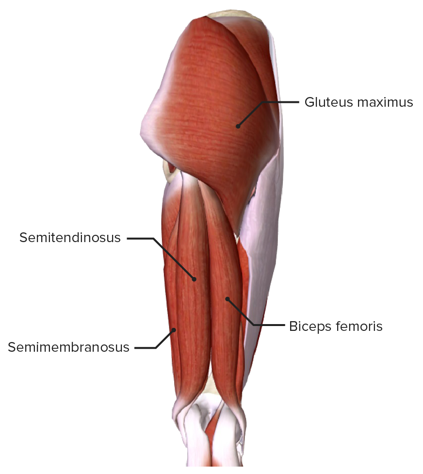
Posterior view of the thigh showing the muscles of the posterior compartment of the thigh and their relationship to other muscles and each other
Image by BioDigital, edited by Lecturio.| Muscle | Origin | Insertion | Innervation | Function |
|---|---|---|---|---|
| Adductor magnus |
|
|
|
|
| Adductor longus | Body of pubis | Middle ⅓ of linea aspera | Obturator nerve (L3) | Adducts the hip |
| Adductor brevis | Body and inferior ramus of pubis | Proximal linea aspera | ||
| Gracilis | Medial surface of the proximal tibia Tibia The second longest bone of the skeleton. It is located on the medial side of the lower leg, articulating with the fibula laterally, the talus distally, and the femur proximally. Knee Joint: Anatomy | Obturator nerve (L2) |
|
|
| Pectineus | Superior pubic ramus | Pectineal line of femur | Femoral nerve Femoral Nerve A nerve originating in the lumbar spinal cord (usually L2 to L4) and traveling through the lumbar plexus to provide motor innervation to extensors of the thigh and sensory innervation to parts of the thigh, lower leg, and foot, and to the hip and knee joints. Femoral Region and Hernias: Anatomy (L2) | Adducts, flexes, and supports medial rotation Rotation Motion of an object in which either one or more points on a line are fixed. It is also the motion of a particle about a fixed point. X-rays of the hip |
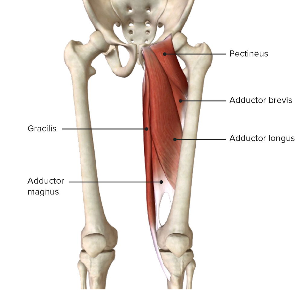
Anterior view of the thighs showing the origin and insertion of the muscles of the medial compartment
Image by BioDigital, edited by Lecturio.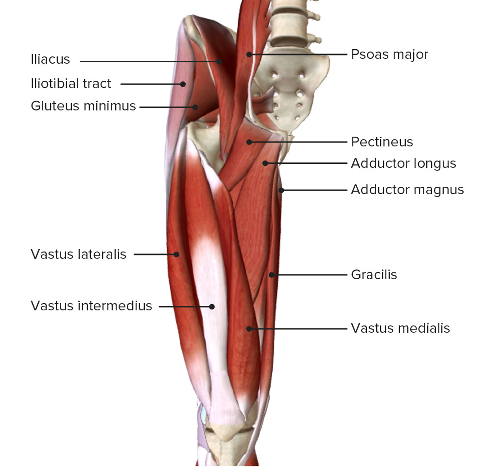
Anterior view of the thigh featuring the intermediate muscular layer:
Note the muscles of the medial compartment (the pectineus, adductor magnus, longus, and brevis, and gracilis) and their relationship to other muscles and each other
The distal end of the medial region of the thigh is the location of the pes anserinus or “goose foot,” which comprises the conjoined tendons of the sartorius, gracilis, and semitendinosus muscles inserting into the tibia Tibia The second longest bone of the skeleton. It is located on the medial side of the lower leg, articulating with the fibula laterally, the talus distally, and the femur proximally. Knee Joint: Anatomy.
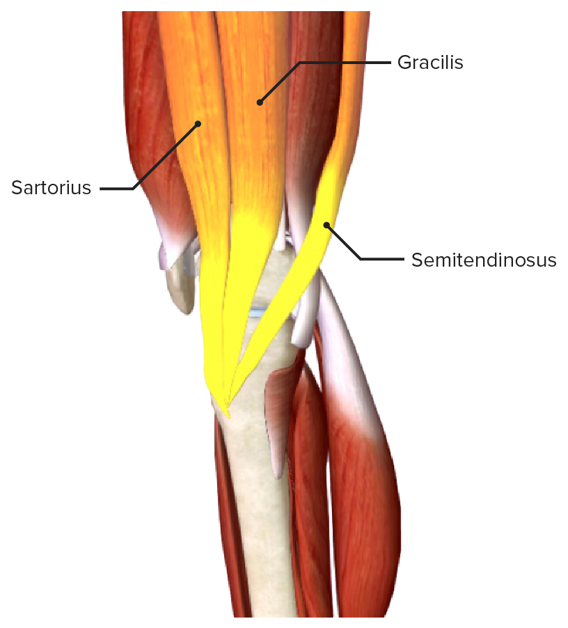
The conjoined tendon of the gracilis, sartorius, and semitendinosus muscles, which form the pes anserinus (goose foot)
Image by BioDigital, edited by Lecturio.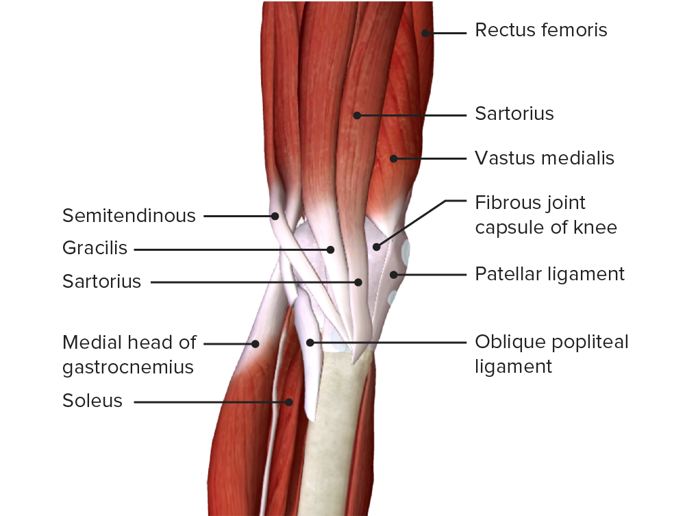
Medial view of the lower thigh and knee joint showing the insertion of the sartorius, gracilis, and semitendinosus muscles, which form the pes anserinus (goose foot)
Image by BioDigital, edited by Lecturio.Three arteries Arteries Arteries are tubular collections of cells that transport oxygenated blood and nutrients from the heart to the tissues of the body. The blood passes through the arteries in order of decreasing luminal diameter, starting in the largest artery (the aorta) and ending in the small arterioles. Arteries are classified into 3 types: large elastic arteries, medium muscular arteries, and small arteries and arterioles. Arteries: Histology exit the pelvis Pelvis The pelvis consists of the bony pelvic girdle, the muscular and ligamentous pelvic floor, and the pelvic cavity, which contains viscera, vessels, and multiple nerves and muscles. The pelvic girdle, composed of 2 “hip” bones and the sacrum, is a ring-like bony structure of the axial skeleton that links the vertebral column with the lower extremities. Pelvis: Anatomy: the femoral, obturator, and inferior gluteal arteries Arteries Arteries are tubular collections of cells that transport oxygenated blood and nutrients from the heart to the tissues of the body. The blood passes through the arteries in order of decreasing luminal diameter, starting in the largest artery (the aorta) and ending in the small arterioles. Arteries are classified into 3 types: large elastic arteries, medium muscular arteries, and small arteries and arterioles. Arteries: Histology. However, the femoral and obturator arteries Arteries Arteries are tubular collections of cells that transport oxygenated blood and nutrients from the heart to the tissues of the body. The blood passes through the arteries in order of decreasing luminal diameter, starting in the largest artery (the aorta) and ending in the small arterioles. Arteries are classified into 3 types: large elastic arteries, medium muscular arteries, and small arteries and arterioles. Arteries: Histology continue toward the thigh, while the inferior gluteal supplies and ends in the gluteal region Gluteal region The gluteal region is located posterior to the pelvic girdle and extends distally into the upper leg as the posterior thigh. The gluteal region consists of the gluteal muscles and several clinically important arteries, veins, and nerves. The muscles of the gluteal region help to move the hip joint during walking, running, standing, and sitting. Gluteal Region: Anatomy.
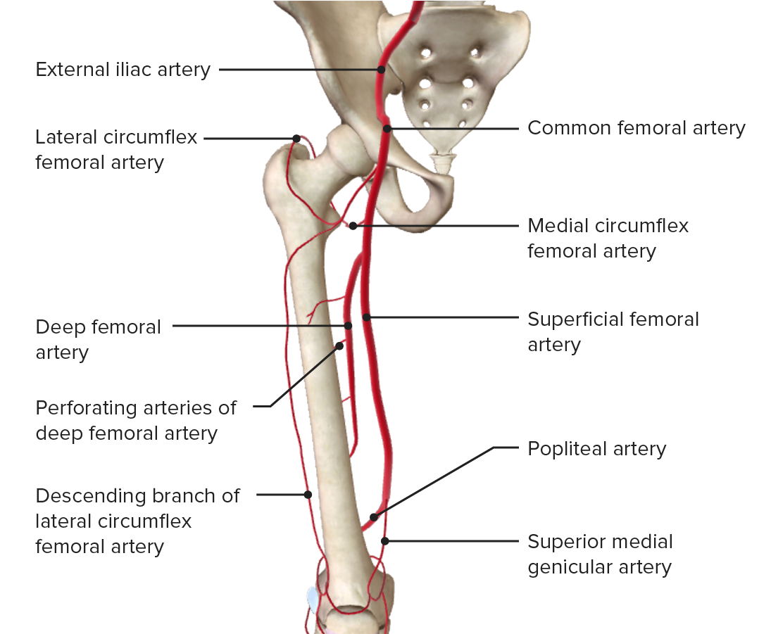
Arterial supply of the thigh showing the trajectory and main branches of the femoral artery
Image by BioDigital, edited by Lecturio.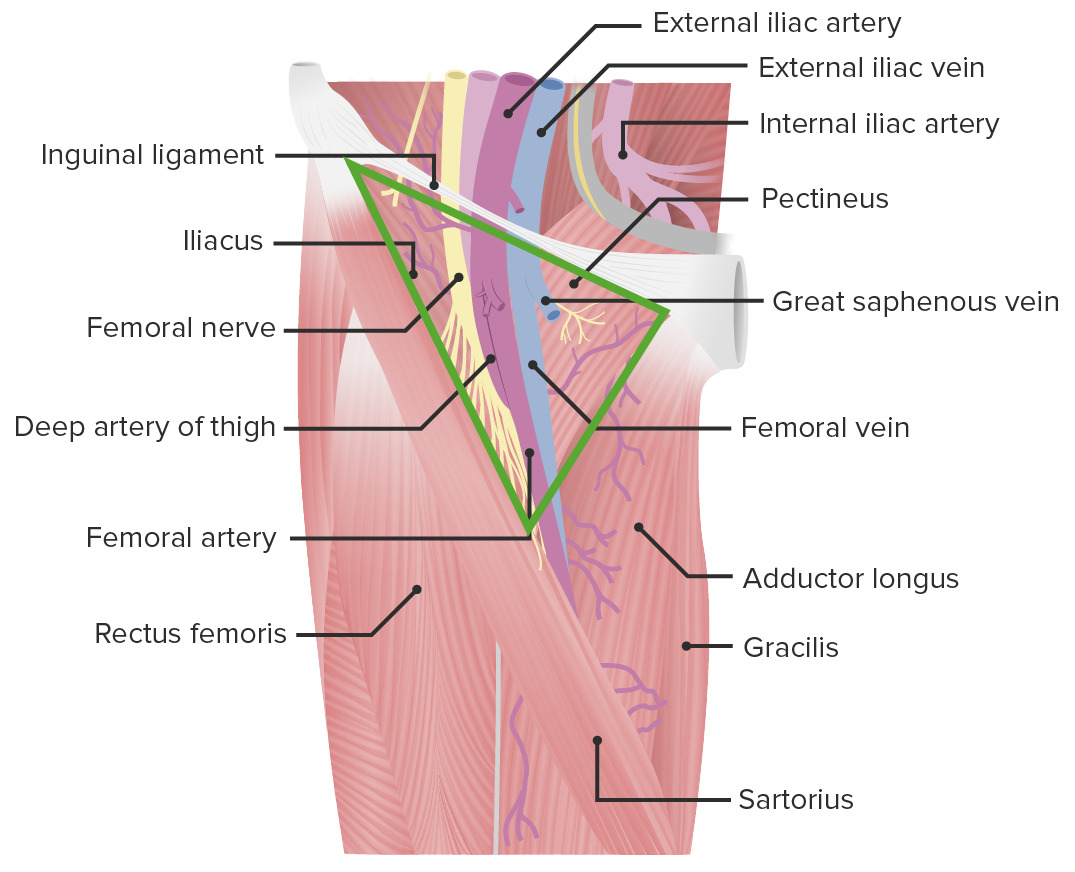
Anterior view of the thigh showing the passage of the femoral artery through the femoral triangle
Image by Lecturio.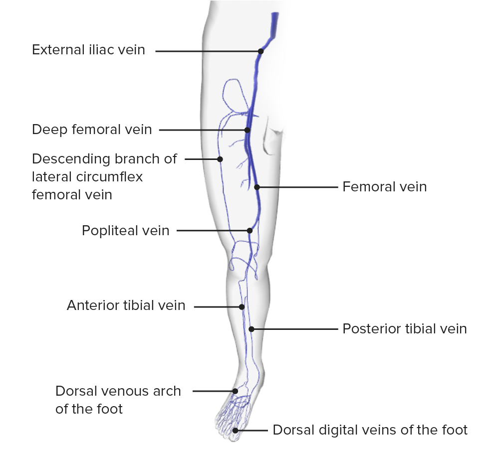
Anterior view of the venous drainage of the lower limb
Image by BioDigital, edited by Lecturio.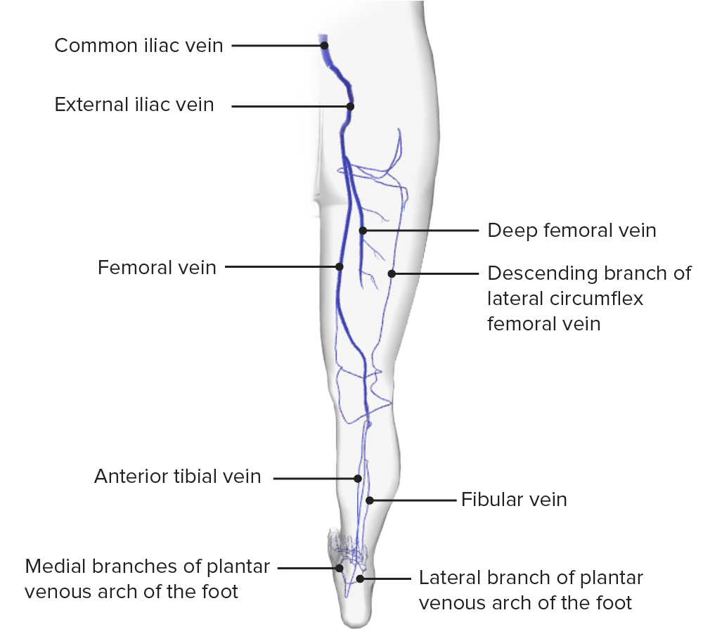
Posterior view of the venous drainage of the lower limb
Image by BioDigital, edited by Lecturio.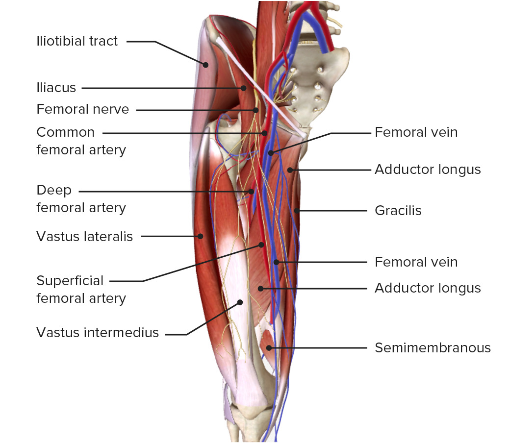
Anterior view of the thigh:
Note the course and branches of the common femoral artery, the superficial venous network, and the spatial relation of the vessels and muscles.
The thigh is innervated by branches of the lumbar and sacral plexus Sacral plexus Pelvis: Anatomy.
| Nerve | Origin | Motor Motor Neurons which send impulses peripherally to activate muscles or secretory cells. Nervous System: Histology supply | Sensory Sensory Neurons which conduct nerve impulses to the central nervous system. Nervous System: Histology supply |
|---|---|---|---|
| Lateral femoral cutaneous nerve | Lumbar plexus (L2–L3) | Skin Skin The skin, also referred to as the integumentary system, is the largest organ of the body. The skin is primarily composed of the epidermis (outer layer) and dermis (deep layer). The epidermis is primarily composed of keratinocytes that undergo rapid turnover, while the dermis contains dense layers of connective tissue. Skin: Structure and Functions of the anterolateral thigh | |
| Posterior femoral cutaneous nerve Posterior femoral cutaneous nerve Gluteal Region: Anatomy | Sacral plexus Sacral plexus Pelvis: Anatomy ( S2 S2 Heart Sounds– S3 S3 Heart Sounds) | Skin Skin The skin, also referred to as the integumentary system, is the largest organ of the body. The skin is primarily composed of the epidermis (outer layer) and dermis (deep layer). The epidermis is primarily composed of keratinocytes that undergo rapid turnover, while the dermis contains dense layers of connective tissue. Skin: Structure and Functions of the gluteal region Gluteal region The gluteal region is located posterior to the pelvic girdle and extends distally into the upper leg as the posterior thigh. The gluteal region consists of the gluteal muscles and several clinically important arteries, veins, and nerves. The muscles of the gluteal region help to move the hip joint during walking, running, standing, and sitting. Gluteal Region: Anatomy, posterior perineum Perineum The body region lying between the genital area and the anus on the surface of the trunk, and to the shallow compartment lying deep to this area that is inferior to the pelvic diaphragm. The surface area is between the vulva and the anus in the female, and between the scrotum and the anus in the male. Vagina, Vulva, and Pelvic Floor: Anatomy, and posterior thigh | |
| Femoral nerve Femoral Nerve A nerve originating in the lumbar spinal cord (usually L2 to L4) and traveling through the lumbar plexus to provide motor innervation to extensors of the thigh and sensory innervation to parts of the thigh, lower leg, and foot, and to the hip and knee joints. Femoral Region and Hernias: Anatomy |
|
|
|
| Saphenous nerve Saphenous nerve Foot: Anatomy | Branch of the femoral nerve Femoral Nerve A nerve originating in the lumbar spinal cord (usually L2 to L4) and traveling through the lumbar plexus to provide motor innervation to extensors of the thigh and sensory innervation to parts of the thigh, lower leg, and foot, and to the hip and knee joints. Femoral Region and Hernias: Anatomy | Skin Skin The skin, also referred to as the integumentary system, is the largest organ of the body. The skin is primarily composed of the epidermis (outer layer) and dermis (deep layer). The epidermis is primarily composed of keratinocytes that undergo rapid turnover, while the dermis contains dense layers of connective tissue. Skin: Structure and Functions of lower 2/3 of the medial thigh, medial lower leg Leg The lower leg, or just “leg” in anatomical terms, is the part of the lower limb between the knee and the ankle joint. The bony structure is composed of the tibia and fibula bones, and the muscles of the leg are grouped into the anterior, lateral, and posterior compartments by extensions of fascia. Leg: Anatomy, and foot Foot The foot is the terminal portion of the lower limb, whose primary function is to bear weight and facilitate locomotion. The foot comprises 26 bones, including the tarsal bones, metatarsal bones, and phalanges. The bones of the foot form longitudinal and transverse arches and are supported by various muscles, ligaments, and tendons. Foot: Anatomy | |
| Obturator nerve |
|
Muscles of the medial compartment | Skin Skin The skin, also referred to as the integumentary system, is the largest organ of the body. The skin is primarily composed of the epidermis (outer layer) and dermis (deep layer). The epidermis is primarily composed of keratinocytes that undergo rapid turnover, while the dermis contains dense layers of connective tissue. Skin: Structure and Functions of upper 1/3 of the medial thigh |
| Sciatic nerve Sciatic Nerve A nerve which originates in the lumbar and sacral spinal cord (l4 to s3) and supplies motor and sensory innervation to the lower extremity. The sciatic nerve, which is the main continuation of the sacral plexus, is the largest nerve in the body. It has two major branches, the tibial nerve and the peroneal nerve. Gluteal Region: Anatomy (thickest nerve in the human body) |
|
Tibial branch: Muscles of the posterior compartment | None in the thigh |
Innervation of the lower limbs
Image by Lecturio.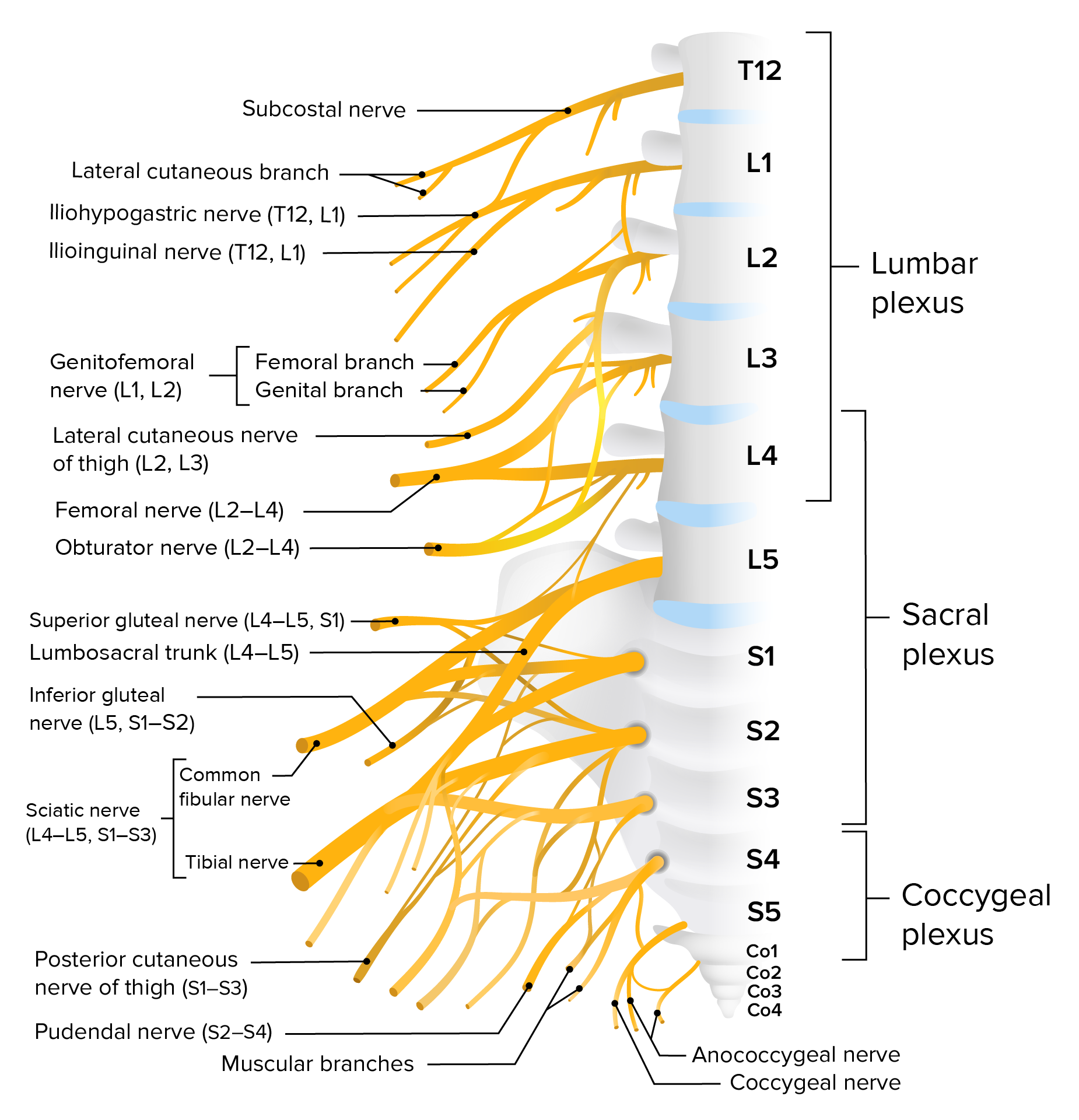
Lumbosacral plexus
Image by Lecturio.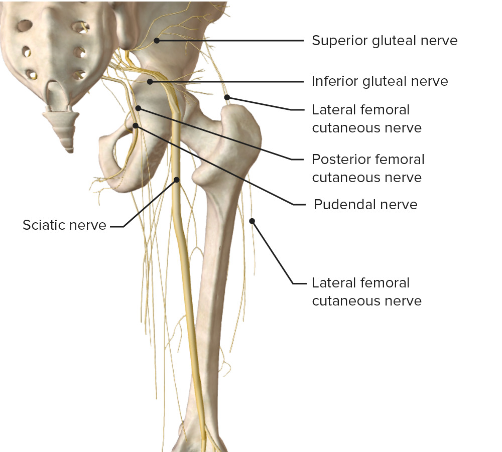
Schematic diagram of the course and main branches of the lumbosacral plexus
Image by BioDigital, edited by Lecturio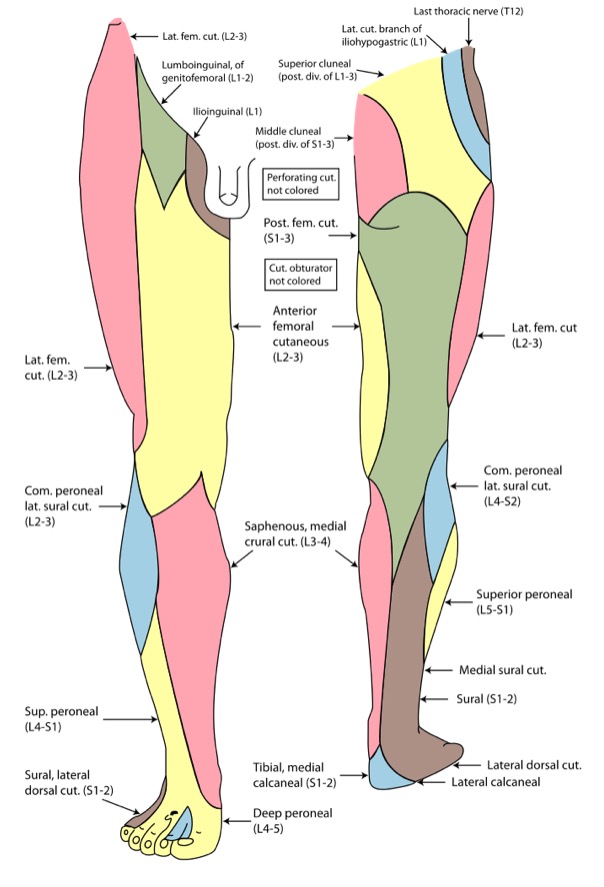
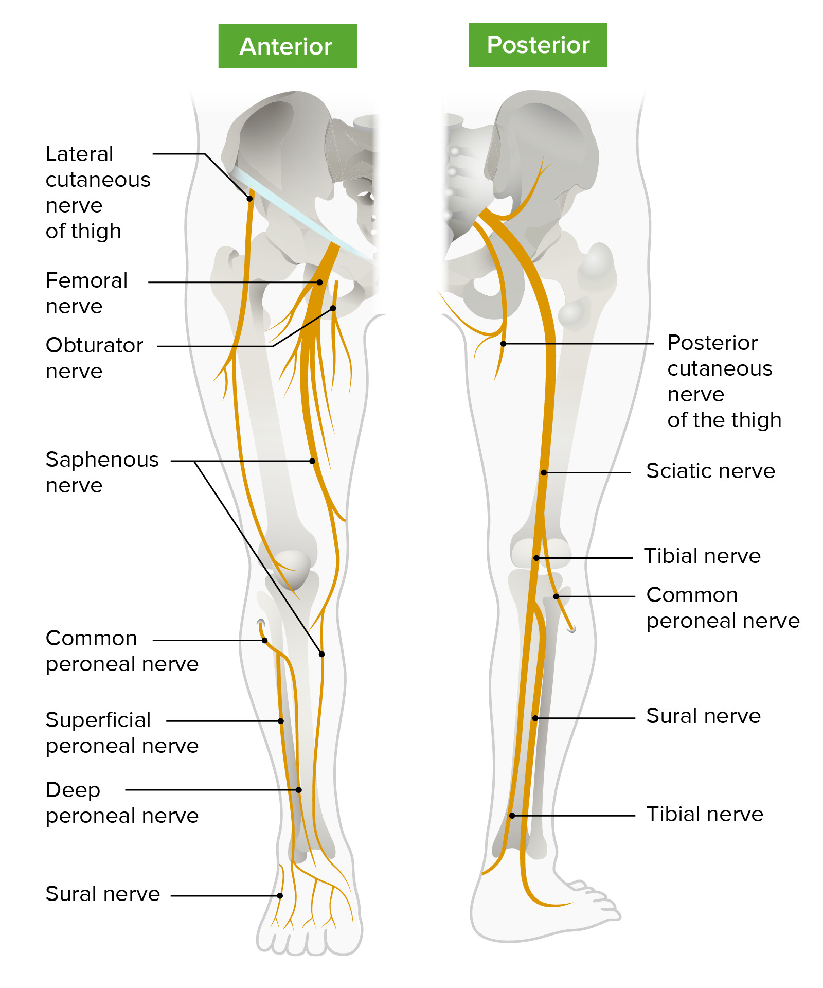
Cutaneous innervation of the lower limb
Image: “Gray826and831” by Henry Vandyke Carter. License: Public DomainThe following conditions are clinically relevant to the thigh: