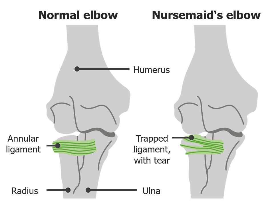
Radial head subluxation, also known as nursemaid’s elbow or babysitter’s elbow, is a frequent injury seen in children under 4 years of age and describes the subluxation of the radial head under the annularAnnularDermatologic Examination ligament at the elbow. The injury primarily occurs when a child is pulled, swung, or lifted by 1 armArmThe arm, or "upper arm" in common usage, is the region of the upper limb that extends from the shoulder to the elbow joint and connects inferiorly to the forearm through the cubital fossa. It is divided into 2 fascial compartments (anterior and posterior).Arm: Anatomy. PatientsPatientsIndividuals participating in the health care system for the purpose of receiving therapeutic, diagnostic, or preventive procedures.Clinician–Patient Relationship present holding their injured upper limb in a guarded and pronated position. Diagnosis is made clinically and the condition is managed by a closed-reduction maneuver. PrognosisPrognosisA prediction of the probable outcome of a disease based on a individual's condition and the usual course of the disease as seen in similar situations.Non-Hodgkin Lymphomas is excellent when diagnosed in a timely manner.
Radial head subluxation, also known as nursemaid’s elbow or babysitter’s elbow, describes the subluxation of the radial head under the annularAnnularDermatologic Examination ligament due to longitudinal traction on the forearmForearmThe forearm is the region of the upper limb between the elbow and the wrist. The term “forearm” is used in anatomy to distinguish this area from the arm, a term that is commonly used to describe the entire upper limb. The forearm consists of 2 long bones (the radius and the ulna), the interosseous membrane, and multiple arteries, nerves, and muscles. Forearm: Anatomy.
Results in over 20,000 emergency department visits annually in the United States.
Most commonly occurs in children aged 1–4 years
Represents more than 20% of upper extremity injuries in children.
The left armArmThe arm, or “upper arm” in common usage, is the region of the upper limb that extends from the shoulder to the elbow joint and connects inferiorly to the forearm through the cubital fossa. It is divided into 2 fascial compartments (anterior and posterior).Arm: Anatomy tends to be injured more often.
Radial head subluxation occurs when the child is lifted up by the arms with resultant axialAxialComputed Tomography (CT) traction of the forearmForearmThe forearm is the region of the upper limb between the elbow and the wrist. The term “forearm” is used in anatomy to distinguish this area from the arm, a term that is commonly used to describe the entire upper limb. The forearm consists of 2 long bones (the radius and the ulna), the interosseous membrane, and multiple arteries, nerves, and muscles. Forearm: Anatomy; e.g., while the child is being swung, lifted, or pulled by 1 armArmThe arm, or “upper arm” in common usage, is the region of the upper limb that extends from the shoulder to the elbow joint and connects inferiorly to the forearm through the cubital fossa. It is divided into 2 fascial compartments (anterior and posterior).Arm: Anatomy.
Pronated forearmForearmThe forearm is the region of the upper limb between the elbow and the wrist. The term “forearm” is used in anatomy to distinguish this area from the arm, a term that is commonly used to describe the entire upper limb. The forearm consists of 2 long bones (the radius and the ulna), the interosseous membrane, and multiple arteries, nerves, and muscles. Forearm: Anatomy undergoes axialAxialComputed Tomography (CT) traction while the elbow is extended.
As children age, the annularAnnularDermatologic Examination ligament thickens and nursemaid’s elbow becomes less likely to occur.
Other possible mechanisms:
Falling onto outstretched armArmThe arm, or “upper arm” in common usage, is the region of the upper limb that extends from the shoulder to the elbow joint and connects inferiorly to the forearm through the cubital fossa. It is divided into 2 fascial compartments (anterior and posterior).Arm: Anatomy
Twisting of forearmForearmThe forearm is the region of the upper limb between the elbow and the wrist. The term “forearm” is used in anatomy to distinguish this area from the arm, a term that is commonly used to describe the entire upper limb. The forearm consists of 2 long bones (the radius and the ulna), the interosseous membrane, and multiple arteries, nerves, and muscles. Forearm: Anatomy
Difference between a normal elbow and a nursemaid’s elbow
History and presentation often lead to the diagnosis:
Young toddler refusing to use armArmThe arm, or “upper arm” in common usage, is the region of the upper limb that extends from the shoulder to the elbow joint and connects inferiorly to the forearm through the cubital fossa. It is divided into 2 fascial compartments (anterior and posterior).Arm: Anatomy
Often associated with history of longitudinal traction:
Young child moves suddenly in opposite direction while holding adult’s handHandThe hand constitutes the distal part of the upper limb and provides the fine, precise movements needed in activities of daily living. It consists of 5 metacarpal bones and 14 phalanges, as well as numerous muscles innervated by the median and ulnar nerves. Hand: Anatomy.
Young child is lifted up by arms.
Physical examination
The entire affected upper limb/clavicleClavicleA bone on the ventral side of the shoulder girdle, which in humans is commonly called the collar bone.Clavicle Fracture should be examined.
Patient is often anxious and protective of injured armArmThe arm, or “upper arm” in common usage, is the region of the upper limb that extends from the shoulder to the elbow joint and connects inferiorly to the forearm through the cubital fossa. It is divided into 2 fascial compartments (anterior and posterior).Arm: Anatomy.
Injured upper limb is held in slightly flexed, pronated position.
PatientsPatientsIndividuals participating in the health care system for the purpose of receiving therapeutic, diagnostic, or preventive procedures.Clinician–Patient Relationship are unable or unwilling to supinate their armArmThe arm, or “upper arm” in common usage, is the region of the upper limb that extends from the shoulder to the elbow joint and connects inferiorly to the forearm through the cubital fossa. It is divided into 2 fascial compartments (anterior and posterior).Arm: Anatomy.
Signs of trauma (e.g., ecchymosisEcchymosisExtravasation of blood into the skin, resulting in a nonelevated, rounded or irregular, blue or purplish patch, larger than a petechia.Orbital Fractures, edemaEdemaEdema is a condition in which excess serous fluid accumulates in the body cavity or interstitial space of connective tissues. Edema is a symptom observed in several medical conditions. It can be categorized into 2 types, namely, peripheral (in the extremities) and internal (in an organ or body cavity). Edema, warmth) or neurovascular compromise are absent; if present, other diagnoses should be considered.
If there is clinical suspicion of child abuseChild abuseChild abuse is an act or failure to act that results in harm to a child’s health or development. The abuse encompasses neglect as well as physical, sexual, and emotional harm. Seen in all subsets of society, child abuse is a cause of significant morbidity and mortality in the pediatric population. Child Abuse, a complete physical examination should be performed.
If radial head spontaneously reduces prior to examination, patientsPatientsIndividuals participating in the health care system for the purpose of receiving therapeutic, diagnostic, or preventive procedures.Clinician–Patient Relationship may be asymptomatic.
Diagnosis
History and physical examination with typical findings are sufficient to diagnose. Imaging may be useful when diagnostic challenges occur with atypical presentations or unknown history.
X-rayX-rayPenetrating electromagnetic radiation emitted when the inner orbital electrons of an atom are excited and release radiant energy. X-ray wavelengths range from 1 pm to 10 nm. Hard x-rays are the higher energy, shorter wavelength x-rays. Soft x-rays or grenz rays are less energetic and longer in wavelength. The short wavelength end of the x-ray spectrum overlaps the gamma rays wavelength range. The distinction between gamma rays and x-rays is based on their radiation source.Pulmonary Function Tests:
Rarely indicated with typical presentation
Useful in evaluation of other diagnoses (e.g., fractureFractureA fracture is a disruption of the cortex of any bone and periosteum and is commonly due to mechanical stress after an injury or accident. Open fractures due to trauma can be a medical emergency. Fractures are frequently associated with automobile accidents, workplace injuries, and trauma.Overview of Bone Fractures, congenital elbow dislocationElbow dislocationElbow dislocation is the displacement of either the radius or the ulna relative to the humerus. The most common mechanism of injury is falling on an outstretched hand. Elbow dislocation presents with joint swelling, pain, and restricted range of motion. Elbow Dislocation, infectious etiology)
A Salter-Harris type I fractureFractureA fracture is a disruption of the cortex of any bone and periosteum and is commonly due to mechanical stress after an injury or accident. Open fractures due to trauma can be a medical emergency. Fractures are frequently associated with automobile accidents, workplace injuries, and trauma.Overview of Bone Fractures of distal humerusHumerusBone in humans and primates extending from the shoulder joint to the elbow joint.Arm: Anatomy may present with normal radiographs.
Management and Prognosis
Management
Closed reduction of a nursemaid’s elbow is the procedure of choice. The clinicianClinicianA physician, nurse practitioner, physician assistant, or another health professional who is directly involved in patient care and has a professional relationship with patients.Clinician–Patient Relationship must be certain there are no fractures prior to manipulation.
This procedure is done while maintaining slight pressure over the radial head; often, the provider will feel a “click” in the elbow.
Typically, the child will be moving the armArmThe arm, or “upper arm” in common usage, is the region of the upper limb that extends from the shoulder to the elbow joint and connects inferiorly to the forearm through the cubital fossa. It is divided into 2 fascial compartments (anterior and posterior).Arm: Anatomy normally within 15 minutes.
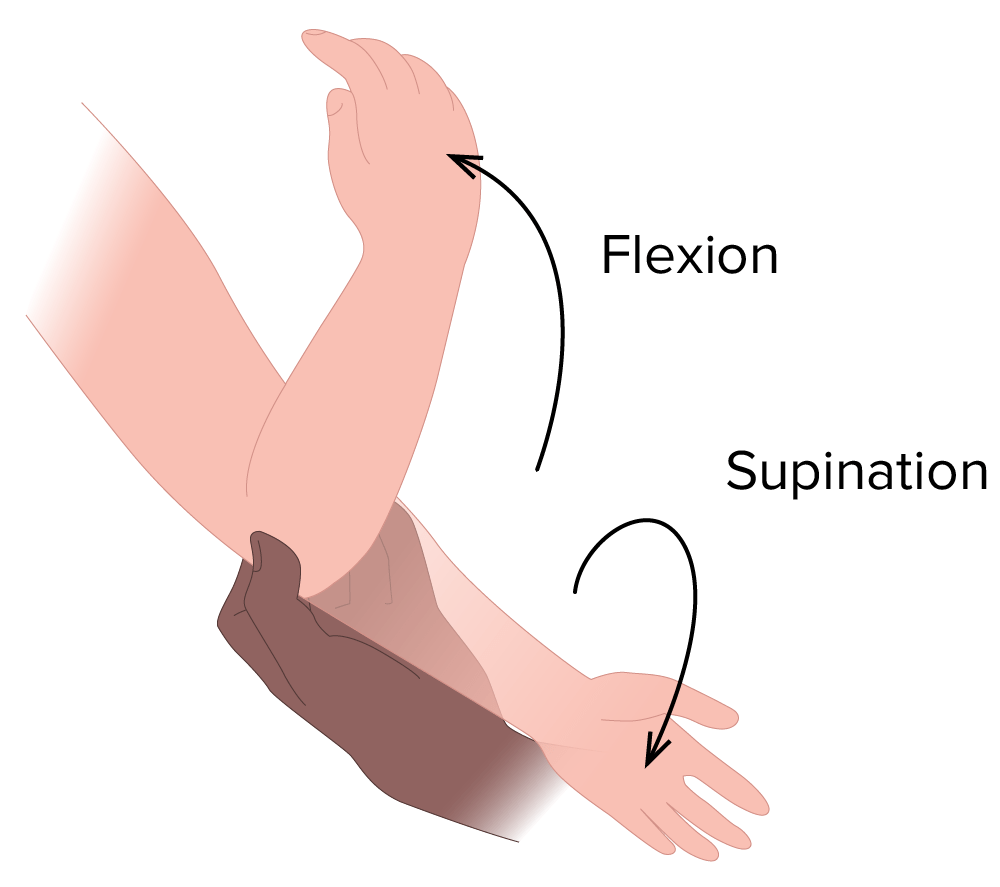
Supination/flexion technique
Image by Lecturio.
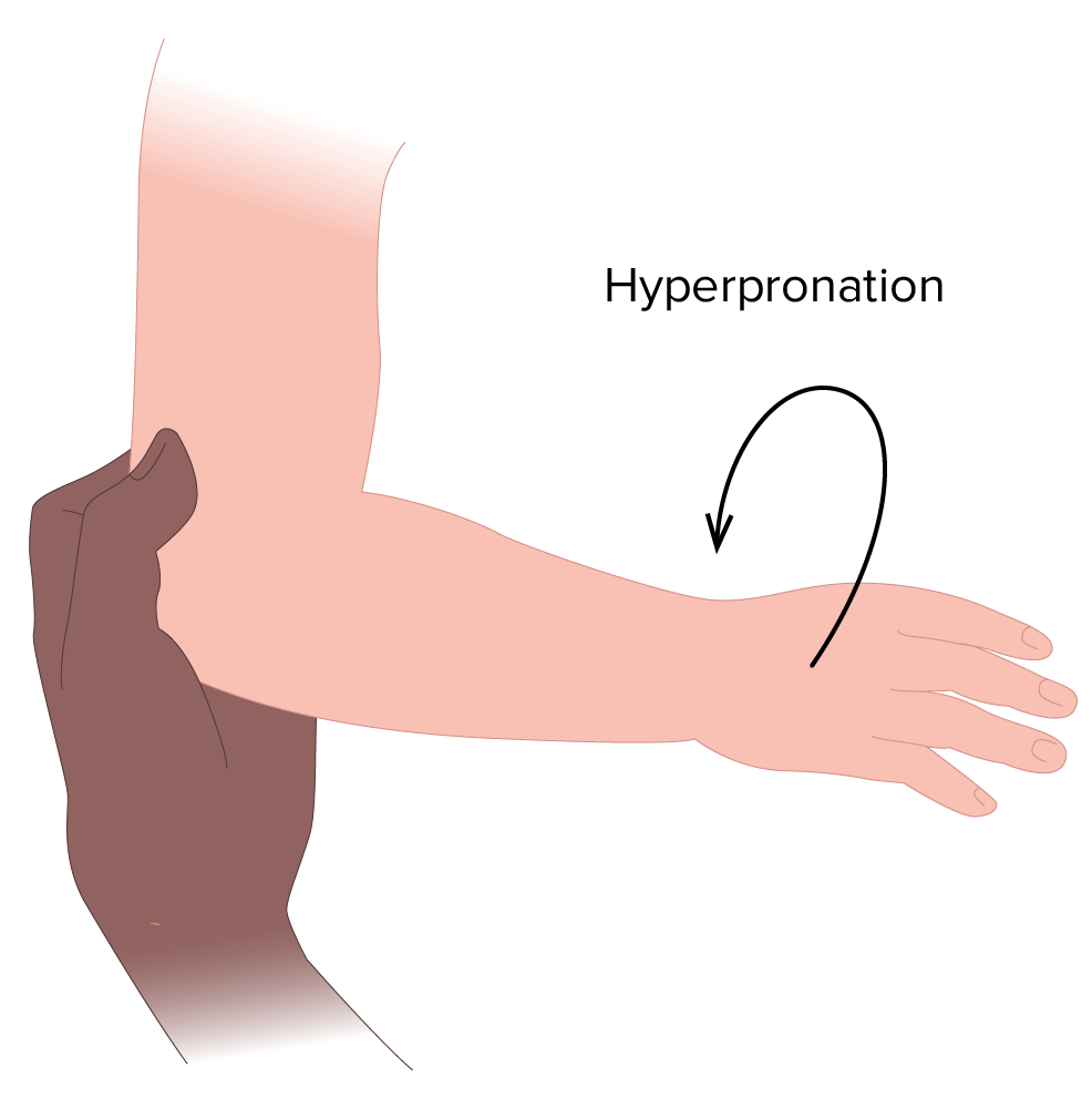
Hyperpronation technique:
Warn caregivers that the maneuver will hurt and the child will likely cry.
Child can be seated in parent’s or caregiver’s lap.
While applying mild pressure over the radial head, the provider holds the elbow in a flexed position and hyperpronates the forearmForearmThe forearm is the region of the upper limb between the elbow and the wrist. The term “forearm” is used in anatomy to distinguish this area from the arm, a term that is commonly used to describe the entire upper limb. The forearm consists of 2 long bones (the radius and the ulna), the interosseous membrane, and multiple arteries, nerves, and muscles. Forearm: Anatomy.
A click may be felt when displacementDisplacementThe process by which an emotional or behavioral response that is appropriate for one situation appears in another situation for which it is inappropriate.Defense Mechanisms is reduced.
Typically, the child will be moving the armArmThe arm, or “upper arm” in common usage, is the region of the upper limb that extends from the shoulder to the elbow joint and connects inferiorly to the forearm through the cubital fossa. It is divided into 2 fascial compartments (anterior and posterior).Arm: Anatomy normally within 15 minutes.
Hyperpronation technique
Image by Lecturio.
PatientsPatientsIndividuals participating in the health care system for the purpose of receiving therapeutic, diagnostic, or preventive procedures.Clinician–Patient Relationship who fail the initial reduction maneuver:
Reconsider the diagnosis.
If there are no signs of fractureFractureA fracture is a disruption of the cortex of any bone and periosteum and is commonly due to mechanical stress after an injury or accident. Open fractures due to trauma can be a medical emergency. Fractures are frequently associated with automobile accidents, workplace injuries, and trauma.Overview of Bone Fractures, reduction attempt may be repeated.
If unable to reduce or if diagnosis is in question, consider a splint and orthopedic referral.
PrognosisPrognosisA prediction of the probable outcome of a disease based on a individual’s condition and the usual course of the disease as seen in similar situations.Non-Hodgkin Lymphomas
PrognosisPrognosisA prediction of the probable outcome of a disease based on a individual’s condition and the usual course of the disease as seen in similar situations.Non-Hodgkin Lymphomas is excellent when reduced in a timely manner.
Recovery is immediate after reduction.
Recurrence rate: approximately 20%
Differential Diagnosis
Child abuseChild abuseChild abuse is an act or failure to act that results in harm to a child’s health or development. The abuse encompasses neglect as well as physical, sexual, and emotional harm. Seen in all subsets of society, child abuse is a cause of significant morbidity and mortality in the pediatric population. Child Abuse: an act or failure to act that results in harm to a child’s health or development. Child abuseChild abuseChild abuse is an act or failure to act that results in harm to a child’s health or development. The abuse encompasses neglect as well as physical, sexual, and emotional harm. Seen in all subsets of society, child abuse is a cause of significant morbidity and mortality in the pediatric population. Child Abuse encompasses neglectNeglectChild Abuse as well as physical, sexual, and emotional harm. Seen in all subsets of society, child abuseChild abuseChild abuse is an act or failure to act that results in harm to a child’s health or development. The abuse encompasses neglect as well as physical, sexual, and emotional harm. Seen in all subsets of society, child abuse is a cause of significant morbidity and mortality in the pediatric population. Child Abuse is a cause of significant morbidityMorbidityThe proportion of patients with a particular disease during a given year per given unit of population.Measures of Health Status and mortalityMortalityAll deaths reported in a given population.Measures of Health Status in the pediatric population.
Greenstick fractureGreenstick fractureThe bones of growing children exhibit unique characteristics, which, combined with the unique mechanisms of injury seen in children, result in fracture patterns differing significantly from those common in adults. The greenstick fracture is an incomplete fracture usually seen in long bones. Greenstick Fracture: partial-thickness fractureFractureA fracture is a disruption of the cortex of any bone and periosteum and is commonly due to mechanical stress after an injury or accident. Open fractures due to trauma can be a medical emergency. Fractures are frequently associated with automobile accidents, workplace injuries, and trauma.Overview of Bone Fractures that involves a complete break of cortex and periosteumPeriosteumThin outer membrane that surrounds a bone. It contains connective tissue, capillaries, nerves, and a number of cell types.Bones: Structure and Types on only 1 side of the boneBoneBone is a compact type of hardened connective tissue composed of bone cells, membranes, an extracellular mineralized matrix, and central bone marrow. The 2 primary types of bone are compact and spongy. Bones: Structure and Types. Termed “greenstick” as it resembles the break in a live, “green” twig, where 1 side of the stick remains intact. High risk for refracture and therefore should be completely immobilized. Rarely requires reduction, but should be managed cautiously to prevent malunionMalunionHip Fractures or angulationAngulationBuckle or Torus Fracture deformities, and often should be referred for orthopedic follow-up.
Buckle or torus fractureTorus FractureBuckle or Torus Fracture:fractureFractureA fracture is a disruption of the cortex of any bone and periosteum and is commonly due to mechanical stress after an injury or accident. Open fractures due to trauma can be a medical emergency. Fractures are frequently associated with automobile accidents, workplace injuries, and trauma.Overview of Bone Fractures affecting growing metaphyseal boneBoneBone is a compact type of hardened connective tissue composed of bone cells, membranes, an extracellular mineralized matrix, and central bone marrow. The 2 primary types of bone are compact and spongy. Bones: Structure and Types secondary to compressionCompressionBlunt Chest Trauma load, where boneBoneBone is a compact type of hardened connective tissue composed of bone cells, membranes, an extracellular mineralized matrix, and central bone marrow. The 2 primary types of bone are compact and spongy. Bones: Structure and Types buckles or compresses. Generally considered a stable fractureFractureA fracture is a disruption of the cortex of any bone and periosteum and is commonly due to mechanical stress after an injury or accident. Open fractures due to trauma can be a medical emergency. Fractures are frequently associated with automobile accidents, workplace injuries, and trauma.Overview of Bone Fractures. Treated by immobilizationImmobilizationDelirium and has good a prognosisPrognosisA prediction of the probable outcome of a disease based on a individual’s condition and the usual course of the disease as seen in similar situations.Non-Hodgkin Lymphomas.
Supracondylar fractureSupracondylar fractureSupracondylar fractures are the most common elbow fractures in the pediatric population. The most common mechanism of injury involves a fall on an outstretched hand, resulting in a fracture of the distal humerus. Patients frequently present with pain, visible deformity, and limited range of motion of the injured elbow. Supracondylar Fracture:complete fractureComplete FractureOverview of Bone Fractures affecting distal humerusHumerusBone in humans and primates extending from the shoulder joint to the elbow joint.Arm: Anatomy after falling on outstretched handHandThe hand constitutes the distal part of the upper limb and provides the fine, precise movements needed in activities of daily living. It consists of 5 metacarpal bones and 14 phalanges, as well as numerous muscles innervated by the median and ulnar nerves. Hand: Anatomy. Commonly fractures of the elbow in children. Requires immediate orthopedic consultation, as many cases are associated with neurovascular injury and require surgical intervention.
Welch, R., Chounthirath, T., Smith, G. A. (2017). Radial Head Subluxation Among Young Children in the United States Associated With Consumer Products and Recreational Activities. Clin Pediatr (Phila). 56(8), 707–715. https://pubmed.ncbi.nlm.nih.gov/28589762/