El virus Virus Viruses are infectious, obligate intracellular parasites composed of a nucleic acid core surrounded by a protein capsid. Viruses can be either naked (non-enveloped) or enveloped. The classification of viruses is complex and based on many factors, including type and structure of the nucleoid and capsid, the presence of an envelope, the replication cycle, and the host range. Virology del herpes simple (VHS) es un virus Virus Viruses are infectious, obligate intracellular parasites composed of a nucleic acid core surrounded by a protein capsid. Viruses can be either naked (non-enveloped) or enveloped. The classification of viruses is complex and based on many factors, including type and structure of the nucleoid and capsid, the presence of an envelope, the replication cycle, and the host range. Virology de ADN de doble cadena que pertenece a la familia Herpesviridae Herpesviridae A family of enveloped, linear, double-stranded DNA viruses infecting a wide variety of animals. Subfamilies, based on biological characteristics, include: alphaherpesvirinae; betaherpesvirinae; and gammaherpesvirinae. Herpes Simplex Virus 1 and 2. El virus Virus Viruses are infectious, obligate intracellular parasites composed of a nucleic acid core surrounded by a protein capsid. Viruses can be either naked (non-enveloped) or enveloped. The classification of viruses is complex and based on many factors, including type and structure of the nucleoid and capsid, the presence of an envelope, the replication cycle, and the host range. Virology del herpes simple suele causar infecciones recurrentes que afectan a la piel y a las superficies mucosas, como la boca, los LOS Neisseria labios, los LOS Neisseria ojos y los LOS Neisseria genitales. Las infecciones mucocutáneas típicas se caracterizan por una aparición aguda localizada de grupos de vesículas pequeñas y dolorosas sobre una base eritematosa. Aunque existe un solapamiento, el VHS-1 se asocia clásicamente a las lesiones orofaríngeas, mientras que el VHS-2 es el principal responsable del herpes genital, una ITS. También pueden producirse infecciones sistémicas y graves, como encefalitis, meningitis Meningitis Meningitis is inflammation of the meninges, the protective membranes of the brain, and spinal cord. The causes of meningitis are varied, with the most common being bacterial or viral infection. The classic presentation of meningitis is a triad of fever, altered mental status, and nuchal rigidity. Meningitis y herpes neonatal. El diagnóstico se realiza con base en EN Erythema nodosum is an immune-mediated panniculitis (inflammation of the subcutaneous fat) caused by a type IV (delayed-type) hypersensitivity reaction. It commonly manifests in young women as tender, erythematous nodules on the shins. Erythema Nodosum la presentación y los LOS Neisseria antecedentes clínicos, que puede confirmarse mediante el examen microscópico de un frotis teñido de una vesícula fresca, la prueba de amplificación nucleica mediante PCR PCR Polymerase chain reaction (PCR) is a technique that amplifies DNA fragments exponentially for analysis. The process is highly specific, allowing for the targeting of specific genomic sequences, even with minuscule sample amounts. The PCR cycles multiple times through 3 phases: denaturation of the template DNA, annealing of a specific primer to the individual DNA strands, and synthesis/elongation of new DNA molecules. Polymerase Chain Reaction (PCR), la inmunofluorescencia directa o las pruebas serológicas. El tratamiento de las lesiones mucocutáneas suele ser sintomático, pero las terapias antivirales con aciclovir, valaciclovir o famciclovir Famciclovir An aminopurine derivative and prodrug of penciclovir which is a competitive inhibitor of herpes simplex 2 DNA polymerase. It is used to treat herpes simplex virus infection. Antivirals for Herpes Virus son útiles si se administran rápidamente y siempre forman parte del tratamiento de las infecciones sistémicas graves.
Last updated: Dec 15, 2025
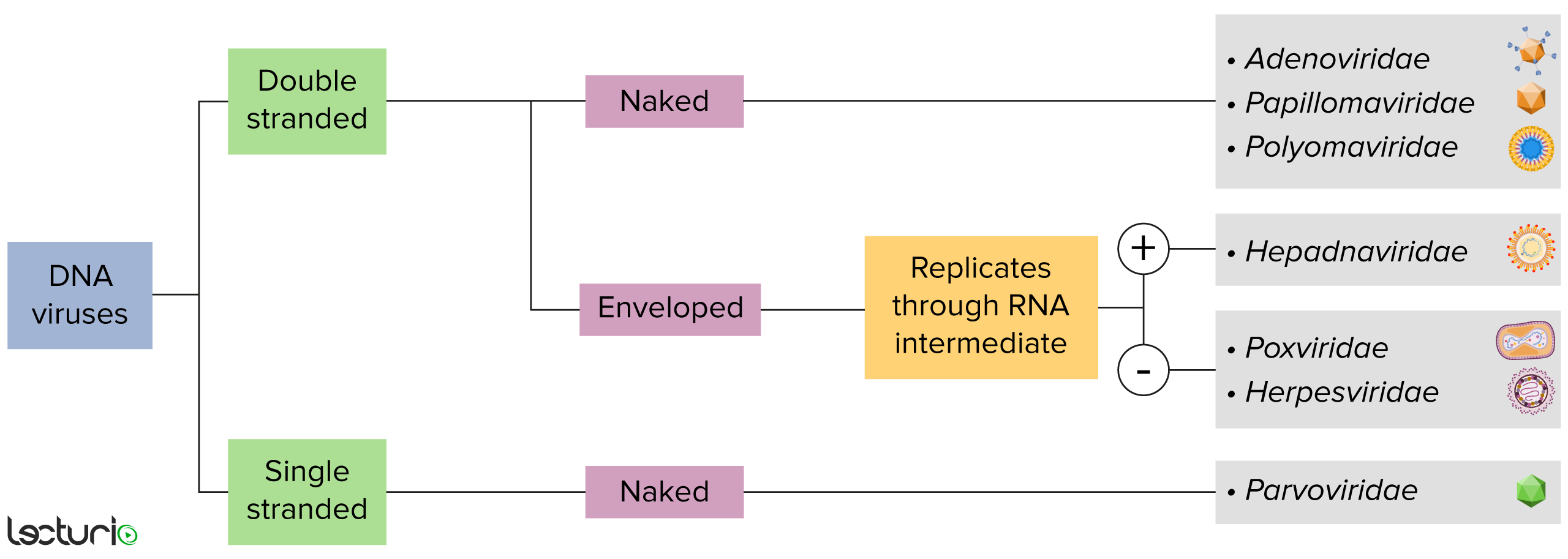
Identificación de virus de ADN:
Los virus pueden clasificarse de muchas maneras. Sin embargo, la mayoría de los virus tienen un genoma formado por ADN o ARN. Los virus con un genoma de ADN pueden caracterizarse además por el hecho de que ese ADN sea de cadena simple o doble. Si los virus están cubiertos por una fina capa de membrana celular (generalmente tomada de la célula huésped), se denominan virus “envueltos”. Si esa capa está ausente, los virus se denominan “desnudos”. Algunos de los virus con envoltura traducen su ADN en ARN antes de incorporarlo al genoma de la célula huésped.
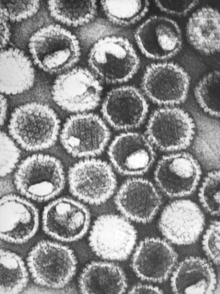
Micrografía electrónica de transmisión con tinción negativa que muestra numerosos viriones de herpes simple, miembros de la familia Herpesviridae:
En el núcleo de su cápside proteica icosaédrica, el virus del herpes simple contiene un genoma lineal de ADN de doble cadena.
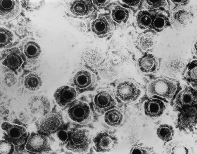
Micrografía electrónica de transmisión de viriones de herpes simple
Imagen: “Transmission electron micrograph of herpes simplex virions” por CDC/Dr. Erskine Palmer. Licencia: Dominio Público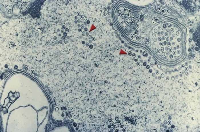
Imagen de microscopía electrónica de transmisión que muestra numerosos viriones redondos de herpes simple dentro del núcleo de una célula (flechas)
Imagen: “Transmission electron microscopic image demonstrating numerous, round herpes simplexvirions inside the nucleus of a cell (arrows)” por CDC. Licencia: Dominio PúblicoSe han reconocido dos tipos que causan infecciones:
VHS-1:
VHS-2:
Infección neonatal por herpes:
Los LOS Neisseria humanos son el principal reservorio.
VHS-1:
VHS-2:
Infección primaria:
Latencia de por vida:
Reactivación:
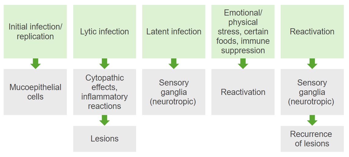
Diagrama que resume la patogénesis de las infecciones por VHS-1 y -2
Imagen de Lecturio. Licencia: CC BY-NC-SA 4.0Los LOS Neisseria virus Virus Viruses are infectious, obligate intracellular parasites composed of a nucleic acid core surrounded by a protein capsid. Viruses can be either naked (non-enveloped) or enveloped. The classification of viruses is complex and based on many factors, including type and structure of the nucleoid and capsid, the presence of an envelope, the replication cycle, and the host range. Virology del herpes simple causan infecciones citolíticas que constituyen la base de todos los LOS Neisseria cambios patológicos: necrosis Necrosis The death of cells in an organ or tissue due to disease, injury or failure of the blood supply. Ischemic Cell Damage de las células infectadas junto con la respuesta inflamatoria.
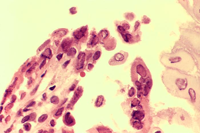
Biopsia de una úlcera esofágica causada por una infección activa por el virus del herpes simple (VHS) en un paciente con SIDA:
El espécimen fue cosechado en el borde ulceroso y revela la presencia de inclusiones intranucleares y células con núcleos múltiples
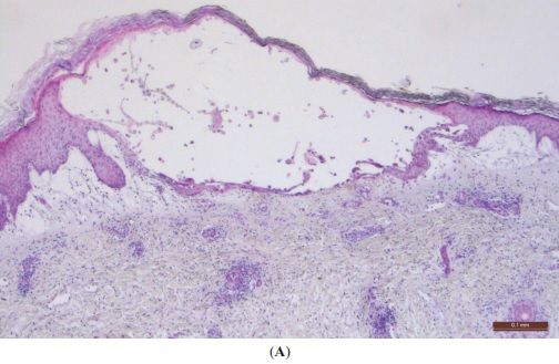
Fotomicrografía de baja potencia de una vesícula típica del virus del herpes:
La vesícula se llenará de neutrófilos (y cambiará de un líquido claro a un aspecto amarillo y turbio) antes de que se rompa y deje una úlcera superficial. Obsérvese cómo la lesión citopática vírica permanece dentro de la epidermis, lo que explica que se cure sin cicatrización, lo que puede ocurrir con una inflamación dérmica grave.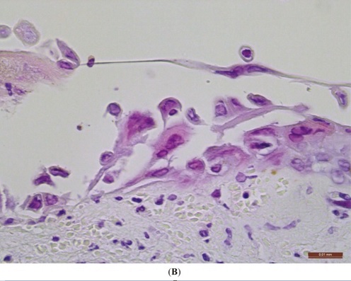
Fotomicrografía de alta potencia de una lesión del virus del herpes en la piel:
Obsérvese la acantólisis (queratinocitos separados entre sí), que es la base de la formación de ampollas. Los queratinocitos infectados están agrandados y presentan inclusiones intranucleares vítreas (“inclusiones Cowdry tipo A”). Unos pocos queratinocitos muestran multinucleación.
El VHS provoca una serie de afecciones. Las infecciones que se enumeran a continuación son las más comunes causadas por el VHS-1. Nota: El VHS-2 también puede estar asociado (aunque con menor frecuencia) a muchos de estos diagnósticos.
Solo el 20%‒25% de los LOS Neisseria pacientes con anticuerpos contra el VHS-1 tienen antecedentes clínicos positivos de infecciones orales-labiales o genitales.
Gingivoestomatitis:
Faringitis:
Herpes labial:
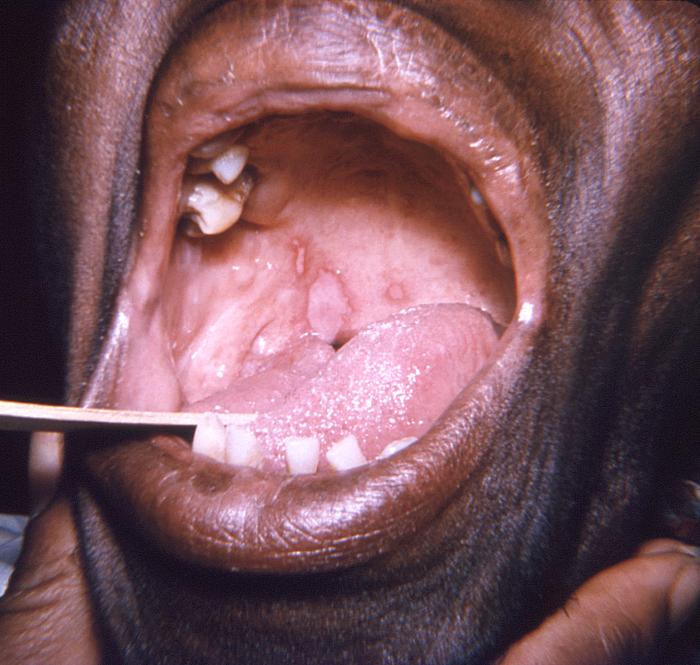
Lesiones en el paladar blando y la lengua causadas por el virus del herpes simple
Imagen: “This photograph depicts a close view of an elderly African American female patient’s oral cavity” por CDC/ Robert E. Sumpter. Licencia: Dominio Público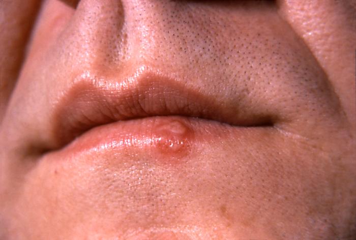
Lesión de herpes simple en el labio inferior, en el borde del bermellón, al segundo día de su aparición
Imagen: “This photograph depicts a close-up of the lips of a patient with a herpes simplex lesion on the lower lip” por CDC/ Dr. Hermann. Licencia: Dominio PúblicoPanadizo herpético:
Herpes del gladiador:
Eritema multiforme:
Eccema herpético:
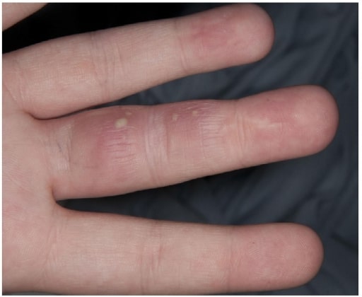
Un paciente con vesículas en el dedo por panadizo herpético
Imagen: “Herpetic Whitlow” por Salford Royal Foundation Trust, Manchester, UK. Licencia: CC BY 3.0
Lesiones vesiculares difusas en la cara de un paciente que padece eccema herpético mientras se somete a una terapia UV
Imagen: “Multiple pustules on the face surface” por AUTHOR. Licencia: CC BY 2.0Las infecciones oculares se producen en EN Erythema nodosum is an immune-mediated panniculitis (inflammation of the subcutaneous fat) caused by a type IV (delayed-type) hypersensitivity reaction. It commonly manifests in young women as tender, erythematous nodules on the shins. Erythema Nodosum < 5% de los LOS Neisseria pacientes con infecciones por VHS-1, lo que conlleva la pérdida de visión y/o la ceguera.
Queratitis:
Necrosis Necrosis The death of cells in an organ or tissue due to disease, injury or failure of the blood supply. Ischemic Cell Damage retiniana aguda:
Conjuntivitis y blefaritis:
Coriorretinitis (uveítis posterior):
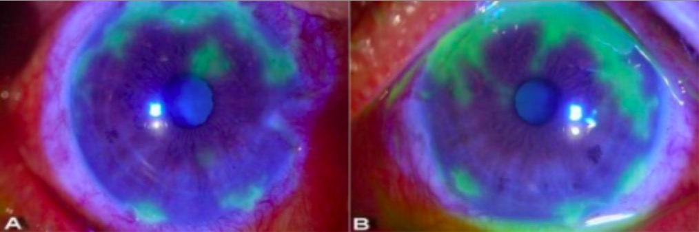
Examen con lámpara de hendidura que muestra lesiones dendríticas en queratitis herpética:
Aquí se ven patrones geográficos irregulares de ulceración resaltados en verde tras la aplicación de un colorante amarillo-naranja de fluoresceína. El tinte es absorbido por la córnea dañada (donde la superficie ha sido alterada) y la zona aparece verde bajo la luz azul cobalto.
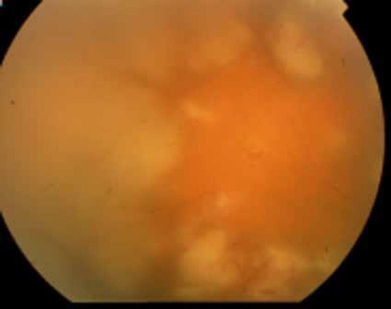
Imagen fundoscópica de un paciente con necrosis retiniana aguda:
Se pueden observar las características áreas necróticas confluentes. La retinitis aparece como manchas profundas, multifocales, de color blanco-amarillento, que suelen comenzar en el fondo de ojo periférico y luego se vuelven concéntricamente confluentes y se extienden hacia el polo posterior.
Encefalitis:
Meningitis Meningitis Meningitis is inflammation of the meninges, the protective membranes of the brain, and spinal cord. The causes of meningitis are varied, with the most common being bacterial or viral infection. The classic presentation of meningitis is a triad of fever, altered mental status, and nuchal rigidity. Meningitis aséptica:
Otras manifestaciones:
Epiglotitis o laringitis (crup herpético):
Neumonitis por VHS:
Esofagitis por VHS:
Hepatitis fulminante:
Las afecciones que se enumeran a continuación son las más comúnmente asociadas a las infecciones por VHS-2. Una vez más, hay que tener en EN Erythema nodosum is an immune-mediated panniculitis (inflammation of the subcutaneous fat) caused by a type IV (delayed-type) hypersensitivity reaction. It commonly manifests in young women as tender, erythematous nodules on the shins. Erythema Nodosum cuenta que el VHS-1 también puede estar asociado a muchos de estos diagnósticos.
Infecciones genitales primarias por VHS-2:
Infección no primaria de primer episodio (reactivación):
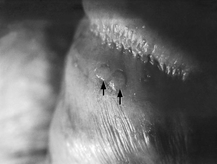
Ampollas en el pene (flechas) debidas a una infección recurrente por el virus del herpes simple-2 (VHS-2)
Imagen: “Penile blisters (arrows), due to a recurring herpes simplex-2 (HSV-2) virus infection” por CDC/ Susan Lindsley. Licencia: Dominio Público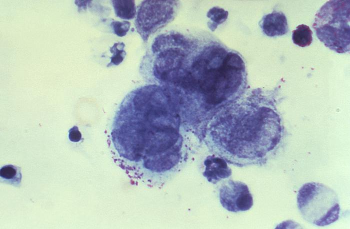
Imagen de un frotis de Tzanck obtenido de una lesión de pene:
Se observan células gigantes multinucleadas, lo que indica una infección por herpes.
Antivirales:
Analgésicos:
La tabla que se presenta a continuación compara y contrasta el VHS-1 y el VHS-2:
| Serotipo | VHS-1 | VHS-2 |
|---|---|---|
| Transmisión |
|
|
| Infección lítica | Células mucoepiteliales | Células mucoepiteliales |
| Latencia | Ganglios del trigémino | Ganglios sacros |
| Enfermedades |
|
|
La siguiente tabla compara los LOS Neisseria 9 herpesvirus considerados endémicos en EN Erythema nodosum is an immune-mediated panniculitis (inflammation of the subcutaneous fat) caused by a type IV (delayed-type) hypersensitivity reaction. It commonly manifests in young women as tender, erythematous nodules on the shins. Erythema Nodosum humanos. En EN Erythema nodosum is an immune-mediated panniculitis (inflammation of the subcutaneous fat) caused by a type IV (delayed-type) hypersensitivity reaction. It commonly manifests in young women as tender, erythematous nodules on the shins. Erythema Nodosum total se conocen 115 especies diferentes de herpesvirus, que se agrupan en EN Erythema nodosum is an immune-mediated panniculitis (inflammation of the subcutaneous fat) caused by a type IV (delayed-type) hypersensitivity reaction. It commonly manifests in young women as tender, erythematous nodules on the shins. Erythema Nodosum 3 familias:
| HHV | Nombre común | Células objetivo primarias | Sitio de latencia | Presentación clínica* |
|---|---|---|---|---|
|
1 (grupo alfa) |
VHS-1 | Células mucoepiteliales | Ganglios de la raíz dorsal |
|
|
2 (grupo alfa) |
VHS-2 |
|
||
|
3 (grupo alfa) |
VZV |
|
||
|
4 (grupo gamma) |
EBV EBV Epstein-barr virus (EBV) is a linear, double-stranded DNA virus belonging to the herpesviridae family. This highly prevalent virus is mostly transmitted through contact with oropharyngeal secretions from an infected individual. The virus can infect epithelial cells and B lymphocytes, where it can undergo lytic replication or latency. Epstein-Barr Virus |
|
Linfocitos B de memoria |
|
|
5 (grupo beta) |
CMV |
|
Células progenitoras hematopoyéticas en EN Erythema nodosum is an immune-mediated panniculitis (inflammation of the subcutaneous fat) caused by a type IV (delayed-type) hypersensitivity reaction. It commonly manifests in young women as tender, erythematous nodules on the shins. Erythema Nodosum la médula ósea |
|
|
6A, 6B (grupo beta) |
HHV-6 HHV-6 Human herpesvirus (HHV)-6 and HHV-7 are similar double-stranded DNA viruses belonging to the Herpesviridae family. Human herpesviruses are ubiquitous and infections are commonly contracted during childhood. Human Herpesvirus 6 and 7 | Linfocitos T | Monocitos | Roséola |
|
7 (grupo beta) |
HHV-7 HHV-7 Human herpesvirus (HHV)-6 and HHV-7 are similar double-stranded DNA viruses belonging to the Herpesviridae family. Human herpesviruses are ubiquitous and infections are commonly contracted during childhood. Human Herpesvirus 6 and 7 | Linfocitos T | ||
|
8 (grupo gamma) |
KSHV |
|
Linfocitos B | Sarcoma de Kaposi Kaposi A multicentric, malignant neoplastic vascular proliferation characterized by the development of bluish-red cutaneous nodules, usually on the lower extremities, most often on the toes or feet, and slowly increasing in size and number and spreading to more proximal areas. The tumors have endothelium-lined channels and vascular spaces admixed with variably sized aggregates of spindle-shaped cells, and often remain confined to the skin and subcutaneous tissue, but widespread visceral involvement may occur. Hhv-8 is the suspected cause. There is also a high incidence in AIDS patients. AIDS-defining Conditions |