La infección del sitio operatorio (ISO) es un tipo de infección quirúrgica que se produce en EN Erythema nodosum is an immune-mediated panniculitis (inflammation of the subcutaneous fat) caused by a type IV (delayed-type) hypersensitivity reaction. It commonly manifests in young women as tender, erythematous nodules on the shins. Erythema Nodosum la incisión quirúrgica o cerca de la misma en EN Erythema nodosum is an immune-mediated panniculitis (inflammation of the subcutaneous fat) caused by a type IV (delayed-type) hypersensitivity reaction. It commonly manifests in young women as tender, erythematous nodules on the shins. Erythema Nodosum los LOS Neisseria 30 días siguientes a la intervención o en EN Erythema nodosum is an immune-mediated panniculitis (inflammation of the subcutaneous fat) caused by a type IV (delayed-type) hypersensitivity reaction. It commonly manifests in young women as tender, erythematous nodules on the shins. Erythema Nodosum los LOS Neisseria 90 días siguientes si material protésico ha HA Hemolytic anemia (HA) is the term given to a large group of anemias that are caused by the premature destruction/hemolysis of circulating red blood cells (RBCs). Hemolysis can occur within (intravascular hemolysis) or outside the blood vessels (extravascular hemolysis). Hemolytic Anemia sido implantado. Las infecciones del sitio operatorio se clasifican según la profundidad del compromiso de los LOS Neisseria tejidos, como superficiales, profundas o de órgano/espacio. El diagnóstico se realiza basándose en EN Erythema nodosum is an immune-mediated panniculitis (inflammation of the subcutaneous fat) caused by a type IV (delayed-type) hypersensitivity reaction. It commonly manifests in young women as tender, erythematous nodules on the shins. Erythema Nodosum los LOS Neisseria hallazgos clínicos y puede requerir imagenología diagnostica. El tratamiento consiste en EN Erythema nodosum is an immune-mediated panniculitis (inflammation of the subcutaneous fat) caused by a type IV (delayed-type) hypersensitivity reaction. It commonly manifests in young women as tender, erythematous nodules on the shins. Erythema Nodosum la administración de antibióticos, así como del drenaje/desbridamiento quirúrgico si es necesario.
Last updated: Dec 15, 2025
La infección del sitio operatorio (ISO) es un tipo de infección quirúrgica que se produce en EN Erythema nodosum is an immune-mediated panniculitis (inflammation of the subcutaneous fat) caused by a type IV (delayed-type) hypersensitivity reaction. It commonly manifests in young women as tender, erythematous nodules on the shins. Erythema Nodosum la incisión quirúrgica o cerca de la misma en EN Erythema nodosum is an immune-mediated panniculitis (inflammation of the subcutaneous fat) caused by a type IV (delayed-type) hypersensitivity reaction. It commonly manifests in young women as tender, erythematous nodules on the shins. Erythema Nodosum los LOS Neisseria 30 días siguientes a la intervención o en EN Erythema nodosum is an immune-mediated panniculitis (inflammation of the subcutaneous fat) caused by a type IV (delayed-type) hypersensitivity reaction. It commonly manifests in young women as tender, erythematous nodules on the shins. Erythema Nodosum los LOS Neisseria 90 días siguientes si material protésico ha HA Hemolytic anemia (HA) is the term given to a large group of anemias that are caused by the premature destruction/hemolysis of circulating red blood cells (RBCs). Hemolysis can occur within (intravascular hemolysis) or outside the blood vessels (extravascular hemolysis). Hemolytic Anemia sido implantado.
Las heridas quirúrgicas se clasifican de limpias a sucias en EN Erythema nodosum is an immune-mediated panniculitis (inflammation of the subcutaneous fat) caused by a type IV (delayed-type) hypersensitivity reaction. It commonly manifests in young women as tender, erythematous nodules on the shins. Erythema Nodosum función del riesgo creciente de infección.
| Clase | Descripción |
|---|---|
| Limpia |
|
| Limpia-contaminada |
|
| Contaminada |
|
| Sucia |
|
La contaminación de las heridas quirúrgicas se produce en EN Erythema nodosum is an immune-mediated panniculitis (inflammation of the subcutaneous fat) caused by a type IV (delayed-type) hypersensitivity reaction. It commonly manifests in young women as tender, erythematous nodules on the shins. Erythema Nodosum algún grado en EN Erythema nodosum is an immune-mediated panniculitis (inflammation of the subcutaneous fat) caused by a type IV (delayed-type) hypersensitivity reaction. It commonly manifests in young women as tender, erythematous nodules on the shins. Erythema Nodosum todos los LOS Neisseria procedimientos quirúrgicos. Sin embargo, en EN Erythema nodosum is an immune-mediated panniculitis (inflammation of the subcutaneous fat) caused by a type IV (delayed-type) hypersensitivity reaction. It commonly manifests in young women as tender, erythematous nodules on the shins. Erythema Nodosum la mayoría de los LOS Neisseria casos, los LOS Neisseria agentes infecciosos no superan las defensas del huésped. Para que se produzca una ISO, la colonización bacteriana debe desencadenar una respuesta inmunitaria.
Factores relacionados con el procedimiento:
Factores del paciente:
Cuando los LOS Neisseria factores de virulencia de un microorganismo son suficientes para superar las respuestas inmunitarias innata y adaptativa, se produce una infección clínicamente importante de la siguiente manera:
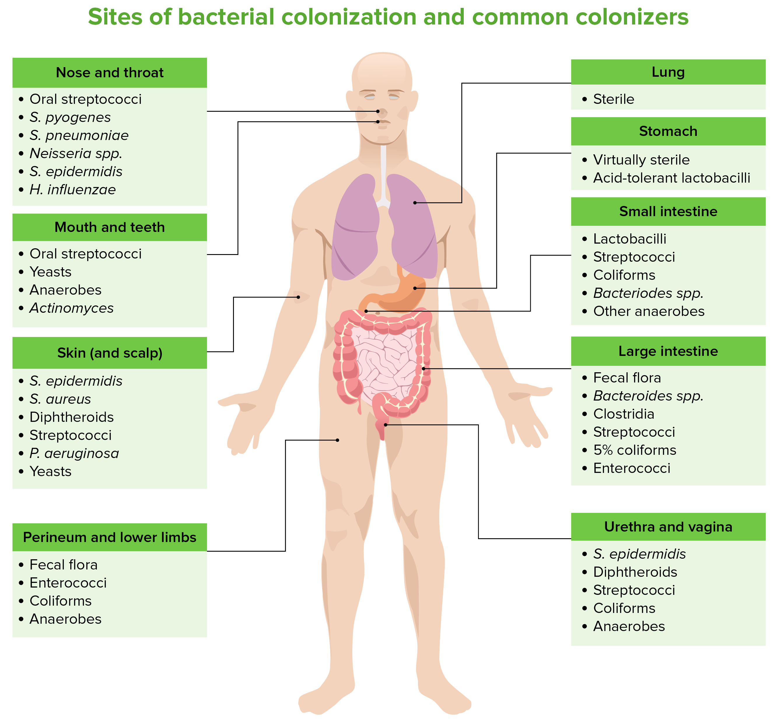
Lugares de colonización bacteriana y colonizadores comunes
Imagen por Lecturio. Licencia: CC BY-NC-SA 4.0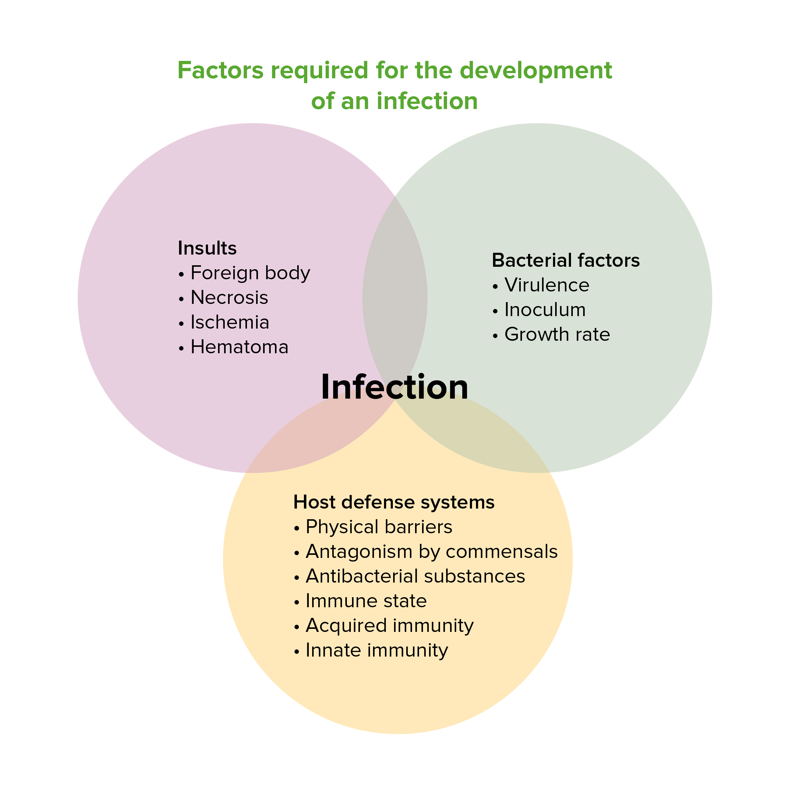
Factores necesarios para el desarrollo de una infección
Imagen por Lecturio. Licencia: CC BY-NC-SA 4.0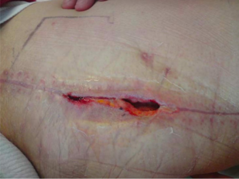
ISO superficial:
Haydehiscencia y apertura de los bordes de la herida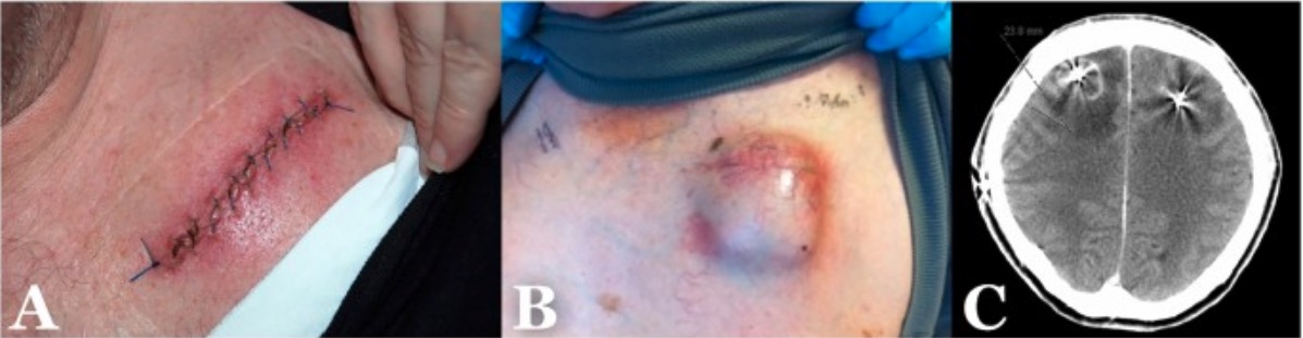
Ejemplos de los diferentes tipos de ISO:
A: ISO superficial
B: ISO profunda
C: ISO de órganos/espacios
Inflamación sistémica:
Inflamación local:
Shock Shock Shock is a life-threatening condition associated with impaired circulation that results in tissue hypoxia. The different types of shock are based on the underlying cause: distributive (↑ cardiac output (CO), ↓ systemic vascular resistance (SVR)), cardiogenic (↓ CO, ↑ SVR), hypovolemic (↓ CO, ↑ SVR), obstructive (↓ CO), and mixed. Types of Shock séptico:
El diagnóstico de la ISO se basa principalmente en EN Erythema nodosum is an immune-mediated panniculitis (inflammation of the subcutaneous fat) caused by a type IV (delayed-type) hypersensitivity reaction. It commonly manifests in young women as tender, erythematous nodules on the shins. Erythema Nodosum los LOS Neisseria hallazgos clínicos. La imagenología diagnostica y los LOS Neisseria estudios de laboratorio son útiles para identificar las infecciones de los LOS Neisseria tejidos profundos y de los LOS Neisseria órganos/espacios.
Hallazgos clínicos (locales):
Hallazgos de sepsis Sepsis Systemic inflammatory response syndrome with a proven or suspected infectious etiology. When sepsis is associated with organ dysfunction distant from the site of infection, it is called severe sepsis. When sepsis is accompanied by hypotension despite adequate fluid infusion, it is called septic shock. Sepsis and Septic Shock (sistémicos):
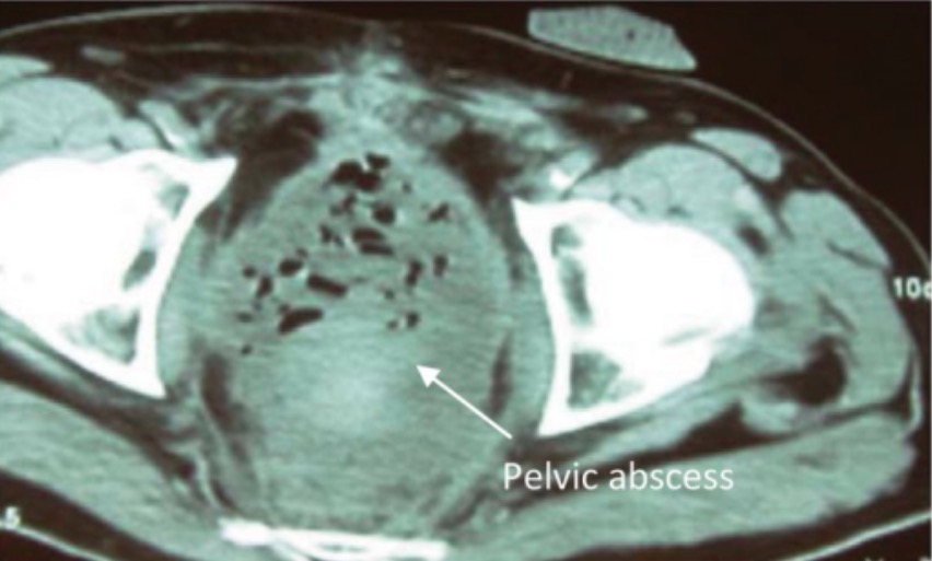
Absceso pélvico posquirúrgico
Imagen: Axial computed tomography of the pelvis showing pelvic abscess” por Plastic and Reconstructive Surgery Department, Cork University Hospital, Wilton, Cork, Ireland. Licencia: CC BY 3.0El tratamiento de las ISO puede incluir antibióticos, drenaje del absceso (abierto o percutáneo) y retirada del material protésico infectado.
| Microorganismo | Primera opción | Alterativa |
|---|---|---|
| Staphylococcus aureus Staphylococcus aureus Potentially pathogenic bacteria found in nasal membranes, skin, hair follicles, and perineum of warm-blooded animals. They may cause a wide range of infections and intoxications. Brain Abscess sensible a la meticilina | Cefazolina | Clindamicina o penicilina antiestafilocócica (nafcilina, oxacilina, dicloxacilina) |
| Staphylococcus aureus Staphylococcus aureus Potentially pathogenic bacteria found in nasal membranes, skin, hair follicles, and perineum of warm-blooded animals. They may cause a wide range of infections and intoxications. Brain Abscess resistente a la meticilina | Vancomicina | Linezolid Linezolid An oxazolidinone and acetamide derived anti-bacterial agent and protein synthesis inhibitor that is used in the treatment of gram-positive bacterial infections of the skin and respiratory tract. Oxazolidinones o daptomicina |
| Estafilococos coagulasa-negativos | Vancomicina | Linezolid Linezolid An oxazolidinone and acetamide derived anti-bacterial agent and protein synthesis inhibitor that is used in the treatment of gram-positive bacterial infections of the skin and respiratory tract. Oxazolidinones o daptomicina |
| Streptococcus Streptococcus Streptococcus is one of the two medically important genera of gram-positive cocci, the other being Staphylococcus. Streptococci are identified as different species on blood agar on the basis of their hemolytic pattern and sensitivity to optochin and bacitracin. There are many pathogenic species of streptococci, including S. pyogenes, S. agalactiae, S. pneumoniae, and the viridans streptococci. Streptococcus pneumoniae | Bencilpenicilina | Claritromicina |
| Streptococcus Streptococcus Streptococcus is one of the two medically important genera of gram-positive cocci, the other being Staphylococcus. Streptococci are identified as different species on blood agar on the basis of their hemolytic pattern and sensitivity to optochin and bacitracin. There are many pathogenic species of streptococci, including S. pyogenes, S. agalactiae, S. pneumoniae, and the viridans streptococci. Streptococcus pyogenes (estreptococo β-hemolítico del grupo A) | Bencilpenicilina, clindamicina | Claritromicina |
| Enterococos | Amoxicilina | Vancomicina |
| Bacteroides Bacteroides Bacteroides is a genus of opportunistic, anaerobic, gram-negative bacilli. Bacteroides fragilis is the most common species involved in human disease and is part of the normal flora of the large intestine. Bacteroides spp. | Metronidazol | Amoxicilina-clavulanato |
| Escherichia coli Escherichia coli The gram-negative bacterium Escherichia coli is a key component of the human gut microbiota. Most strains of E. coli are avirulent, but occasionally they escape the GI tract, infecting the urinary tract and other sites. Less common strains of E. coli are able to cause disease within the GI tract, most commonly presenting as abdominal pain and diarrhea. Escherichia coli | Piperacilina-tazobactam | Meropenem Meropenem A thienamycin derivative antibacterial agent that is more stable to renal dehydropeptidase I than imipenem, but does not need to be given with an enzyme inhibitor such as cilastatin. It is used in the treatment of bacterial infections, including infections in immunocompromised patients. Carbapenems and Aztreonam |
| Haemophilus influenzae Haemophilus Influenzae A species of Haemophilus found on the mucous membranes of humans and a variety of animals. The species is further divided into biotypes I through viii. Haemophilus | Amoxicilina | Amoxicilina-clavulanato |
| Klebsiella Klebsiella Klebsiella are encapsulated gram-negative, lactose-fermenting bacilli. They form pink colonies on MacConkey agar due to lactose fermentation. The main virulence factor is a polysaccharide capsule. Klebsiella pneumoniae is the most important pathogenic species. Klebsiella spp. | Ceftriaxona o piperacilina-tazobactam | Meropenem Meropenem A thienamycin derivative antibacterial agent that is more stable to renal dehydropeptidase I than imipenem, but does not need to be given with an enzyme inhibitor such as cilastatin. It is used in the treatment of bacterial infections, including infections in immunocompromised patients. Carbapenems and Aztreonam |
| Proteus Proteus Proteus spp. are gram-negative, facultatively anaerobic bacilli. Different types of infection result from Proteus, but the urinary tract is the most common site. The majority of cases are caused by Proteus mirabilis (P. mirabilis). The bacteria are part of the normal intestinal flora and are also found in the environment. Proteus spp. | Amoxicilina-clavulanato | Meropenem Meropenem A thienamycin derivative antibacterial agent that is more stable to renal dehydropeptidase I than imipenem, but does not need to be given with an enzyme inhibitor such as cilastatin. It is used in the treatment of bacterial infections, including infections in immunocompromised patients. Carbapenems and Aztreonam |
| Pseudomonas aeruginosa Pseudomonas aeruginosa A species of gram-negative, aerobic, rod-shaped bacteria commonly isolated from clinical specimens (wound, burn, and urinary tract infections). It is also found widely distributed in soil and water. P. Aeruginosa is a major agent of nosocomial infection. Pseudomonas | Piperacilina-tazobactam | Meropenem Meropenem A thienamycin derivative antibacterial agent that is more stable to renal dehydropeptidase I than imipenem, but does not need to be given with an enzyme inhibitor such as cilastatin. It is used in the treatment of bacterial infections, including infections in immunocompromised patients. Carbapenems and Aztreonam |
| Clostridium spp. |
|
Metronidazol |