Domina Conceptos Médicos

Estudia para la escuela de medicina y tus examenes con Lecturio.

Diarrea

La diarrea se define como ≥ 3 deposiciones acuosas o sueltas en EN Erythema nodosum is an immune-mediated panniculitis (inflammation of the subcutaneous fat) caused by a type IV (delayed-type) hypersensitivity reaction. It commonly manifests in young women as tender, erythematous nodules on the shins. Erythema Nodosum un periodo de 24 horas. Existen múltiples etiologías, que pueden clasificarse en EN Erythema nodosum is an immune-mediated panniculitis (inflammation of the subcutaneous fat) caused by a type IV (delayed-type) hypersensitivity reaction. It commonly manifests in young women as tender, erythematous nodules on the shins. Erythema Nodosum función del mecanismo subyacente de la enfermedad. La duración de los LOS Neisseria síntomas (agudos o crónicos) y las características de las heces (e.g., acuosas, sanguinolentas, esteatorreicas, mucosas) pueden ayudar a orientar la evaluación diagnóstica. También es importante obtener de los LOS Neisseria antecedentes  los LOS Neisseria síntomas asociados, como fiebre, náuseas y vómitos, pérdida de peso y heces con sangre. La mayoría de las causas de la diarrea aguda son infecciosas y no requieren exámenes adicionales. Dado que la diarrea suele ser una afección autolimitada, el tratamiento suele ser de soporte. Sin embargo, la diarrea crónica puede requerir estudios de laboratorio, estudios de heces, imagenología o procedimientos para determinar la causa. En EN Erythema nodosum is an immune-mediated panniculitis (inflammation of the subcutaneous fat) caused by a type IV (delayed-type) hypersensitivity reaction. It commonly manifests in young women as tender, erythematous nodules on the shins. Erythema Nodosum última instancia, el manejo depende del tratamiento de la patología subyacente, aunque pueden utilizarse terapias sintomáticas y empíricas en EN Erythema nodosum is an immune-mediated panniculitis (inflammation of the subcutaneous fat) caused by a type IV (delayed-type) hypersensitivity reaction. It commonly manifests in young women as tender, erythematous nodules on the shins. Erythema Nodosum las circunstancias adecuadas.

Last updated: Dec 15, 2025

Editorial responsibility: Stanley Oiseth, Lindsay Jones, Evelin Maza

Descripción General

Definición

La diarrea es la evacuación de ≥ 3 heces acuosas o sueltas en EN Erythema nodosum is an immune-mediated panniculitis (inflammation of the subcutaneous fat) caused by a type IV (delayed-type) hypersensitivity reaction. It commonly manifests in young women as tender, erythematous nodules on the shins. Erythema Nodosum 24 horas.

Clasificación

La diarrea puede clasificarse según la duración de los LOS Neisseria síntomas:

  • Diarrea aguda: ≤ 2 semanas
  • Diarrea persistente: > 2 semanas pero < 4 semanas
  • Diarrea crónica: ≥ 4 semanas

Adicionalmente, la diarrea puede clasificarse en EN Erythema nodosum is an immune-mediated panniculitis (inflammation of the subcutaneous fat) caused by a type IV (delayed-type) hypersensitivity reaction. It commonly manifests in young women as tender, erythematous nodules on the shins. Erythema Nodosum función de la etiología y la fisiopatología subyacentes:

  • Diarrea de etiología infecciosa:
    • Inflamatoria (invasión por un organismo infeccioso)
    • No inflamatoria (no hay invasión de la mucosa por el organismo)
  • Diarrea de etiología no infecciosa:
    • Secretora (por salida de electrolitos y agua)
    • Osmótica (el agua es arrastrada al AL Amyloidosis lumen intestinal)
    • Por malabsorción (alteración de la absorción de nutrientes)
    • Inflamatoria (proceso inflamatorio que provoca daños en EN Erythema nodosum is an immune-mediated panniculitis (inflammation of the subcutaneous fat) caused by a type IV (delayed-type) hypersensitivity reaction. It commonly manifests in young women as tender, erythematous nodules on the shins. Erythema Nodosum la mucosa)
    • Por alteración de la motilidad (tránsito intestinal rápido)

Etiología

Diarrea infecciosa

Inflamatoria/invasiva:

  • Por bacterias:
    • Shigella Shigella Shigella is a genus of gram-negative, non-lactose-fermenting facultative intracellular bacilli. Infection spreads most commonly via person-to-person contact or through contaminated food and water. Humans are the only known reservoir. Shigella
    • Salmonella Salmonella Salmonellae are gram-negative bacilli of the family Enterobacteriaceae. Salmonellae are flagellated, non-lactose-fermenting, and hydrogen sulfide-producing microbes. Salmonella enterica, the most common disease-causing species in humans, is further classified based on serotype as typhoidal (S. typhi and paratyphi) and nontyphoidal (S. enteritidis and typhimurium). Salmonella
    • Campylobacter jejuni Campylobacter jejuni A species of bacteria that resemble small tightly coiled spirals. Its organisms are known to cause abortion in sheep and fever and enteritis in man and may be associated with enteric diseases of calves, lambs, and other animals. Campylobacter
    • Yersinia enterocolitica Yersinia enterocolitica A species of the genus yersinia, isolated from both man and animal. It is a frequent cause of bacterial gastroenteritis in children. Yersinia spp./Yersiniosis
    • Escherichia coli Escherichia coli The gram-negative bacterium Escherichia coli is a key component of the human gut microbiota. Most strains of E. coli are avirulent, but occasionally they escape the GI tract, infecting the urinary tract and other sites. Less common strains of E. coli are able to cause disease within the GI tract, most commonly presenting as abdominal pain and diarrhea. Escherichia coli (enterohemorrágica y enteroinvasiva)
    • Clostridioides difficile
    • Listeria Listeria Listeria spp. are motile, flagellated, gram-positive, facultative intracellular bacilli. The major pathogenic species is Listeria monocytogenes. Listeria are part of the normal gastrointestinal flora of domestic mammals and poultry and are transmitted to humans through the ingestion of contaminated food, especially unpasteurized dairy products. Listeria Monocytogenes/Listeriosis monocytogenes
    • Vibrio parahaemolyticus Vibrio parahaemolyticus A species of bacteria found in the marine environment, sea foods, and the feces of patients with acute enteritis. Vibrio
  • Por protozoos:
    • Entameoba histolytica
    • Strongyloides

No inflamatoria/no invasiva:

  • Por bacterias:
    • Staphylococcus aureus Staphylococcus aureus Potentially pathogenic bacteria found in nasal membranes, skin, hair follicles, and perineum of warm-blooded animals. They may cause a wide range of infections and intoxications. Brain Abscess
    • Clostridium perfringens Clostridium perfringens The most common etiologic agent of gas gangrene. It is differentiable into several distinct types based on the distribution of twelve different toxins. Gas Gangrene
    • Bacillus cereus Bacillus cereus A species of rod-shaped bacteria that is a common soil saprophyte. Its spores are widespread and multiplication has been observed chiefly in foods. Contamination may lead to food poisoning. Bacillus
    • E. coli (enterotoxigénica y enteroagregante)
    • Vibrio cholerae Vibrio cholerae The etiologic agent of cholera. Vibrio
  • Por protozoos:
    • Giardia Giardia A genus of flagellate intestinal eukaryotes parasitic in various vertebrates, including humans. Characteristics include the presence of four pairs of flagella arising from a complicated system of axonemes and cysts that are ellipsoidal to ovoidal in shape. Nitroimidazoles lamblia
    • Cryptosporidium Cryptosporidium A genus of coccidian parasites of the family cryptosporidiidae, found in the intestinal epithelium of many vertebrates including humans. Hyper-IgM Syndrome
  • Por virus Virus Viruses are infectious, obligate intracellular parasites composed of a nucleic acid core surrounded by a protein capsid. Viruses can be either naked (non-enveloped) or enveloped. The classification of viruses is complex and based on many factors, including type and structure of the nucleoid and capsid, the presence of an envelope, the replication cycle, and the host range. Virology:
    • Rotavirus Rotavirus A genus of Reoviridae, causing acute gastroenteritis in birds and mammals, including humans. Transmission is horizontal and by environmental contamination. Seven species (rotaviruses A through G) are recognized. Rotavirus
    • Norovirus Norovirus Norovirus is a nonenveloped, single-stranded, positive-sense RNA virus belonging to the Caliciviridae family. Norovirus infections are transmitted via the fecal-oral route or by aerosols from vomiting. The virus is one of the most common causes of nonbacterial gastroenteritis epidemic worldwide. Symptoms include watery and nonbloody diarrhea, nausea, vomiting, and low-grade fever. Norovirus
    • Adenovirus Adenovirus Adenovirus (member of the family Adenoviridae) is a nonenveloped, double-stranded DNA virus. Adenovirus is transmitted in a variety of ways, and it can have various presentations based on the site of entry. Presentation can include febrile pharyngitis, conjunctivitis, acute respiratory disease, atypical pneumonia, and gastroenteritis. Adenovirus
    • Citomegalovirus

Factores de riesgo:

  • Productos alimentarios contaminados:
    • Mariscos
    • Aves de corral
    • Pavo
    • Huevos
    • Carne de vacuno
  • Agua contaminada
  • Exposición a animales
  • Entornos de alto riesgo de transmisión:
    • Guarderías
    • Residencias de ancianos
    • Hospitales

Diarrea no infecciosa

Secretora:

  • Laxantes:
    • Hierba sen
    • Docusato
  • Tumores productores de hormonas:
    • Carcinoide
    • Tumor Tumor Inflammation secretor de péptidos intestinales vasoactivos
    • Gastrinoma Gastrinoma A gastrinoma is a tumor that secretes excessive levels of the hormone gastrin and is responsible for Zollinger-Ellison syndrome (ZES). Gastrinomas are frequently associated with multiple endocrine neoplasia 1 (MEN 1) and can arise from the pancreas, stomach, duodenum, jejunum, and/or even from the lymph nodes. Gastrinoma
  • Malabsorción de ácidos biliares
  • Enfermedad endocrina:
    • Enfermedad de Addison
    • Diabetes Diabetes Diabetes mellitus (DM) is a metabolic disease characterized by hyperglycemia and dysfunction of the regulation of glucose metabolism by insulin. Type 1 DM is diagnosed mostly in children and young adults as the result of autoimmune destruction of β cells in the pancreas and the resulting lack of insulin. Type 2 DM has a significant association with obesity and is characterized by insulin resistance. Diabetes Mellitus
  • Medicamentos:
    • Quinina
    • Colchicina
    • Antibióticos
    • Digoxina
    • Misoprostol Misoprostol A synthetic analog of natural prostaglandin e1. It produces a dose-related inhibition of gastric acid and pepsin secretion, and enhances mucosal resistance to injury. It is an effective anti-ulcer agent and also has oxytocic properties. Eicosanoids

Osmótica:

  • Laxantes osmóticos:
    • Sulfato de magnesio o hidróxido de magnesio
    • Polietilenglicol
  • Deficiencia de lactasa (intolerancia a la lactosa)
  • Hidratos de carbono no absorbibles:
    • Sorbitol Sorbitol A polyhydric alcohol with about half the sweetness of sucrose. Sorbitol occurs naturally and is also produced synthetically from glucose. It was formerly used as a diuretic and may still be used as a laxative and in irrigating solutions for some surgical procedures. It is also used in many manufacturing processes, as a pharmaceutical aid, and in several research applications. Laxatives
    • Lactulosa
    • Xilitol

Por malabsorción:

  • Maldigestión intraluminal:
    • Insuficiencia exocrina pancreática (e.g., pancreatitis Pancreatitis Inflammation of the pancreas. Pancreatitis is classified as acute unless there are computed tomographic or endoscopic retrograde cholangiopancreatographic findings of chronic pancreatitis. The two most common forms of acute pancreatitis are alcoholic pancreatitis and gallstone pancreatitis. Acute Pancreatitis crónica)
    • Sobrecrecimiento bacteriano
    • Cirugía bariátrica
  • Malabsorción de la mucosa:
    • Enfermedad celíaca
    • Enfermedad de Whipple
    • Isquemia mesentérica
  • Medicamentos:
    • Orlistat
    • Acarbosa

Inflamatoria/exudativa:

  • Enfermedades inflamatorias intestinales:
    • Enfermedad de Crohn
    • Colitis Colitis Inflammation of the colon section of the large intestine, usually with symptoms such as diarrhea (often with blood and mucus), abdominal pain, and fever. Pseudomembranous Colitis ulcerosa
  • Colitis Colitis Inflammation of the colon section of the large intestine, usually with symptoms such as diarrhea (often with blood and mucus), abdominal pain, and fever. Pseudomembranous Colitis microscópica
  • Enfermedad de la mucosa relacionada con la inmunidad:
    • Inmunodeficiencia
    • Alergia alimentaria
    • Gastroenteritis Gastroenteritis Gastroenteritis is inflammation of the stomach and intestines, commonly caused by infections from bacteria, viruses, or parasites. Transmission may be foodborne, fecal-oral, or through animal contact. Common clinical features include abdominal pain, diarrhea, vomiting, fever, and dehydration. Gastroenteritis eosinofílica
  • Infecciones
  • Lesión por radiación
  • Neoplasias gastrointestinales:
    • Linfoma
    • Cáncer de colon Colon The large intestines constitute the last portion of the digestive system. The large intestine consists of the cecum, appendix, colon (with ascending, transverse, descending, and sigmoid segments), rectum, and anal canal. The primary function of the colon is to remove water and compact the stool prior to expulsion from the body via the rectum and anal canal. Colon, Cecum, and Appendix: Anatomy

Por alteración de la motilidad:

  • Síndrome de intestino irritable
  • Hipertiroidismo
  • Síndrome de vaciamiento o evacuación rápida
  • Medicamentos
    • Medicamentos que contienen magnesio
    • Inhibidores de la colinesterasa
    • Inhibidores selectivos de la recaptación de serotonina

Related videos

Fisiopatología

Diarrea infecciosa

Inflamatoria/invasiva:

  • Invasión de la mucosa por un organismo infeccioso → respuesta inflamatoria y daño del epitelio → ↓ absorción y exudación de suero y sangre en EN Erythema nodosum is an immune-mediated panniculitis (inflammation of the subcutaneous fat) caused by a type IV (delayed-type) hypersensitivity reaction. It commonly manifests in young women as tender, erythematous nodules on the shins. Erythema Nodosum el lumen
  • En EN Erythema nodosum is an immune-mediated panniculitis (inflammation of the subcutaneous fat) caused by a type IV (delayed-type) hypersensitivity reaction. It commonly manifests in young women as tender, erythematous nodules on the shins. Erythema Nodosum las heces pueden detectarse mucosidad, sangre y leucocitos.

No inflamatoria/no invasiva:

  • Los LOS Neisseria patógenos no invaden la mucosa.
  • Asociado con mayor frecuencia a las enterotoxinas bacterianas → altera el transporte de iones → salida de iones y agua hacia la luz del intestino
  • La mucosa permanece normal o está mínimamente alterada.
  • Las heces son acuosas y carecen de leucocitos fecales y sangre.

Diarrea no infecciosa

Secretora:

  • Secreción activa de agua en EN Erythema nodosum is an immune-mediated panniculitis (inflammation of the subcutaneous fat) caused by a type IV (delayed-type) hypersensitivity reaction. It commonly manifests in young women as tender, erythematous nodules on the shins. Erythema Nodosum la luz intestinal debido a la activación de los LOS Neisseria sistemas de transporte de iones
  • La diarrea se produce durante todo el día y la noche.
Diarrea secretora

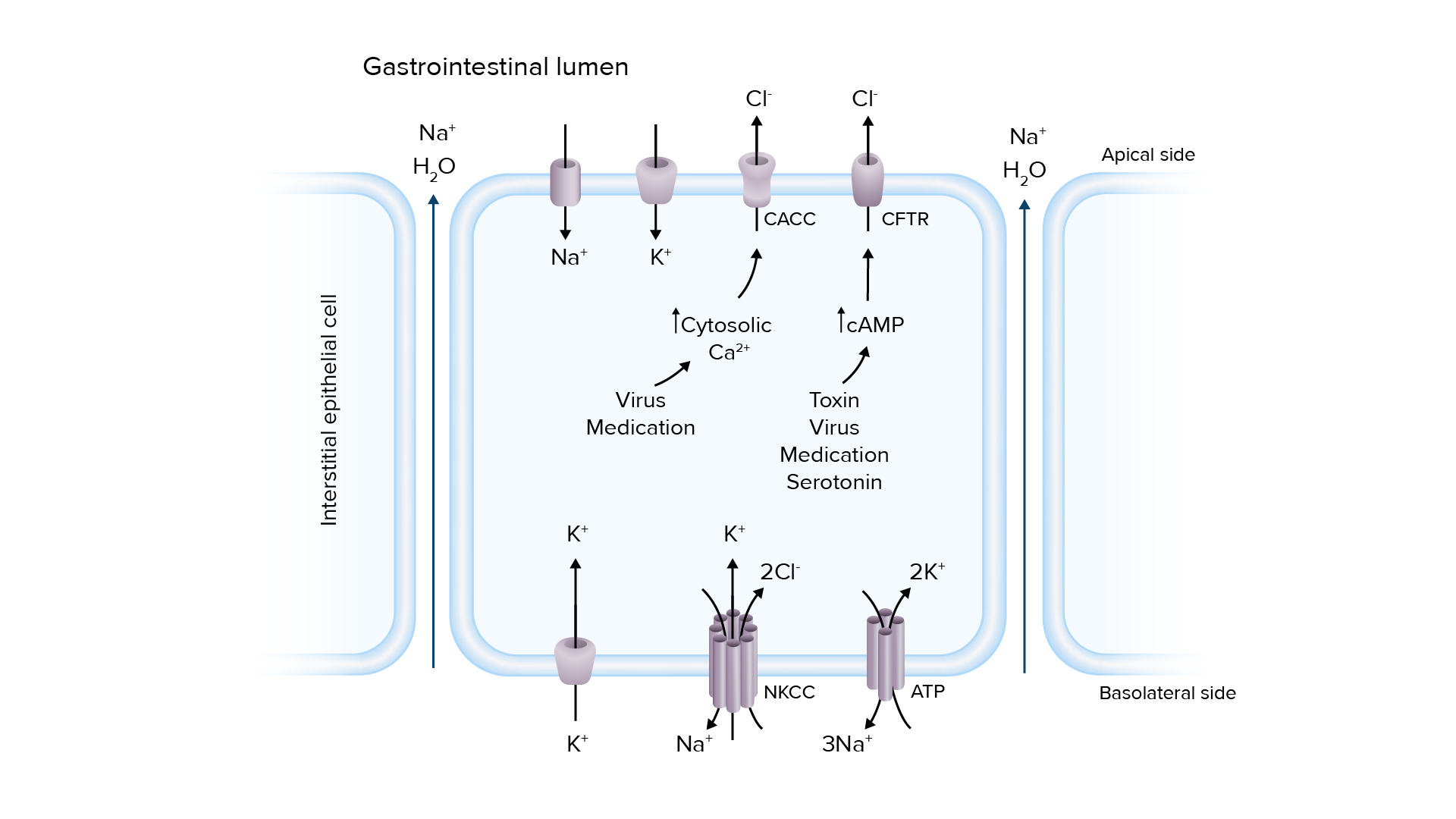
Patogénesis de la diarrea secretora:
La sobreactivación de los canales de transporte de iones puede conducir a la secreción de electrolitos y agua en el lumen intestinal, lo que provoca diarrea.
Ca2+: calcio
CaCC: canales de cloruro activados por el calcio
AMPc: monofosfato de adenosina cíclico
CFTR: regulador de la conductancia transmembrana de la fibrosis quística
Cl-: cloruro
K+: potasio
Na+: sodio
NKCC: cotransportador sodio-cloruro de potasio

Imagen por Lecturio.

Osmótica:

  • El agua es arrastrada al AL Amyloidosis lumen intestinal por las sustancias mal absorbidas.
  • La producción de heces es consistente con la cantidad de sustancia no absorbible que se ingiere.
  • Ocurre solo durante el día, y las heces ↓ con la interrupción de la sustancia ofensiva
  • La brecha osmolar será evidente.
Diarrea osmótica

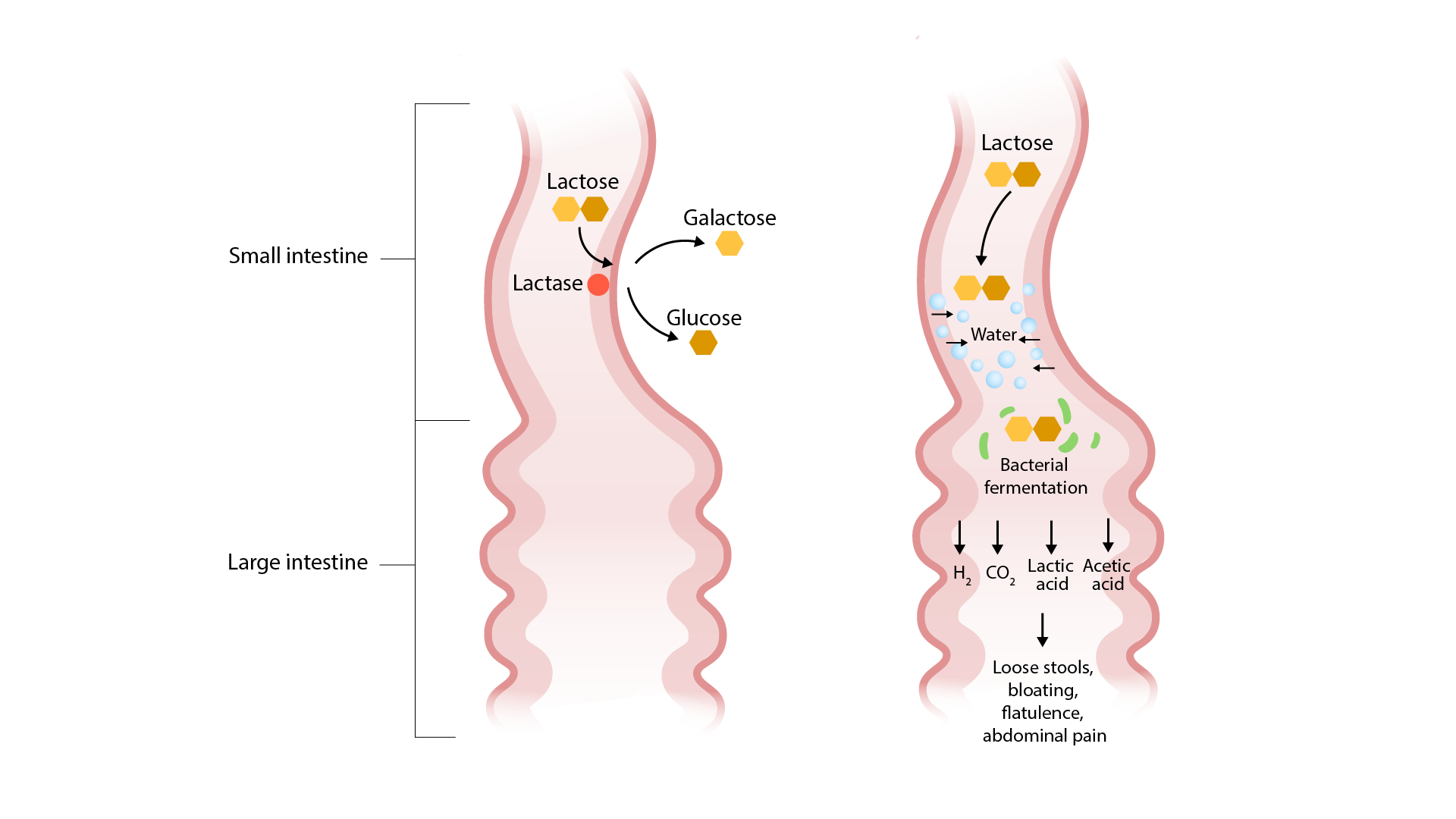
Patogénesis de la deficiencia de lactasa (una etiología de la diarrea osmótica):
La lactosa no se descompone y permanece en la luz del intestino delgado, atrayendo agua y provocando una diarrea osmótica. La fermentación bacteriana de la lactosa provoca los síntomas de hinchazón, flatulencia y dolor abdominal.

Imagen por Lecturio.

Por malabsorción:

  • Deterioro de la absorción o digestión de nutrientes
  • A menudo da lugar a heces grasas (esteatorrea)

Inflamatoria/exudativa:

  • Inflamación → daño de la mucosa intestinal → deterioro de la absorción
  • Mucosidad, sangre y leucocitos están presentes en EN Erythema nodosum is an immune-mediated panniculitis (inflammation of the subcutaneous fat) caused by a type IV (delayed-type) hypersensitivity reaction. It commonly manifests in young women as tender, erythematous nodules on the shins. Erythema Nodosum las heces.

Por alteración de la motilidad: paso intestinal rápido → ↓ tiempo de absorción de líquidos.

Presentación Clínica

  • Determinar la duración:
    • Aguda
    • Crónica
  • Caracterizar el tipo de diarrea:
    • Acuosa (puede ser secretora u osmótica)
    • Esteatorreica (probablemente por mala absorción)
    • Sanguinolenta (probablemente inflamatoria)
    • Mucoide (probablemente inflamatoria)
  • Síntomas asociados:
    • Fiebre
    • Dolor Dolor Inflammation abdominal y cólicos
    • Flatulencia e hinchazón
    • Náuseas y vómitos (especialmente debido a una etiología infecciosa o mediada por toxinas)
    • Tenesmo
  • Signos de deshidratación:
    • Piel y mucosas secas
    • Disminución de la turgencia de la piel
    • Fatiga
    • Taquicardia
    • Respiración rápida

Diagnóstico

Diarrea aguda

La mayoría de los LOS Neisseria casos son de etiología infecciosa.

La mayoría de los LOS Neisseria pacientes tendrán síntomas autolimitados y no requerirán pruebas.

Indicaciones para los LOS Neisseria estudios de heces:

  • Deshidratación severa
  • Heces con sangre
  • > 6 deposiciones no formadas en EN Erythema nodosum is an immune-mediated panniculitis (inflammation of the subcutaneous fat) caused by a type IV (delayed-type) hypersensitivity reaction. It commonly manifests in young women as tender, erythematous nodules on the shins. Erythema Nodosum 24 horas
  • Fiebre ≥ 38,5°C (≥ 101°F)
  • Duración > 48 horas sin mejora
  • Uso reciente de antibióticos
  • Dolor Dolor Inflammation abdominal intenso
  • Población de alto riesgo (ancianos, pacientes inmunocomprometidos, aquellos con enfermedad inflamatoria intestinal conocida)

Análisis de heces:

  • Leucocitos fecales
  • Huevos y parásitos
  • Cultivo de heces y reacción en EN Erythema nodosum is an immune-mediated panniculitis (inflammation of the subcutaneous fat) caused by a type IV (delayed-type) hypersensitivity reaction. It commonly manifests in young women as tender, erythematous nodules on the shins. Erythema Nodosum cadena de la polimerasa
  • Sangre oculta
  • Lactoferrina
  • Inmunoanálisis de la toxina de C. difficile (especialmente si hay un uso reciente de antibióticos)

Evaluación de laboratorios de apoyo:

  • Generalmente se hace HACE Altitude Sickness solo en EN Erythema nodosum is an immune-mediated panniculitis (inflammation of the subcutaneous fat) caused by a type IV (delayed-type) hypersensitivity reaction. It commonly manifests in young women as tender, erythematous nodules on the shins. Erythema Nodosum pacientes con enfermedad grave y evidencia de deshidratación
  • Hemograma
    • Leucocitosis
    • Eosinofilia → puede indicar una infección parasitaria
    • Bandemia significativa → común en EN Erythema nodosum is an immune-mediated panniculitis (inflammation of the subcutaneous fat) caused by a type IV (delayed-type) hypersensitivity reaction. It commonly manifests in young women as tender, erythematous nodules on the shins. Erythema Nodosum C. difficile
  • Panel metabólico básico:
    • Anomalías electrolíticas (hipopotasemia)
    • ↑ Creatinina → lesión renal aguda por deshidratación.
    • Acidosis Acidosis A pathologic condition of acid accumulation or depletion of base in the body. The two main types are respiratory acidosis and metabolic acidosis, due to metabolic acid build up. Respiratory Acidosis metabólica sin brecha aniónica

Diarrea crónica

El diagnóstico diferencial de la diarrea crónica es extenso, y la evaluación se guiará por la sospecha clínica de los LOS Neisseria antecedentes y el examen físico. Puede ser necesario consultar a un gastroenterólogo.

Estudios de laboratorio:

  • Se utilizan para afinar el diagnóstico diferencial
  • Hemograma → evaluar la anemia Anemia Anemia is a condition in which individuals have low Hb levels, which can arise from various causes. Anemia is accompanied by a reduced number of RBCs and may manifest with fatigue, shortness of breath, pallor, and weakness. Subtypes are classified by the size of RBCs, chronicity, and etiology. Anemia: Overview and Types (malabsorción, malignidad)
  • Panel metabólico básico → evaluar la deshidratación y las alteraciones electrolíticas
  • Hormona estimulante de la tiroides → tamizaje de hipertiroidismo
  • Velocidad de eritrosedimentación y proteína C reactiva:
    • No específicas
    • Pueden estar elevadas debido a etiologías inflamatorias
  • Serologías celíacas
  • Prueba de aliento → sobrecrecimiento bacteriano
  • Estudios de heces:
    • Electrolitos de las heces → calcular la brecha osmolar de las heces
      • Osmolalidad normal de las heces = 290 mmol/L (igual que el suero)
      • La brecha osmótica = 290 – 2 × (sodio en EN Erythema nodosum is an immune-mediated panniculitis (inflammation of the subcutaneous fat) caused by a type IV (delayed-type) hypersensitivity reaction. It commonly manifests in young women as tender, erythematous nodules on the shins. Erythema Nodosum heces + potasio en EN Erythema nodosum is an immune-mediated panniculitis (inflammation of the subcutaneous fat) caused by a type IV (delayed-type) hypersensitivity reaction. It commonly manifests in young women as tender, erythematous nodules on the shins. Erythema Nodosum heces)
      • Diarrea osmótica: > 125 mmol/L
      • Diarrea secretora: < 50 mmol/L
    • Sangre oculta → se observa en EN Erythema nodosum is an immune-mediated panniculitis (inflammation of the subcutaneous fat) caused by a type IV (delayed-type) hypersensitivity reaction. It commonly manifests in young women as tender, erythematous nodules on the shins. Erythema Nodosum la enfermedad inflamatoria intestinal, en EN Erythema nodosum is an immune-mediated panniculitis (inflammation of the subcutaneous fat) caused by a type IV (delayed-type) hypersensitivity reaction. It commonly manifests in young women as tender, erythematous nodules on the shins. Erythema Nodosum las neoplasias y en EN Erythema nodosum is an immune-mediated panniculitis (inflammation of the subcutaneous fat) caused by a type IV (delayed-type) hypersensitivity reaction. It commonly manifests in young women as tender, erythematous nodules on the shins. Erythema Nodosum las infecciones crónicas
    • Calprotectina fecal o lactoferrina → causas inflamatorias
    • Evaluación de la infección (especialmente si la diarrea persiste después de un viaje)
      • Cultivo de heces
      • Huevos y parásitos
      • Inmunoensayo de toxina de C. difficile (si hay antecedentes de uso de antibióticos)
    • Grasa fecal → malabsorción de grasas
    • Quimotripsina y elastasa fecales → posible insuficiencia pancreática
    • Tamizaje de laxantes
  • Pruebas de malabsorción:
    • Folato
    • Estudios de hierro
    • Vitamina B12
    • Albúmina
    • 25-Hidroxivitamina D

Imagenología y procedimientos:

  • El uso de estos métodos está guiado por la historia del paciente, los LOS Neisseria síntomas, la presencia de signos de alarma y la sospecha clínica.
  • Endoscopia y/o colonoscopia:
    • Debe realizarse con prontitud en EN Erythema nodosum is an immune-mediated panniculitis (inflammation of the subcutaneous fat) caused by a type IV (delayed-type) hypersensitivity reaction. It commonly manifests in young women as tender, erythematous nodules on the shins. Erythema Nodosum pacientes con signos de alarma:
      • Inicio de los LOS Neisseria síntomas después de los LOS Neisseria 50 años
      • Melena Melena The black, tarry, foul-smelling feces that contain degraded blood. Gastrointestinal Bleeding o hematoquecia
      • Síntomas nocturnos
      • Dolor Dolor Inflammation abdominal progresivo
      • Pérdida de peso
      • Síntomas sistémicos
      • Evidencia de malabsorción
      • Antecedentes de enfermedad maligna o inflamatoria intestinal
    • La visualización y la biopsia de la mucosa pueden ayudar a diagnosticar:
      • Enfermedad inflamatoria intestinal
      • Enfermedad celíaca
      • Colitis Colitis Inflammation of the colon section of the large intestine, usually with symptoms such as diarrhea (often with blood and mucus), abdominal pain, and fever. Pseudomembranous Colitis microscópica
      • Malignidad
  • TC o RM:
    • Incluyen pruebas especializadas, como la enterografía y la colangiopancreatografía
    • Deben considerarse en EN Erythema nodosum is an immune-mediated panniculitis (inflammation of the subcutaneous fat) caused by a type IV (delayed-type) hypersensitivity reaction. It commonly manifests in young women as tender, erythematous nodules on the shins. Erythema Nodosum pacientes con:
    • Es útil para detectar:
      • Malignidad
      • Patología del páncreas
      • Enfermedad de Crohn

Tratamiento

Diarrea aguda

Tratamiento de soporte:

  • La mayoría de los LOS Neisseria pacientes solo necesitarán terapia de rehidratación oral.
  • La hidratación de fluidos por vía intravenosa debe utilizarse en EN Erythema nodosum is an immune-mediated panniculitis (inflammation of the subcutaneous fat) caused by a type IV (delayed-type) hypersensitivity reaction. It commonly manifests in young women as tender, erythematous nodules on the shins. Erythema Nodosum caso de enfermedad grave.
  • Las soluciones orales e intravenosas deben contener electrolitos para su sustitución.
  • Si es posible, retire cualquier medicamento potencialmente nocivo.

Agentes antidiarreicos:

  • Opciones: loperamida, subsalicilato de bismuto
  • Reducir la duración de la diarrea
  • Tenga en EN Erythema nodosum is an immune-mediated panniculitis (inflammation of the subcutaneous fat) caused by a type IV (delayed-type) hypersensitivity reaction. It commonly manifests in young women as tender, erythematous nodules on the shins. Erythema Nodosum cuenta que el tratamiento puede retrasar la extracción de patógenos y toxinas.
  • Contraindicaciones:
    • Diarrea con fiebre
    • Heces sanguinolentas o mucoides
    • Diarrea causada por C. difficile y Shigella Shigella Shigella is a genus of gram-negative, non-lactose-fermenting facultative intracellular bacilli. Infection spreads most commonly via person-to-person contact or through contaminated food and water. Humans are the only known reservoir. Shigella

Terapia con antibióticos:

  • No se requiere de forma rutinaria
  • La terapia empírica puede ser considerada para:
    • Heces con sangre
    • Fiebre
    • Síntomas graves que requieren hospitalización
    • Pacientes de alto riesgo (e.g. lactantes, ancianos, inmunodeprimidos)
    • Sospecha de infección por C. difficile
  • Antibióticos de uso frecuente:
    • Fluoroquinolonas
    • Azitromicina
    • Trimetoprima–sulfametoxazol
    • Cefalosporinas de 3ra generación
Análisis de la diarrea aguda

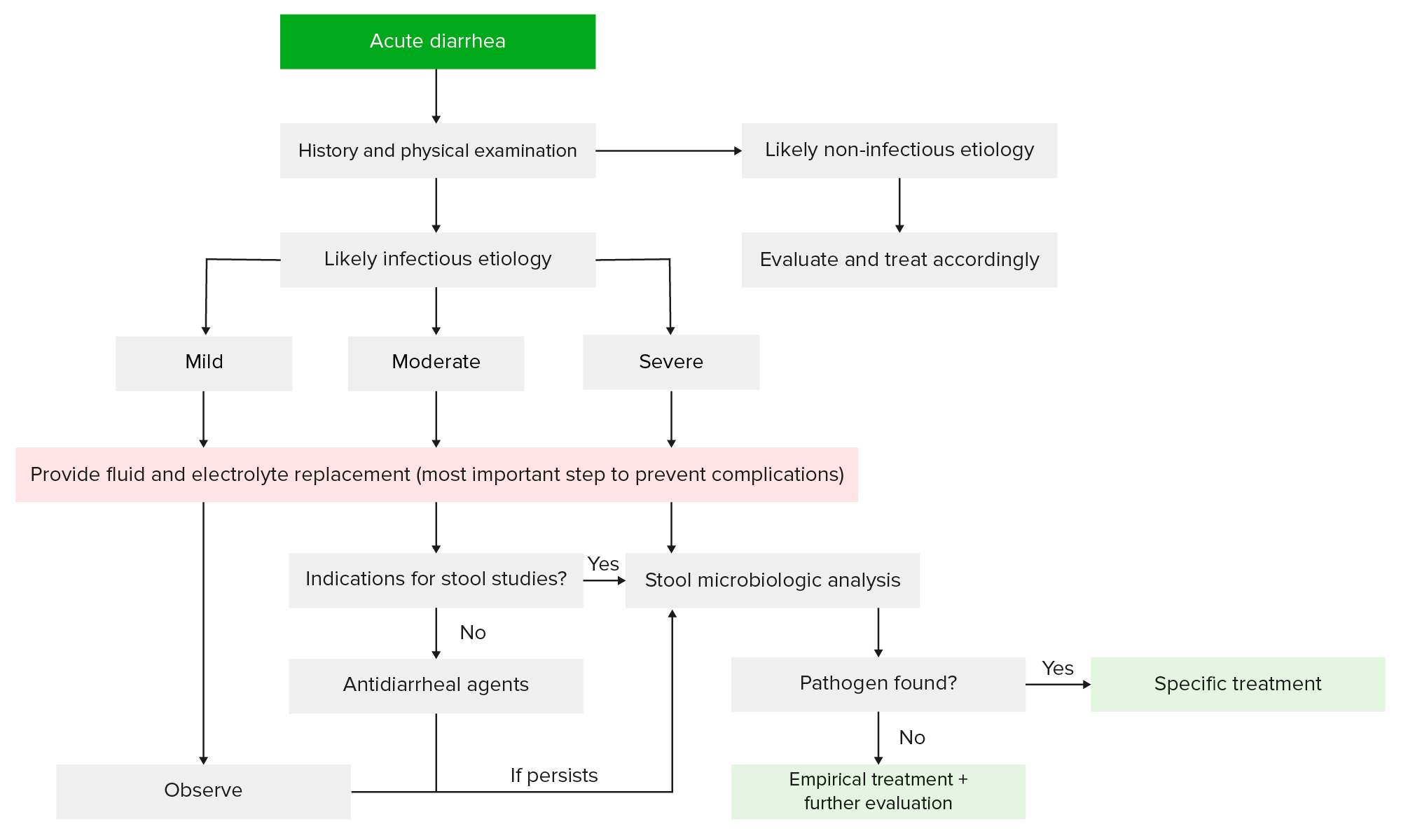
Evaluación y tratamiento de pacientes con diarrea aguda:
Basándose en los antecedentes y el examen físico, se puede determinar si la diarrea está relacionada con una etiología infecciosa o no infecciosa (e.g., medicamentos). La mayoría de los pacientes no necesitarán más que cuidados de soporte. Sin embargo, los que tienen indicaciones para un estudio más profundo pueden someterse a pruebas de laboratorio y de heces, que pueden ayudar a guiar la terapia posterior.

Imagen por Lecturio.

Diarrea crónica

El tratamiento de la diarrea crónica pasa por diagnosticar y tratar la etiología subyacente.

Terapia sintomática:

  • Indicada:
    • Para los LOS Neisseria pacientes que no toleran la terapia definitiva
    • Cuando no se ha HA Hemolytic anemia (HA) is the term given to a large group of anemias that are caused by the premature destruction/hemolysis of circulating red blood cells (RBCs). Hemolysis can occur within (intravascular hemolysis) or outside the blood vessels (extravascular hemolysis). Hemolytic Anemia encontrado un diagnóstico definitivo
    • Para el alivio temporal durante el trabajo
  • Opciones:
    • Loperamida
    • Anticolinérgicos
    • Bismuto
    • Fibra
    • Arcillas

Terapia empírica:

  • Puede utilizarse en EN Erythema nodosum is an immune-mediated panniculitis (inflammation of the subcutaneous fat) caused by a type IV (delayed-type) hypersensitivity reaction. It commonly manifests in young women as tender, erythematous nodules on the shins. Erythema Nodosum pacientes en EN Erythema nodosum is an immune-mediated panniculitis (inflammation of the subcutaneous fat) caused by a type IV (delayed-type) hypersensitivity reaction. It commonly manifests in young women as tender, erythematous nodules on the shins. Erythema Nodosum los LOS Neisseria que se sospecha mucho el diagnóstico pero que no toleran las pruebas
  • Restricción de lactosa → sospecha de intolerancia a la lactosa
  • Colestiramina:
    • Resección ileal reciente
    • Radioterapia abdominal
    • Después de la colecistectomía
  • Antibióticos → por sospecha de sobrecrecimiento bacteriano

Subtipos Especiales

Diarrea del viajero

  • Diarrea que se desarrolla después de que individuos de entornos ricos en EN Erythema nodosum is an immune-mediated panniculitis (inflammation of the subcutaneous fat) caused by a type IV (delayed-type) hypersensitivity reaction. It commonly manifests in young women as tender, erythematous nodules on the shins. Erythema Nodosum recursos regresan de un viaje a regiones con recursos limitados:
    • Ocurre en EN Erythema nodosum is an immune-mediated panniculitis (inflammation of the subcutaneous fat) caused by a type IV (delayed-type) hypersensitivity reaction. It commonly manifests in young women as tender, erythematous nodules on the shins. Erythema Nodosum 40%–60% de los LOS Neisseria viajeros a zonas con recursos limitados
    • Puede ser inflamatoria o secretora
  • Etiología (la lista no es excluyente):
    • La E. coli enterotoxigénica es la causa más común.
    • Campylobacter jejuni Campylobacter jejuni A species of bacteria that resemble small tightly coiled spirals. Its organisms are known to cause abortion in sheep and fever and enteritis in man and may be associated with enteric diseases of calves, lambs, and other animals. Campylobacter
    • Shigella Shigella Shigella is a genus of gram-negative, non-lactose-fermenting facultative intracellular bacilli. Infection spreads most commonly via person-to-person contact or through contaminated food and water. Humans are the only known reservoir. Shigella
    • Salmonella Salmonella Salmonellae are gram-negative bacilli of the family Enterobacteriaceae. Salmonellae are flagellated, non-lactose-fermenting, and hydrogen sulfide-producing microbes. Salmonella enterica, the most common disease-causing species in humans, is further classified based on serotype as typhoidal (S. typhi and paratyphi) and nontyphoidal (S. enteritidis and typhimurium). Salmonella
    • Giardia Giardia A genus of flagellate intestinal eukaryotes parasitic in various vertebrates, including humans. Characteristics include the presence of four pairs of flagella arising from a complicated system of axonemes and cysts that are ellipsoidal to ovoidal in shape. Nitroimidazoles lamblia
    • Norovirus Norovirus Norovirus is a nonenveloped, single-stranded, positive-sense RNA virus belonging to the Caliciviridae family. Norovirus infections are transmitted via the fecal-oral route or by aerosols from vomiting. The virus is one of the most common causes of nonbacterial gastroenteritis epidemic worldwide. Symptoms include watery and nonbloody diarrhea, nausea, vomiting, and low-grade fever. Norovirus
  • Transmisión:
    • Transmisión de alimentos
    • Agua contaminada
  • Presentación clínica:
    • Los LOS Neisseria síntomas comienzan entre 12–72 horas después de la ingestión de agua o alimentos contaminados.
    • Cólicos abdominales
    • Náuseas y vómitos
    • Fiebre de bajo grado
    • Heces típicamente de color arroz
    • Ruidos intestinales hiperactivos
  • La mayoría de los LOS Neisseria casos son leves y no requieren una evaluación diagnóstica.
  • Tratamiento:
    • Reanimación con líquidos
    • Medicamentos antidiarreicos
    • Antibióticos
      • No es necesario
      • Puede considerarse en EN Erythema nodosum is an immune-mediated panniculitis (inflammation of the subcutaneous fat) caused by a type IV (delayed-type) hypersensitivity reaction. It commonly manifests in young women as tender, erythematous nodules on the shins. Erythema Nodosum caso de enfermedad moderada o grave
      • Opciones: fluoroquinolonas o azitromicina

Diarrea facticia y abuso de laxantes

  • Demografía:
    • Diarrea facticia:
      • > El 90% de los LOS Neisseria casos se dan en EN Erythema nodosum is an immune-mediated panniculitis (inflammation of the subcutaneous fat) caused by a type IV (delayed-type) hypersensitivity reaction. It commonly manifests in young women as tender, erythematous nodules on the shins. Erythema Nodosum mujeres.
      • Una proporción significativa ha HA Hemolytic anemia (HA) is the term given to a large group of anemias that are caused by the premature destruction/hemolysis of circulating red blood cells (RBCs). Hemolysis can occur within (intravascular hemolysis) or outside the blood vessels (extravascular hemolysis). Hemolytic Anemia trabajado en EN Erythema nodosum is an immune-mediated panniculitis (inflammation of the subcutaneous fat) caused by a type IV (delayed-type) hypersensitivity reaction. It commonly manifests in young women as tender, erythematous nodules on the shins. Erythema Nodosum el sector sanitario
      • Muchos pacientes tendrán un historial de ingresos hospitalarios frecuentes.
    • Abuso de laxantes:
      • Pacientes de edad avanzada que siguen tomando laxantes tras la resolución del estreñimiento
      • Pacientes con anorexia Anorexia The lack or loss of appetite accompanied by an aversion to food and the inability to eat. It is the defining characteristic of the disorder anorexia nervosa. Anorexia Nervosa nerviosa o bulimia Bulimia Eating an excess amount of food in a short period of time, as seen in the disorder of bulimia nervosa. It is caused by an abnormal craving for food, or insatiable hunger also known as ‘ox hunger’. Bulimia Nervosa nerviosa
  • Etiología: uso excesivo subrepticio o inadvertido de laxantes
  • Presentación clínica:
    • Diarrea acuosa:
      • Gran volumen
      • Puede alternarse con el estreñimiento
    • Cólicos abdominales
    • Debilidad generalizada
    • Deshidratación
    • Pérdida de peso
  • Diagnóstico:
    • Desequilibrios electrolíticos:
      • Hiponatremia
      • Hipopotasemia
      • Hiperuricemia
      • Alcalosis metabólica (con uso crónico)
      • Hipermagnesemia (con laxantes que contienen magnesio)
    • Las pruebas de detección de laxantes pueden ayudar a determinar la causa.
    • Colonoscopia:
      • Pigmentación marrón oscuro ( melanosis Melanosis Disorders of increased melanin pigmentation that develop without preceding inflammatory disease. Primary Biliary Cholangitis coli)
      • Manchas pálidas
      • La biopsia muestra macrófagos cargados de lipofuscina.
    • Enema de bario:
      • “Colon catártico” (dilatación del intestino grueso con ↓ o ausencia de haustras)
      • Lo más probable es que se vea en EN Erythema nodosum is an immune-mediated panniculitis (inflammation of the subcutaneous fat) caused by a type IV (delayed-type) hypersensitivity reaction. It commonly manifests in young women as tender, erythematous nodules on the shins. Erythema Nodosum el colon Colon The large intestines constitute the last portion of the digestive system. The large intestine consists of the cecum, appendix, colon (with ascending, transverse, descending, and sigmoid segments), rectum, and anal canal. The primary function of the colon is to remove water and compact the stool prior to expulsion from the body via the rectum and anal canal. Colon, Cecum, and Appendix: Anatomy derecho
  • Tratamiento:
    • Corregir la deshidratación y las anomalías electrolíticas.
    • Suspenda los LOS Neisseria laxantes.
    • Tratar los LOS Neisseria problemas psicológicos subyacentes.
Melanosis coli

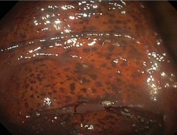
Melanosis coli, debido al abuso de laxantes, como se ve en la colonoscopia

Imagen: “Black pigmentation of colonic mucosa” por University of Sidi Mohammed Ben Abdellah, Faculty of Medicine and Pharmacy, Department of gastroenterology C, Fez, Morocco. Licencia: CC BY 2.0

Related videos

Referencias

  1. Gotfried, J. (2020). Diarrhea. [online] MSD Manual Professional Version. Retrieved December 4, 2020, from https://www.msdmanuals.com/professional/gastrointestinal-disorders/symptoms-of-gastrointestinal-disorders/diarrhea
  2. Guandalini, S., Frye, R.E., Tamer, M.A. (2020). Diarrhea. In Cuffari, C. (Ed.), Medscape. Retrieved December 4, 2020, from https://emedicine.medscape.com/article/928598-overview
  3. LaRocque, R., Harris, J.B. (2020). Approach to the adult with acute diarrhea in resource-rich settings. In Bloom, A. (Ed.), Uptodate. Retrieved December 4, 2020, from https://www.uptodate.com/contents/approach-to-the-adult-with-acute-diarrhea-in-resource-rich-settings
  4. LaRocque, R., Pietroni, M. (2020). Approach to the adult with acute diarrhea in resource-limited countries. In Bloom, A. (Ed.), UpToDate. Retrieved December 4, 2020, from https://www.uptodate.com/contents/approach-to-the-adult-with-acute-diarrhea-in-resource-limited-countries
  5. Bonis, P. A. L., Lamont, J. T. (2020). Approach to the adult with chronic diarrhea in resource-rich settings. In Grover, S. (Ed.), UpToDate. Retrieved December 4, 2020, from https://www.uptodate.com/contents/approach-to-the-adult-with-chronic-diarrhea-in-resource-rich-settings
  6. Juckett, G., Trivedi, R. (2011). Evaluation of chronic diarrhea. American Family Physician, 84(10):1119-26. https://www.aafp.org/afp/2011/1115/p1119.html
  7. Bashir, A., Sizar, O. (2020). Laxatives. StatPearls. Retrieved February 11, 2021, from https://www.ncbi.nlm.nih.gov/books/NBK537246/
  8. Wald, A. (2021). Factitious diarrhea: Clinical manifestations, diagnosis, and management. In Grover, S. (Ed.), UpToDate. Retrieved February 11, 2021, from https://www.uptodate.com/contents/factitious-diarrhea-clinical-manifestations-diagnosis-and-management
  9. LaRocque, R., Harris, J.B. (2018). Travelers’ diarrhea: Clinical manifestations, diagnosis, and treatment. In Bloom, A. (Ed.), UpToDate. Retrieved February 11, 2021, from https://www.uptodate.com/contents/travelers-diarrhea-clinical-manifestations-diagnosis-and-treatment

¡Crea tu cuenta gratis o inicia una sesión para seguir leyendo!

Regístrate ahora y obtén acceso gratuito a Lecturio con páginas de concepto, videos médicos y cuestionarios para tu educación médica.

User Reviews

Que tengas una sesión de estudio alegre y navideña 🎁 Ahorra 50% en todos los planes >>

Details