La inmunidad mediada por células es una parte integral del sistema inmunitario adaptativo, montando una defensa altamente específica contra los LOS Neisseria patógenos. La inmunidad mediada por células se desarrolla durante un período de tiempo más largo que la inmunidad innata. La respuesta adaptativa mediada por células ocurre en EN Erythema nodosum is an immune-mediated panniculitis (inflammation of the subcutaneous fat) caused by a type IV (delayed-type) hypersensitivity reaction. It commonly manifests in young women as tender, erythematous nodules on the shins. Erythema Nodosum células/tejidos (infecciones intracelulares o células aberrantes (e.g., tumores)) e involucra a las células T o linfocitos T. Las células, que surgen de la médula ósea, migran al AL Amyloidosis timo para una mayor maduración. Un receptor Receptor Receptors are proteins located either on the surface of or within a cell that can bind to signaling molecules known as ligands (e.g., hormones) and cause some type of response within the cell. Receptors de células T se ensambla y la activación es facilitada por células presentadoras de antígenos en EN Erythema nodosum is an immune-mediated panniculitis (inflammation of the subcutaneous fat) caused by a type IV (delayed-type) hypersensitivity reaction. It commonly manifests in young women as tender, erythematous nodules on the shins. Erythema Nodosum órganos linfoides secundarios. Bajo la influencia de las citoquinas, los LOS Neisseria linfocitos T activados pueden diferenciarse en EN Erythema nodosum is an immune-mediated panniculitis (inflammation of the subcutaneous fat) caused by a type IV (delayed-type) hypersensitivity reaction. It commonly manifests in young women as tender, erythematous nodules on the shins. Erythema Nodosum subconjuntos (e.g., linfocitos T colaboradores CD4+, linfocitos T citotóxicos CD8+, linfocitos T de memoria). Las células T colaboradoras ayudan en EN Erythema nodosum is an immune-mediated panniculitis (inflammation of the subcutaneous fat) caused by a type IV (delayed-type) hypersensitivity reaction. It commonly manifests in young women as tender, erythematous nodules on the shins. Erythema Nodosum la erradicación de patógenos al AL Amyloidosis ayudar a los LOS Neisseria socios de las células inmunitarias (e.g., macrófagos y granulocitos). Con mecanismos que activan la vía de la caspasa, las células T citotóxicas tienen la capacidad de matar microbios. Las células T de memoria se desarrollan después de la exposición inicial al AL Amyloidosis antígeno y se convierten en EN Erythema nodosum is an immune-mediated panniculitis (inflammation of the subcutaneous fat) caused by a type IV (delayed-type) hypersensitivity reaction. It commonly manifests in young women as tender, erythematous nodules on the shins. Erythema Nodosum células T efectoras en EN Erythema nodosum is an immune-mediated panniculitis (inflammation of the subcutaneous fat) caused by a type IV (delayed-type) hypersensitivity reaction. It commonly manifests in young women as tender, erythematous nodules on the shins. Erythema Nodosum la reinfección. Las células T también activan las células B (un componente principal de la inmunidad humoral), que mejoran la capacidad de eliminar patógenos.
Last updated: Dec 15, 2025
El sistema inmunológico proporciona defensa (inmunidad) contra patógenos invasores que van desde virus Virus Viruses are infectious, obligate intracellular parasites composed of a nucleic acid core surrounded by a protein capsid. Viruses can be either naked (non-enveloped) or enveloped. The classification of viruses is complex and based on many factors, including type and structure of the nucleoid and capsid, the presence of an envelope, the replication cycle, and the host range. Virology hasta parásitos. Los LOS Neisseria componentes están interconectados por la circulación sanguínea y linfática.
Dos líneas de defensa superpuestas:
| Inmunidad innata | Inmunidad adaptativa | |
|---|---|---|
| Genética | Línea germinal codificada | Reordenamientos genéticos implicados en EN Erythema nodosum is an immune-mediated panniculitis (inflammation of the subcutaneous fat) caused by a type IV (delayed-type) hypersensitivity reaction. It commonly manifests in young women as tender, erythematous nodules on the shins. Erythema Nodosum el desarrollo de linfocitos |
| Respuesta inmune | Inespecífica | Altamente específica |
| Tiempo de respuesta | Inmediato (minutos a horas) | Se desarrolla durante un período de tiempo más largo |
| Respuesta de memoria | Ninguna | Responde rápidamente al AL Amyloidosis reconocimiento del antígeno con respuesta de memoria |
| Reconocimiento del patógeno | Los LOS Neisseria receptores de reconocimiento de patrones, como los LOS Neisseria receptores tipo toll, reconocen patrones moleculares asociados a patógenos |
|
| Componentes |
|
|
Responder a los LOS Neisseria invasores microbianos es responsabilidad del sistema inmunológico. A menudo, el sistema inmunológico innato tiene la capacidad de contener los LOS Neisseria patógenos, pero los LOS Neisseria invasores pueden evolucionar para evadir la inmunidad innata. La siguiente línea de defensa es el sistema inmunológico adaptativo.

Inmunidad mediada por células:
La activación de los linfocitos T auxiliares da lugar a la liberación de citoquinas, activando así los linfocitos T citotóxicos y los fagocitos (como los macrófagos).
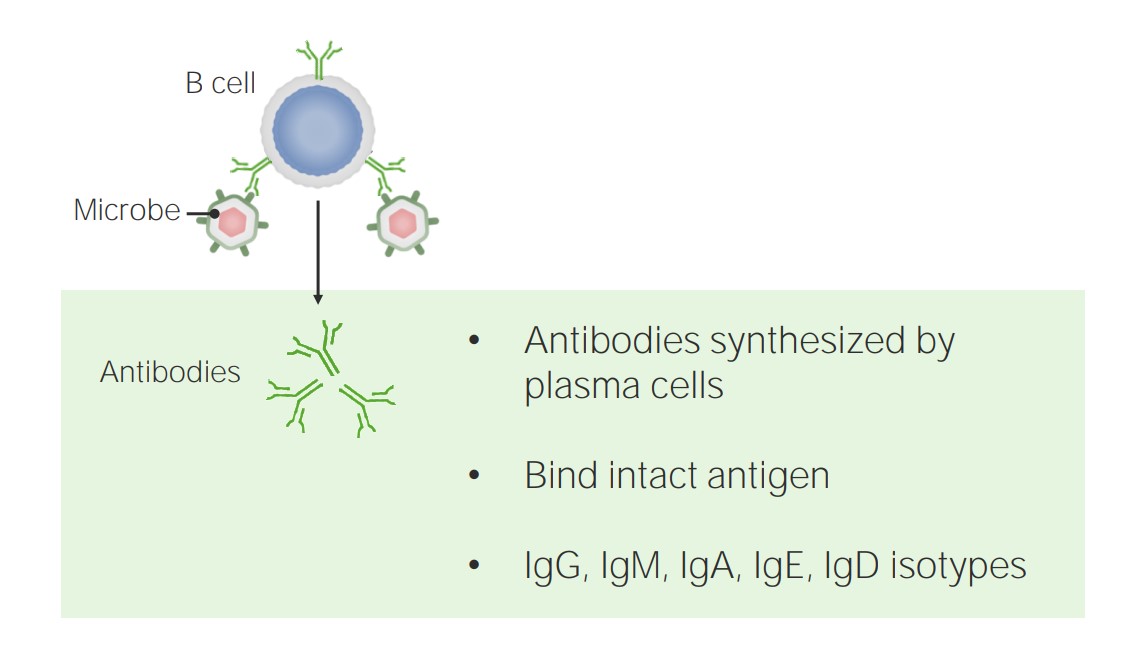
La inmunidad humoral está mediada por las células B y los anticuerpos.
Imagen por Lecturio.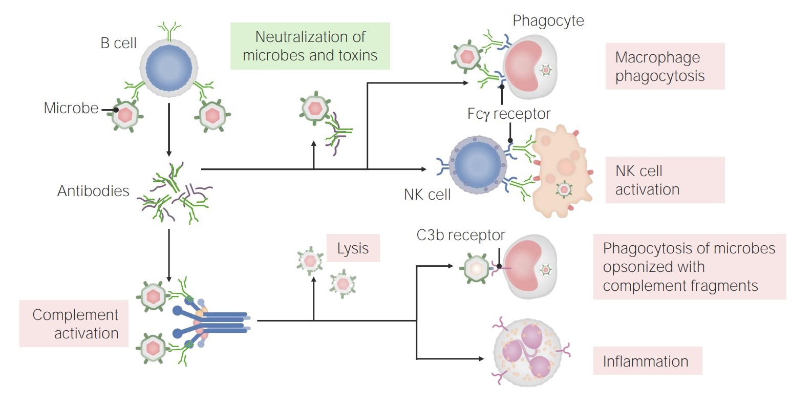
Las funciones de los anticuerpos:
Los anticuerpos tienen múltiples funciones en la inmunidad, incluyendo la neutralización (de microbios y toxinas), la promoción de la fagocitosis y la activación de las células NK. Además, los anticuerpos tienen un papel en la activación del complemento, que puede conducir a la lisis directa de los microbios, la opsonización y la fagocitosis, y el reclutamiento/activación de los neutrófilos.
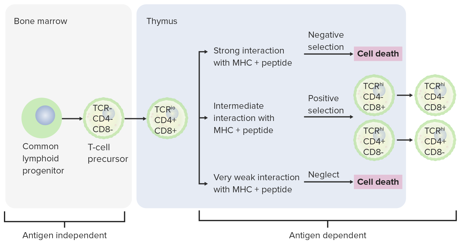
Etapas de diferenciación de la célula T:
Desde la médula ósea, las células progenitoras van al timo para una mayor maduración. Las células DN (sin expresión de CD4/CD8 o CD4–/CD8–) no han desarrollado el receptor de célula T. Las células DN experimentan un reordenamiento del gen receptor de célula T y se convierten en células pro-T, luego en células pre-T. A través de la serie, se expresan CD4 y CD8, y el receptor de célula T se ensambla a través de reordenamientos de genes (células DP). Luego, el timo presenta moléculas del complejo mayor de histocompatibilidad a las células T en desarrollo. Algunas células experimentan una selección positiva (tiene lugar una interacción intermedia entre el complejo mayor de histocompatibilidad y el receptor de célula T) y producen células funcionales. Algunas células experimentan una selección negativa (fuerte interacción entre el complejo mayor de histocompatibilidad y el receptor de células T), lo que resulta en la muerte celular. Se previene la liberación de células T disfuncionales, que pueden activar la autoinmunidad. Algunas células T no logran interactuar, lo que lleva a la apoptosis. Las células T maduras expresan CD4 (células T colaboradoras) o CD8 (células T citotóxicas), no ambas.
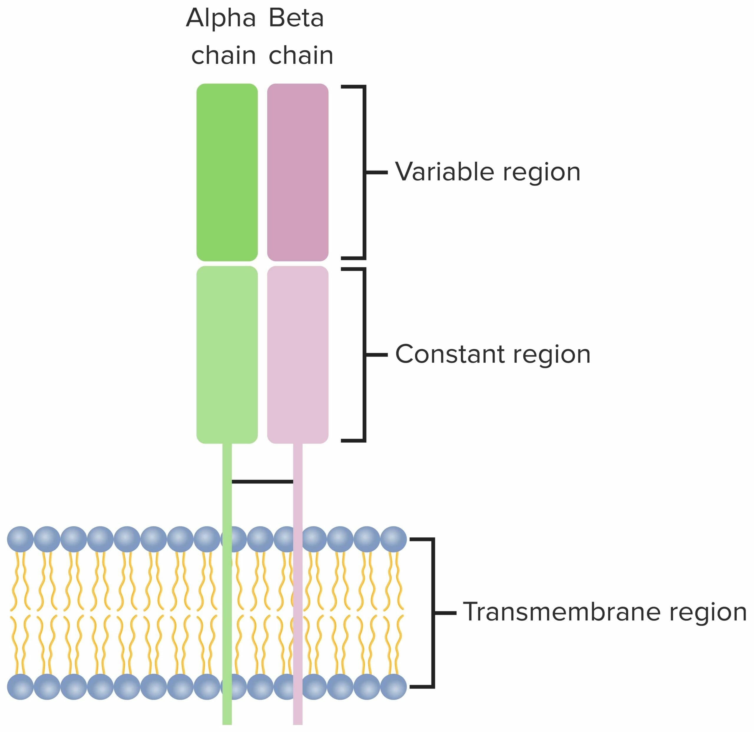
Receptor de células T:
Heterodímero que consta de 2 cadenas polipeptídicas transmembrana, con cadenas α y β que se encuentran en la mayoría de las células T. Ambas cadenas tienen una región transmembrana variable, constante y una región citoplasmática corta.
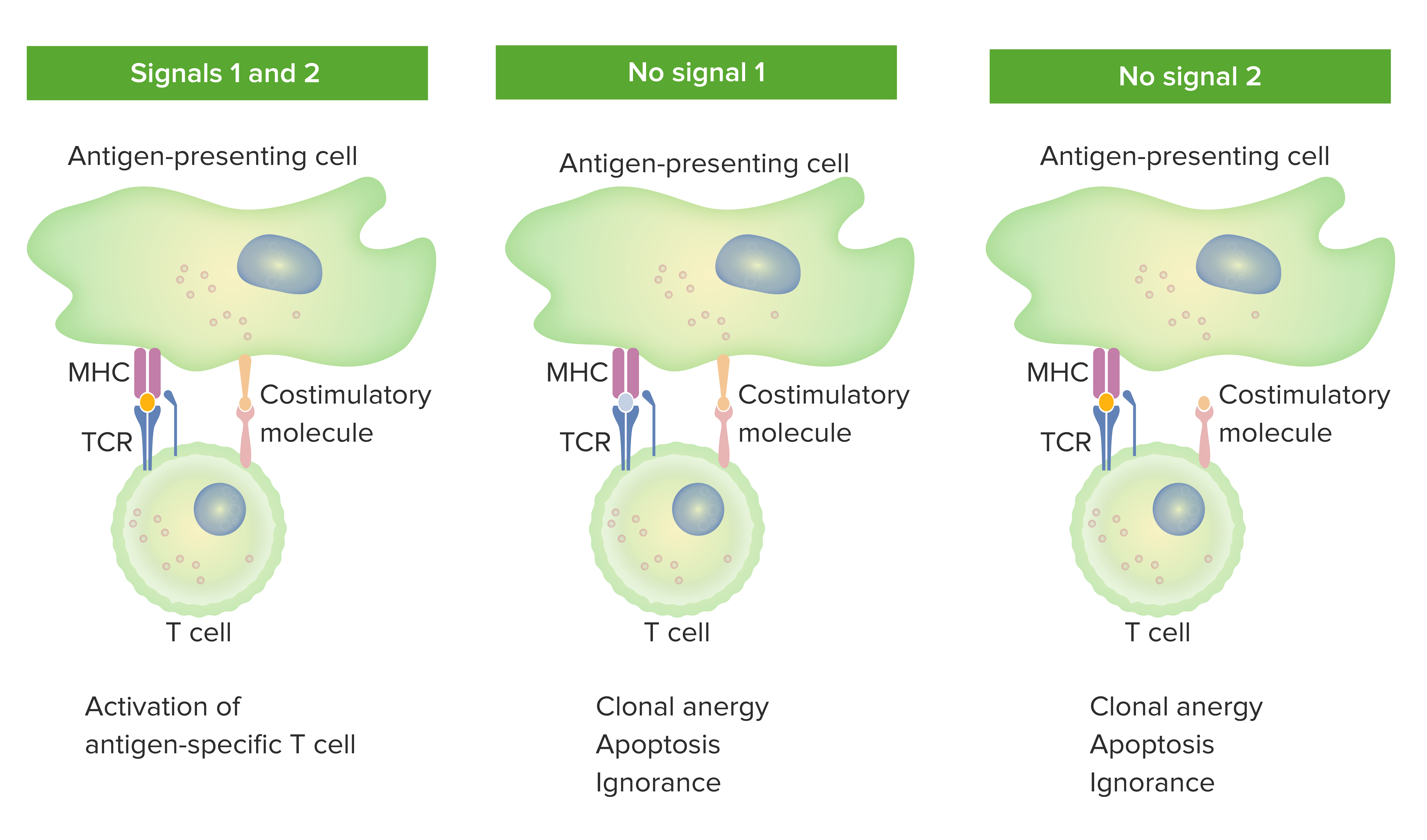
Modelo de 2 señales de dependencia de las células T de la coestimulación:
Cuando están presentes tanto la señal 1 (TCR que se une al antígeno afín presentado por la molécula del complejo mayor de histocompatibilidad en la célula presentadora de antígeno) como la señal 2 (interacción de la molécula coestimuladora entre la célula presentadora de antígeno y la célula T), la célula T madura está completamente activada.
El área naranja en el panel izquierdo indica la unión adecuada entre el antígeno y el TCR. Sin embargo, cuando falta la señal 1 (la imagen del medio no muestra unión entre antígeno y TCR) o la señal 2 (la imagen de la derecha no muestra coestimulación), la célula T no se activará por completo.
Los resultados serían anergia (estado de falta de respuesta), apoptosis (muerte celular) o ignorancia (la célula T no se da cuenta o no se ve afectada por el antígeno).
TCR: receptor de células T
| Células T CD4+ | Estimulado por | Citoquinas producidas | Funciones | Papel en EN Erythema nodosum is an immune-mediated panniculitis (inflammation of the subcutaneous fat) caused by a type IV (delayed-type) hypersensitivity reaction. It commonly manifests in young women as tender, erythematous nodules on the shins. Erythema Nodosum la enfermedad |
|---|---|---|---|---|
| Th1 Th1 A subset of helper-inducer T-lymphocytes which synthesize and secrete interleukin-2; interferon-gamma; and interleukin-12. Due to their ability to kill antigen-presenting cells and their lymphokine-mediated effector activity, th1 cells are associated with vigorous delayed-type hypersensitivity reactions. T cells: Types and Functions | IL-12, IFN-γ | IFN-γ, TNF TNF Tumor necrosis factor (TNF) is a major cytokine, released primarily by macrophages in response to stimuli. The presence of microbial products and dead cells and injury are among the stimulating factors. This protein belongs to the TNF superfamily, a group of ligands and receptors performing functions in inflammatory response, morphogenesis, and cell proliferation. Tumor Necrosis Factor (TNF), IL-2 |
|
|
| Th2 Th2 A subset of helper-inducer T-lymphocytes which synthesize and secrete the interleukins il-4; il-5; il-6; and il-10. These cytokines influence b-cell development and antibody production as well as augmenting humoral responses. T cells: Types and Functions | IL-2, IL-4 | IL-4, IL-5, IL-6, IL-9, IL-10, IL-13 |
|
|
| Th17 Th17 A subset of helper-effector T-lymphocytes which synthesize and secrete interleukins il-17; il-17f; and il-22. These cytokines are involved in host defenses and tissue inflammation in autoimmune diseases. T cells: Types and Functions | IL-1, IL-6, IL-23, TGF-β | IL-17, IL-21, IL-22 | Promover la inflamación neutrofílica |
|
| Tfh | IL-6 | IL-4, IL-21 | Facilitar la activación y maduración de las células B | Producción de anticuerpos |
| Treg | TGF-β, IL-2 | TGF-β, IL-10, IL-35 |
|
↓ Autoinmunidad, alergia, inflamación |
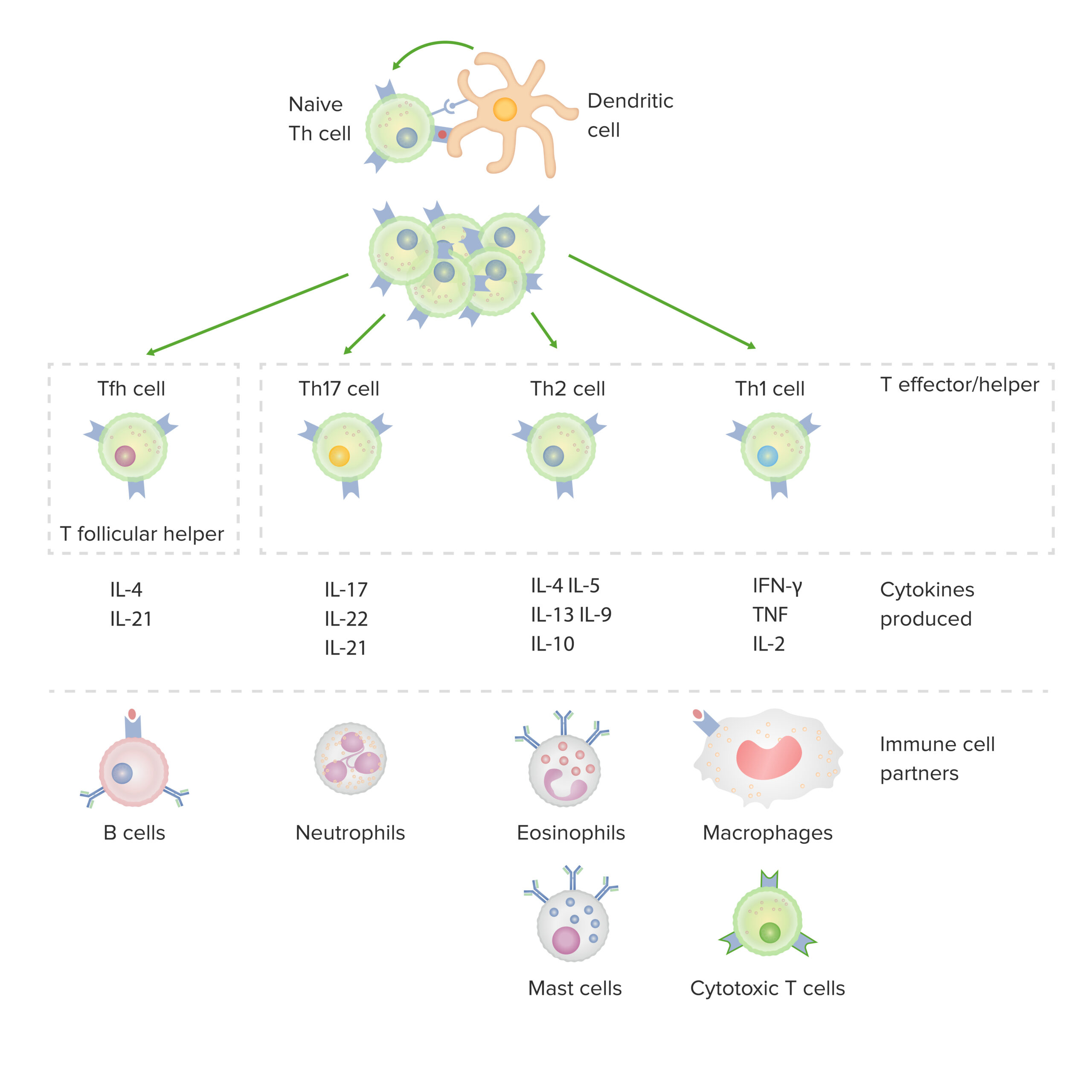
Subconjuntos de células T colaboradoras CD4 positivas:
Después de la activación por una célula dendrítica, en presencia de citoquinas particulares, un linfocito T CD4-positivo virgen se divide y se diferencia en subconjuntos efector/colaborador (Th1, Th2 o Th17) o colaborador folicular (Tfh). Cada tipo de célula produce citoquinas que facilitan la activación de otras células inmunitarias asociadas.
IFN: interferón
TNF: factor de necrosis tumoral
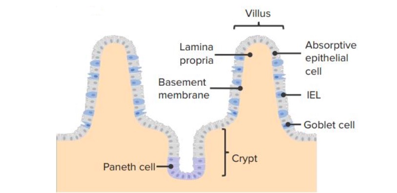
Linfocitos intraepiteliales (IEL) en el epitelio intestinal
Imagen por Lecturio.| Citoquina | Función y actividad |
|---|---|
| IL-2 |
|
| IL-4 |
|
| IL-5 |
|
| IL-10 |
|
| IL-13 |
|
| IL-17 | Liberación de citoquinas inflamatorias (incluyendo IL-6, IL-1, TNF TNF Tumor necrosis factor (TNF) is a major cytokine, released primarily by macrophages in response to stimuli. The presence of microbial products and dead cells and injury are among the stimulating factors. This protein belongs to the TNF superfamily, a group of ligands and receptors performing functions in inflammatory response, morphogenesis, and cell proliferation. Tumor Necrosis Factor (TNF), que median fiebre y sepsis Sepsis Systemic inflammatory response syndrome with a proven or suspected infectious etiology. When sepsis is associated with organ dysfunction distant from the site of infection, it is called severe sepsis. When sepsis is accompanied by hypotension despite adequate fluid infusion, it is called septic shock. Sepsis and Septic Shock) |
| IL-21 | Diferenciación de células B y células T |
| IL-22 |
|
| IFN-ɣ |
|
| TNF TNF Tumor necrosis factor (TNF) is a major cytokine, released primarily by macrophages in response to stimuli. The presence of microbial products and dead cells and injury are among the stimulating factors. This protein belongs to the TNF superfamily, a group of ligands and receptors performing functions in inflammatory response, morphogenesis, and cell proliferation. Tumor Necrosis Factor (TNF) |
|
| Inhibición de citoquinas | Nombre del medicamento | Afecciones tratadas |
|---|---|---|
| Anti-TNF |
|
|
| Anti-interleuquina-1 | Anakinra Anakinra Immunosuppressants | Artritis reumatoide |
| Anti-interleuquina-1b | Canakinumab Canakinumab Immunosuppressants |
|
| Anti-interleuquina-6 | Tocilizumab Tocilizumab Immunosuppressants |
|
| Citoquina terapéutica | Afecciones tratadas |
|---|---|
| Interferón-α |
|
| Interferón-β | Esclerosis múltiple |
| Interferón-γ |
|
| Interleuquina-2 |