La fascitis necrosante es una infección potencialmente mortal que provoca una rápida destrucción y necrosis Necrosis The death of cells in an organ or tissue due to disease, injury or failure of the blood supply. Ischemic Cell Damage de la fascia Fascia Layers of connective tissue of variable thickness. The superficial fascia is found immediately below the skin; the deep fascia invests muscles, nerves, and other organs. Cellulitis y los LOS Neisseria tejidos subcutáneos. Los LOS Neisseria pacientes pueden presentar un dolor Dolor Inflammation importante desproporcionado con respecto a los LOS Neisseria síntomas que presentan y un eritema rápidamente progresivo de la zona afectada. La mayoría de los LOS Neisseria pacientes también presentarán signos sistémicos de infección, como fiebre, hipotensión, alteración del estado mental y fallo orgánico multisistémico. El diagnóstico es principalmente clínico, ya que los LOS Neisseria pacientes pueden evolucionar rápidamente hacia un shock Shock Shock is a life-threatening condition associated with impaired circulation that results in tissue hypoxia. The different types of shock are based on the underlying cause: distributive (↑ cardiac output (CO), ↓ systemic vascular resistance (SVR)), cardiogenic (↓ CO, ↑ SVR), hypovolemic (↓ CO, ↑ SVR), obstructive (↓ CO), and mixed. Types of Shock séptico si no se controla el sitio de infección. Este tipo de infección es una emergencia quirúrgica y requiere un desbridamiento quirúrgico de emergencia, antibióticos parenterales y una estrecha monitorización hemodinámica.
Last updated: Dec 15, 2025
La fascitis necrosante se divide microbiológicamente en EN Erythema nodosum is an immune-mediated panniculitis (inflammation of the subcutaneous fat) caused by a type IV (delayed-type) hypersensitivity reaction. It commonly manifests in young women as tender, erythematous nodules on the shins. Erythema Nodosum función del organismo u organismos causantes en EN Erythema nodosum is an immune-mediated panniculitis (inflammation of the subcutaneous fat) caused by a type IV (delayed-type) hypersensitivity reaction. It commonly manifests in young women as tender, erythematous nodules on the shins. Erythema Nodosum:
Signos tempranos:
Signos tardíos:
Sitios comunes de infección:
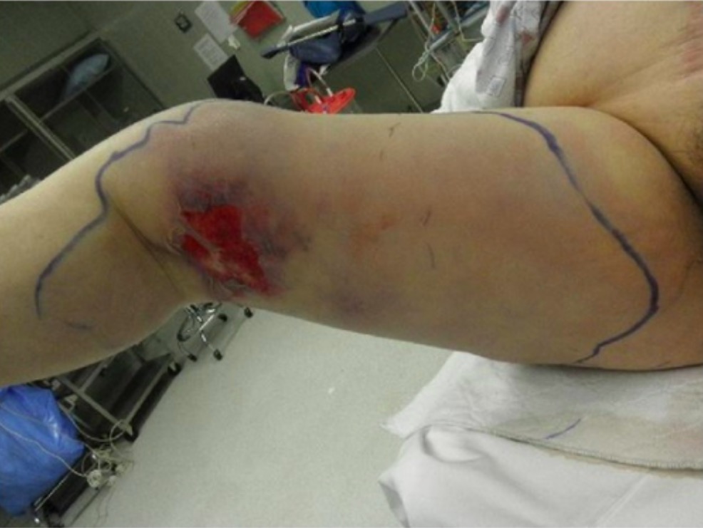
Eritema de rápida evolución, ulceración y edema de pierna derecha debido a fascitis necrosante.
Imagen: “Preoperative photograph” por Department of Family Medicine, Morehouse School of Medicine, 1513 East Cleveland Avenue, Building 100, Suite 300A, Atlanta, GA 30344, USA. Licencia: CC BY 3.0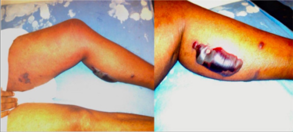
Bulas y eritema resultantes de la fascitis necrosante
Imagen: “Superficial skin manifestations of necrotizing fasciitis” por National Hospital of Sri Lanka, Regent Street, Colombo 10, Sri Lanka. Licencia: CC BY 4.0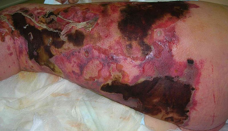
Necrosis cutánea, eritema y cambios bulosos por fascitis necrosante de la pierna
Imagen: “Necrotizing fasciitis” por Piotr Smuszkiewicz et al. Licencia: CC BY 2.0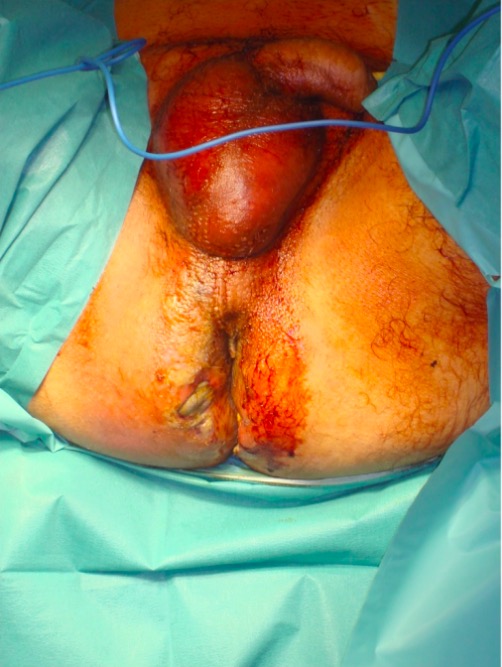
Gangrena de Fournier:
Se observa un importante eritema y edema en todo el escroto y la región glútea.
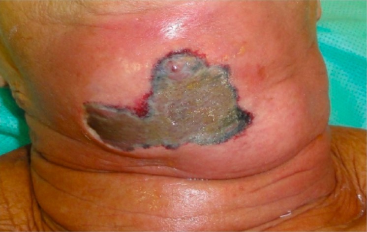
Eritema y tejido necrótico por fascitis necrosante del cuello:
Esto se desarrolló después de una extracción dental.
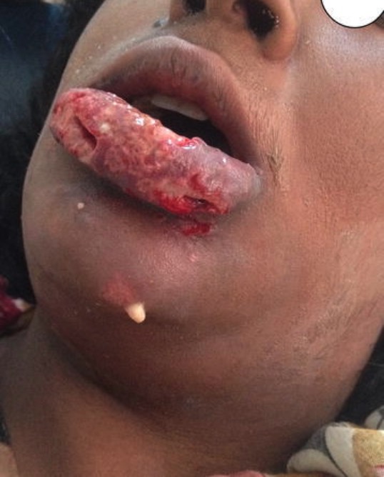
Fascitis necrosante del labio y la cara:
Se observa una importante inflamación, eritema, exfoliación y drenaje purulento.
El diagnóstico definitivo de la fascitis necrosante se realiza mediante la exploración quirúrgica y el desbridamiento. Estos procesos no deben retrasarse para obtener información diagnóstica si la sospecha clínica es alta.
Sin embargo, lo siguiente puede ser útil:
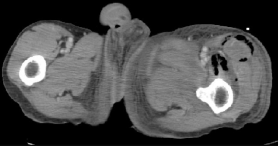
Hallazgos de la TC en la fascitis necrosante:
Se observa gas en el espacio subcutáneo.
Hallazgos de la TC en la fascitis necrosante:
Este corte muestra el edema de la fascia, la trabeculación anormal de la grasa subcutánea y el gas en los compartimentos musculares.

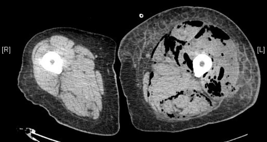
Hallazgos de la TC en la fascitis necrosante:
Este corte muestra la presencia de gas subcutáneo a lo largo de los planos fasciales de la pierna izquierda y la pelvis.
El desbridamiento quirúrgico es la base del tratamiento.
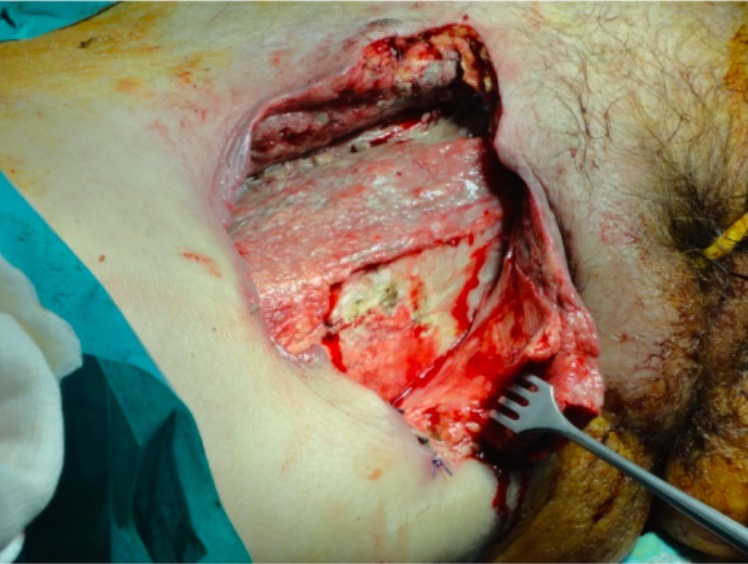
Escisión quirúrgica de los tejidos necróticos en la fascitis necrosante
Imagen: “Management of necrotizing fasciitis” por 3rd Department of Surgery, Attikon University Hospital, University of Athens School of Medicine, Athens, Greece. Licencia: CC BY 4.0