Las anomalías y afecciones del pene pueden ser congénitas o adquiridas y pueden afectar el orificio uretral, el prepucio, el cuerpo o el glande del pene. Algunos ejemplos son la fimosis, la parafimosis, el epispadias Epispadias A birth defect due to malformation of the urethra in which the urethral opening is above its normal location. In the male, the malformed urethra generally opens on the top or the side of the penis, but the urethra can also be open the entire length of the penis. In the female, the malformed urethral opening is often between the clitoris and the labia, or in the abdomen. Penile Anomalies and Conditions, el hipospadias, la balanitis Balanitis Inflammation of the head of the penis, glans penis. Penile Anomalies and Conditions, la enfermedad de Peyronie y el priapismo. La gravedad de los LOS Neisseria síntomas clínicos varía, pero el diagnóstico de cada una de estas condiciones suele basarse en EN Erythema nodosum is an immune-mediated panniculitis (inflammation of the subcutaneous fat) caused by a type IV (delayed-type) hypersensitivity reaction. It commonly manifests in young women as tender, erythematous nodules on the shins. Erythema Nodosum los LOS Neisseria antecedentes y la exploración física. El tratamiento varía desde las terapias médicas hasta la intervención quirúrgica. Es importante conocer estos diagnósticos, ya que algunos (como la fimosis y la balanitis Balanitis Inflammation of the head of the penis, glans penis. Penile Anomalies and Conditions) son relativamente comunes, mientras que otros (como la parafimosis y el priapismo) pueden tener complicaciones graves si no se tratan a tiempo.
Last updated: Dec 15, 2025
El pene se compone de:
Las afecciones penianas pueden clasificarse en EN Erythema nodosum is an immune-mediated panniculitis (inflammation of the subcutaneous fat) caused by a type IV (delayed-type) hypersensitivity reaction. It commonly manifests in young women as tender, erythematous nodules on the shins. Erythema Nodosum función de la región del pene comprometida:
Bebé masculino con epispadias
Imagen: “Male baby with epispadias” por Department of Pediatric Urology, University Medical Center Regensburg, Germany. Licencia: CC BY 2.0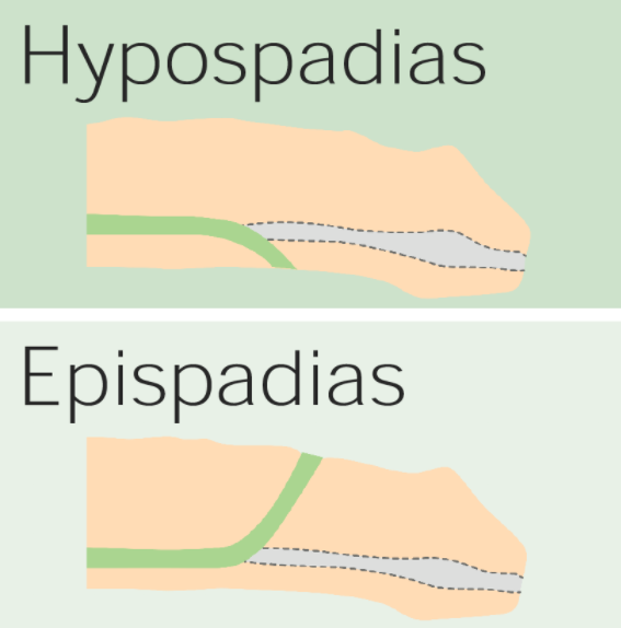
La diferencia entre hipospadias y epispadias: El hipospadias resulta en una abertura uretral anormal en la superficie ventral del pene, mientras que el epispadias se debe a una abertura anormal en la superficie dorsal.
Imagen por Lecturio.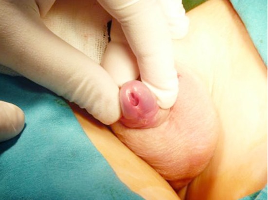
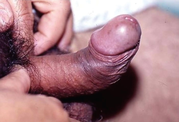
Hipospadias glanular
Imagen: “Glanular hypospadias is detected with the retraction of the foreskin” por Yesildag E et al. Licencia: CC BY 3.0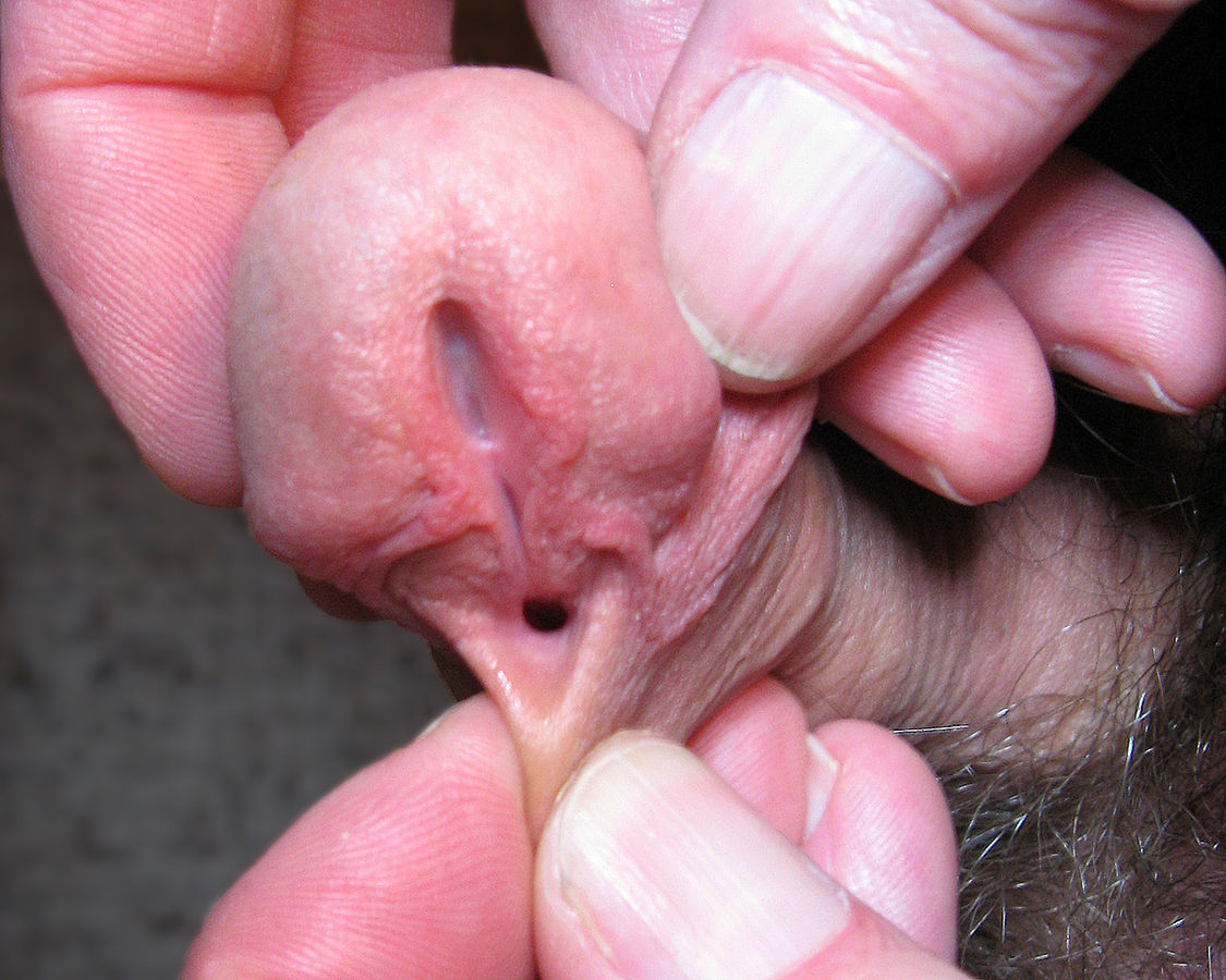
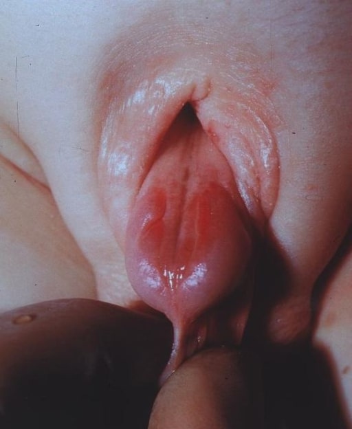
Abertura anormal en la superficie ventral del pene consistente con hipospadias
Imagen: “Depiction of a human penis with hypospadias” por Buddy. Licencia: Dominio Público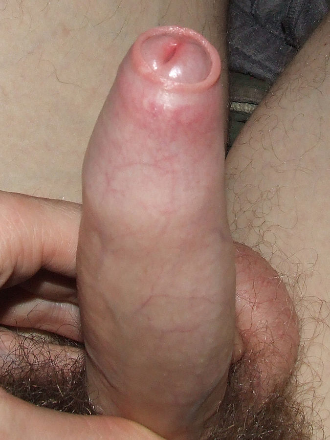
Un pene erecto con fimosis
Imagen: “Example of an erect penis with Phimosis. The foreskin will not retract.” por Andrew1985. Licencia: Dominio Público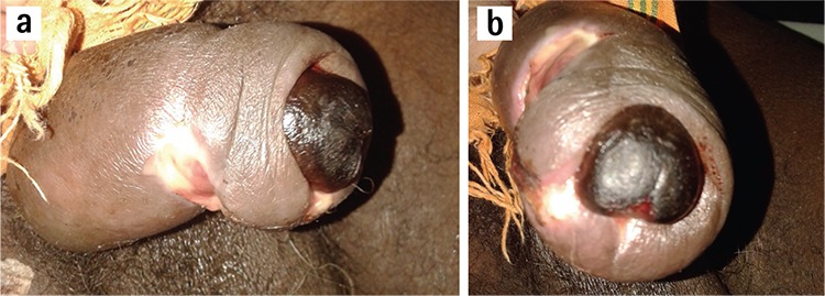
Parafimosis complicada por gangrena del glande:
Esta condición es causada por la alteración del flujo sanguíneo, linfático y venoso, lo que conduce a la inflamación y al compromiso del flujo sanguíneo arterial.
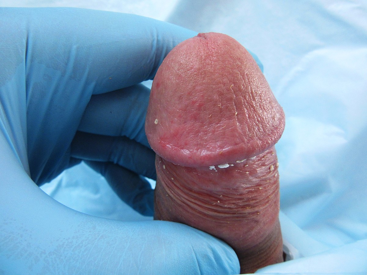
Eritema y edema del glande del pene compatible con balanitis
Imagen: “Balanitis caused by smegma” por MFN24. Licencia: CC0 1.0La enfermedad de Peyronie es una deformidad del pene causada por la fibrosis Fibrosis Any pathological condition where fibrous connective tissue invades any organ, usually as a consequence of inflammation or other injury. Bronchiolitis Obliterans/cicatrización de la túnica albugínea.
Curvatura anormal del pene secundaria a la enfermedad de Peyronie
Imagen: “Penile deformity secondary to Peyronie’s disease” por Tran VQ et al. Licencia: CC BY 3.0El diagnóstico es clínico.
El priapismo es una erección anormal y persistente (generalmente > 4 horas) que no está asociada a la estimulación sexual.
El diagnóstico se sospecha con base en EN Erythema nodosum is an immune-mediated panniculitis (inflammation of the subcutaneous fat) caused by a type IV (delayed-type) hypersensitivity reaction. It commonly manifests in young women as tender, erythematous nodules on the shins. Erythema Nodosum los LOS Neisseria antecedentes y el examen físico. Los LOS Neisseria siguientes hallazgos pueden ayudar a diferenciar el priapismo isquémico del no isquémico:
Las complicaciones del priapismo isquémico incluyen: