Imaging of the Spleen

Introduction Imaging modalities The common radiologic modalities used to evaluate the spleen are the following: Preparation and orientation Ultrasonography Overview Exam technique Interpretation and evaluation Normal findings Normal ultrasound appearance: Computed Tomography Indication Exam technique Standard CT scanning: Interpretation and evaluation Interpretation should follow a systematic and reproducible pattern: Normal findings Normal CT appearance: Magnetic […]
Imaging of the Mediastinum
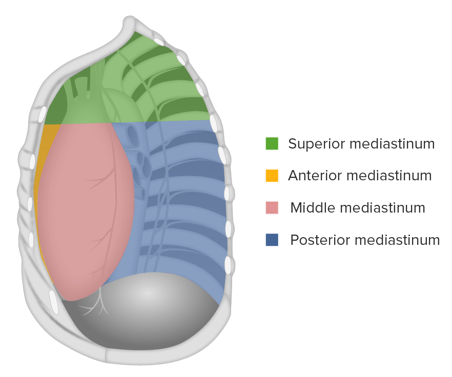
Introduction Mediastinum Imaging modalities Chest Radiograph Overview PA view Lateral view Compartments of the mediastinum Computed Tomography Overview Normal findings Magnetic resonance imaging Overview MRI Normal Findings Abnormal Findings Overview Pneumomediastinum Thyroid-related masses Teratoma Thymoma and thymic carcinoma Lymphoma Bronchogenic cyst LAD Aortic aneurysm Schwannoma References
Pediatric Chest Abnormalities
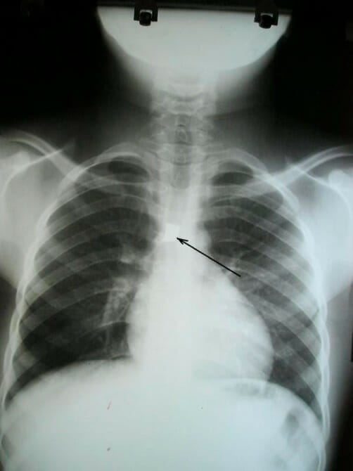
Radiography Indication Exam technique Interpretation/evaluation Systematic approach: Normal findings Anteroposterior (AP) view: Transient tachypnea of the newborn Meconium aspiration syndrome Hyaline membrane disease Epiglottitis Croup Foreign body aspiration Computed Tomography Indications Exam technique Standard CT scanning: Interpretation and evaluation Normal findings Normal chest CT (findings may be dependent on the individual’s age): Cystic fibrosis Multiple […]
Imaging of the Intestines
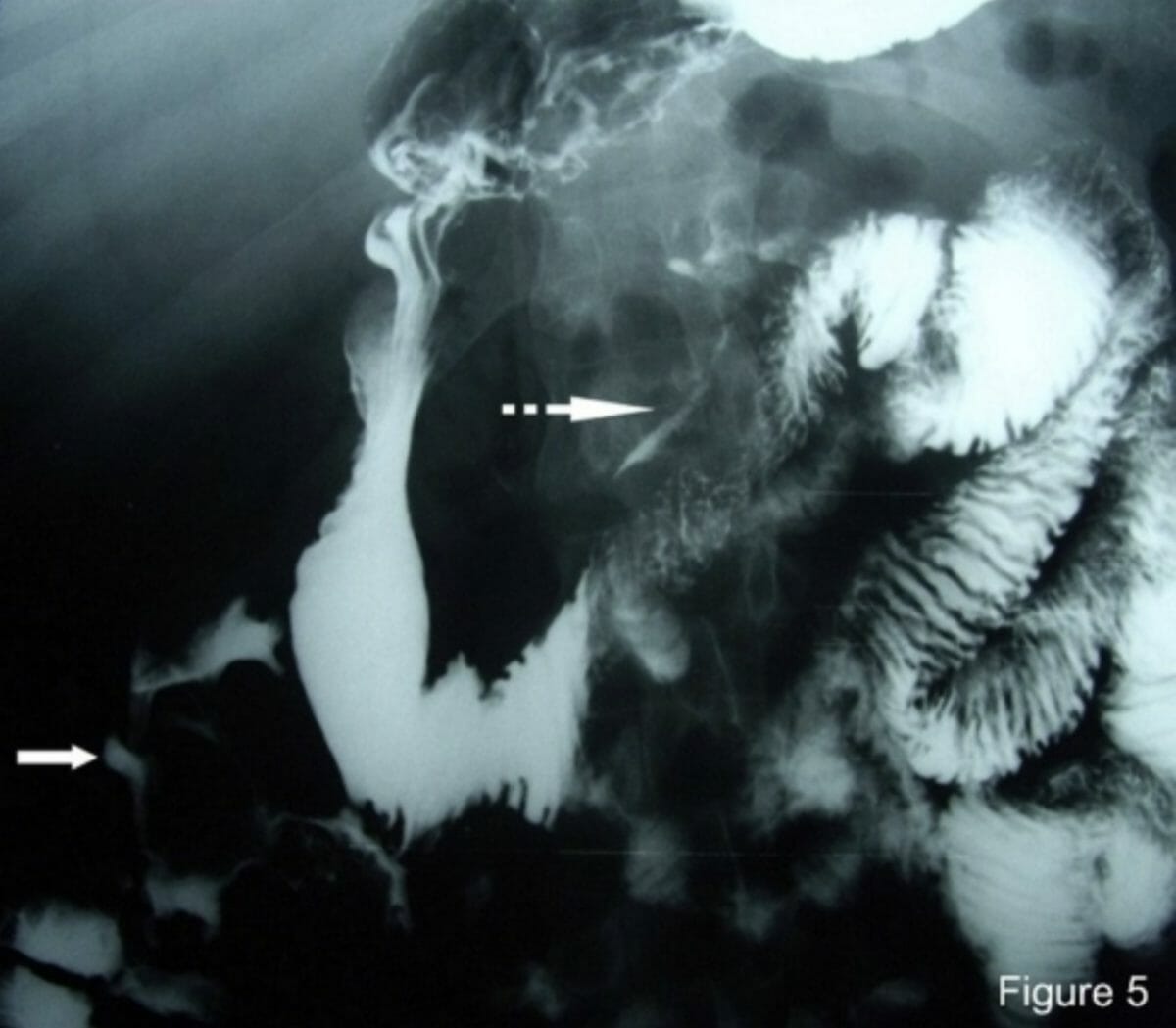
Introduction Imaging modalities The common radiologic modalities used to evaluate the intestines are the following: Preparation and orientation Prior to interpretation of any image, the physician should take certain preparatory steps. The same systematic approach should be followed every time: Determine the orientation of the image: Radiography (X-ray) Overview Exam technique General positioning: Positioning for […]
Imaging of the Liver and Biliary Tract
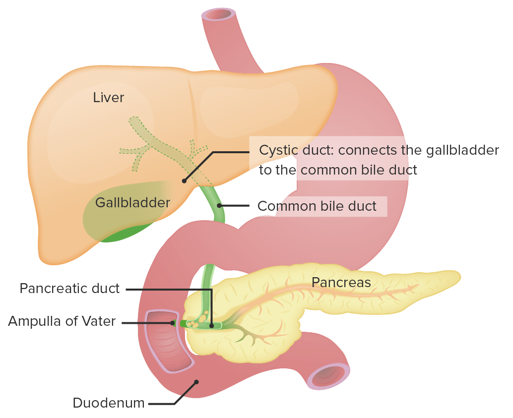
Introduction Imaging methods Preparation and orientation Prior to interpretation of any image, the physician should take certain preparatory steps. The same systematic approach should be followed every time. Ultrasonography Overview Exam technique Interpretation and evaluation Report includes: Normal findings CT Overview Exam technique Interpretation and evaluation Normal findings MRI Overview Exam technique Table: General principles […]
Gynecological Imaging
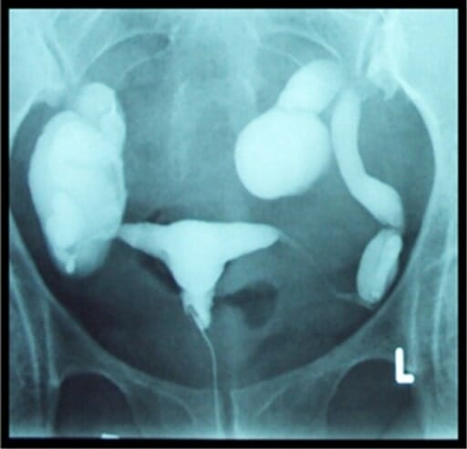
Overview Internal gynecologic organs commonly evaluated on imaging Studies of choice for gynecologic imaging Preparation Prior to the interpretation of any image, the physician should take certain preparatory steps. The same systematic approach should be followed every time: Ultrasonography Indications Ultrasound (i.e., sonography) is almost always the imaging modality of choice when evaluating the internal […]
Imaging of the Breast
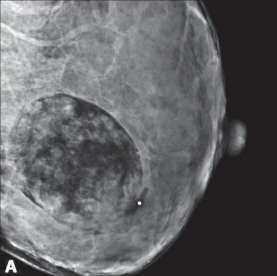
Mammography Mammogram Indications Contraindications There are no absolute contraindications, but there are relative ones (owing to adverse effects of radiation exposure). Mammogram views Table: Mammogram views Views Description Standard views Mediolateral oblique (MLO) view Better view of the superior lateral quadrant of the breast and axilla Craniocaudal view (CC) Medial part of the breast: lower […]
Scrotal Imaging

Ultrasonography Overview Ultrasonography is often the best imaging method for the scrotum. Normal scrotal ultrasonography Epididymitis and orchitis Ultrasound findings include: Hydrocele Ultrasound findings include: Varicocele Ultrasound findings include: Testicular torsion Ultrasound findings: Testicular carcinoma Ultrasound findings include: MRI Normal scrotal MRI Table: Normal scrotal MRI Structure T1-weighted T2-weighted With contrast Testicle (homogeneous oval structure) […]
Obstetric Imaging
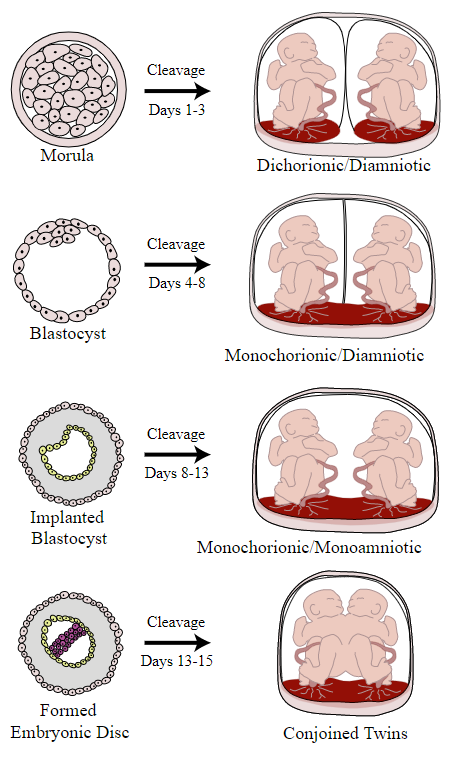
Overview Introduction Obstetric imaging refers to imaging of the female reproductive tract and developing fetus during pregnancy. Types of obstetric imaging exams There are 2 primary types of obstetric imaging exams: Specific studies Several specific types of studies can be performed. All are ultrasound exams and may be either abdominal or transvaginal. Indications for Obstetric […]
Imaging of the Heart and Great Vessels

Chest X-ray Overview Anatomy The following structures must be identified and checked for abnormalities in size or shape: Heart Table: Artifactual causes of cardiac enlargement Cause Observation Rotation The shadow of the heart appears bigger in the projection because of the rotation of the subject to either side. Suboptimal inspiration The diaphragm moves upward and […]