


O vírus Herpes simplex Herpes Simplex A group of acute infections caused by herpes simplex virus type 1 or type 2 that is characterized by the development of one or more small fluid-filled vesicles with a raised erythematous base on the skin or mucous membrane. It occurs as a primary infection or recurs due to a reactivation of a latent infection. Congenital TORCH Infections (VHS) é um vírus de DNA DNA A deoxyribonucleotide polymer that is the primary genetic material of all cells. Eukaryotic and prokaryotic organisms normally contain DNA in a double-stranded state, yet several important biological processes transiently involve single-stranded regions. DNA, which consists of a polysugar-phosphate backbone possessing projections of purines (adenine and guanine) and pyrimidines (thymine and cytosine), forms a double helix that is held together by hydrogen bonds between these purines and pyrimidines (adenine to thymine and guanine to cytosine). DNA Types and Structure de cadeia dupla pertencente à família Herpesviridae Herpesviridae A family of enveloped, linear, double-stranded DNA viruses infecting a wide variety of animals. Subfamilies, based on biological characteristics, include: alphaherpesvirinae; betaherpesvirinae; and gammaherpesvirinae. Herpes Simplex Virus 1 and 2. O vírus do herpes simplex Herpes Simplex A group of acute infections caused by herpes simplex virus type 1 or type 2 that is characterized by the development of one or more small fluid-filled vesicles with a raised erythematous base on the skin or mucous membrane. It occurs as a primary infection or recurs due to a reactivation of a latent infection. Congenital TORCH Infections causa frequentemente infeções recorrentes que envolvem a pele e as superfícies mucosas, incluindo boca, lábios, olhos e genitais. As infeções mucocutâneas típicas são caracterizadas pelo aparecimento súbito localizado de aglomerados de pequenas vesículas dolorosas sobre uma base eritematosa. Embora exista sobreposição, o VHS-1 está tipicamente associado a lesões orofaríngeas, enquanto o VHS-2 é o principal responsável pelo herpes genital, uma IST. Também podem surgir infeções sistémicas e graves, como encefalite, meningite e herpes neonatal. O diagnóstico é estabelecido com base na história clínica, que pode ser confirmada pelo exame microscópico de um esfregaço de uma vesícula, teste de amplificação de ácidos nucleicos por PCR PCR Polymerase chain reaction (PCR) is a technique that amplifies DNA fragments exponentially for analysis. The process is highly specific, allowing for the targeting of specific genomic sequences, even with minuscule sample amounts. The PCR cycles multiple times through 3 phases: denaturation of the template DNA, annealing of a specific primer to the individual DNA strands, and synthesis/elongation of new DNA molecules. Polymerase Chain Reaction (PCR), imunofluorescência direta ou testes Testes Gonadal Hormones serológicos. O tratamento das lesões mucocutâneas é habitualmente sintomático, no entanto, a terapêutica com antivirais com aciclovir, valaciclovir ou famciclovir Famciclovir An aminopurine derivative and prodrug of penciclovir which is a competitive inhibitor of herpes simplex 2 DNA polymerase. It is used to treat herpes simplex virus infection. Antivirals for Herpes Virus está indicada nas infeções sistémicas graves e deverá ser administrada precocemente.
Last updated: Dec 15, 2025
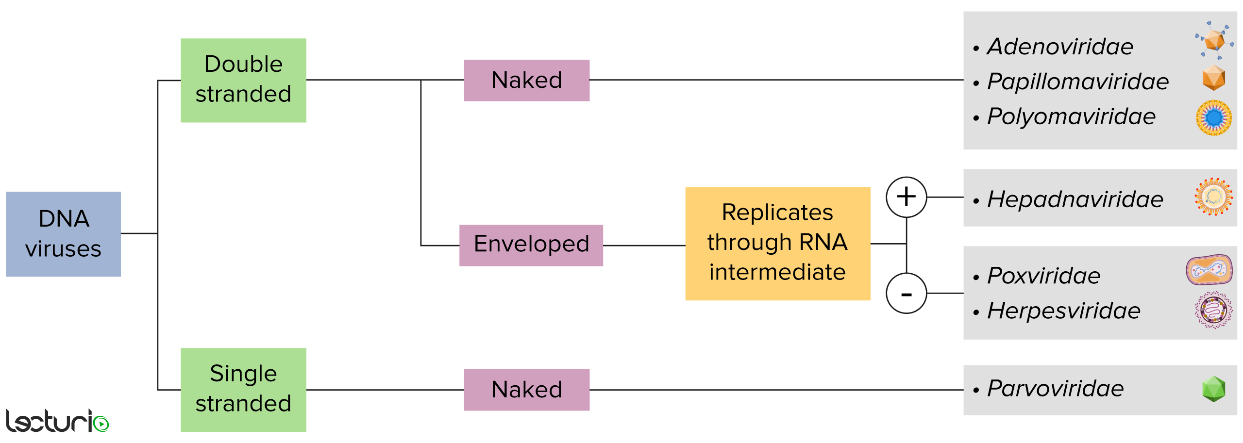
Identificação de vírus de DNA:
Os vírus podem ser classificados de várias formas. Contudo, a maioria dos vírus possui um genoma formado por DNA ou RNA. Os vírus com genoma de DNA podem ainda ser caracterizados como de cadeia simples ou dupla. Os vírus com envelope são revestidos por uma camada fina de membrana celular, que geralmente é retirada da célula hospedeira. Os vírus sem envelope são apelidados de vírus “nus”. Alguns vírus com envelope traduzem DNA em RNA antes de serem incorporados no genoma da célula hospedeira.
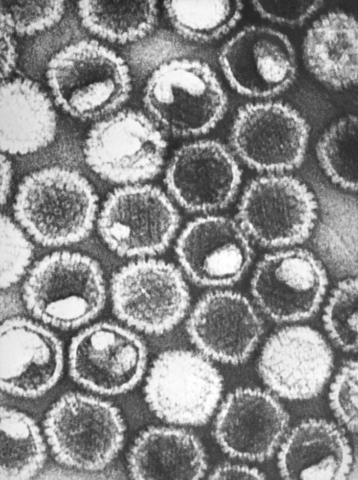
Micrografia eletrónica de transmissão com coloração negativa a revelar vários viriões de herpes simplex, membros da família de vírus Herpesviridae:
No centro da sua capsídeo proteico icosaédrico, o vírus herpes simplex contém um genoma linear de DNA de fita dupla.
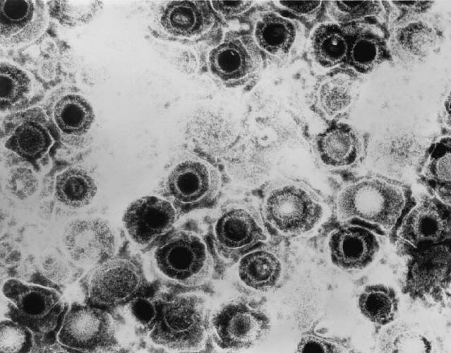
Micrografia eletrónica de transmissão de viriões de herpes simplex
Imagem: “Transmission electron micrograph of herpes simplex virions” pelo CDC/Dr. Erskine Palmer. Licença: Domínio Público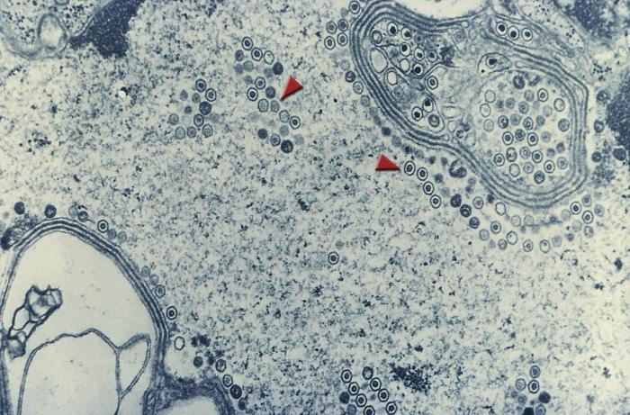
Imagem de microscopia eletrónica de transmissão demonstrando numerosos viriões arredondados de herpes simplex no núcleo de uma célula (setas)
Imagem: “Transmission electron microscopic image demonstrating numerous, round herpes simplex virions inside the nucleus of a cell (arrows)” pelo CDC. Licença: Domínio PúblicoForam reconhecidas duas espécies infeciosas:
VSH-1:
VSH-2:
Infeção por herpes neonatal:
Os humanos são o principal reservatório.
VHS-1:
VHS-2:
Infeção primária:
Período de latência:
Reativação:
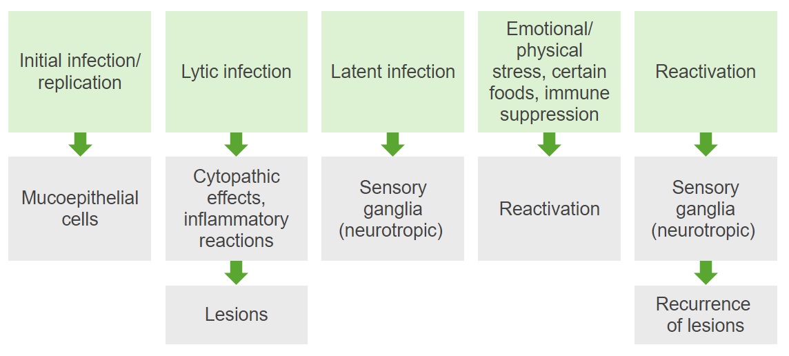
Diagrama resumo da patogénese das infeções por VHS-1 e -2
Imagem por Lecturio. Licença: CC BY-NC-SA 4.0Os vírus do herpes simplex Herpes Simplex A group of acute infections caused by herpes simplex virus type 1 or type 2 that is characterized by the development of one or more small fluid-filled vesicles with a raised erythematous base on the skin or mucous membrane. It occurs as a primary infection or recurs due to a reactivation of a latent infection. Congenital TORCH Infections causam infeções citolíticas que constituem a base de todas as alterações patológicas: necrose das células infetadas associada a resposta inflamatória
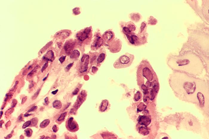
Biópsia de úlcera esofágica causada por uma infeção ativa pelo vírus herpes simplex (HSV) num paciente com SIDA:
A espécie foi colhida no bordo da úlcera e revela a presença de inclusões intranucleares e células com múltiplos núcleos
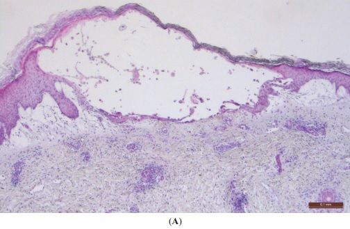
Fotomicrografia de baixa potência de uma vesícula típica do vírus do herpes:
A vesícula contém neutrófilos (e mudará de um líquido claro para uma aparência amarela e turva) antes de romper e deixar uma úlcera superficial. É possível observar a lesão citopática viral que permanece dentro da epiderme, explicando o facto de não deixar cicatriz, o que pode ocorrer com inflamação dérmica grave.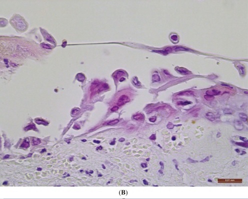
Fotomicrografia de alta potência de uma lesão do vírus do herpes na pele:
É possível observar a acantólise (queratinócitos separados uns dos outros), que é a base da formação das bolhas. Os queratinócitos infetados estão aumentados e contêm inclusões intranucleares vítreas (“corpos tipo A de Cowdry”). Alguns queratinócitos são multinucleados.
Existem diversas condições causadas por VHS. As infeções especificadas abaixo são frequentemente causadas por VHS-1. Nota: O VHS-2 também pode ser (embora com menos frequência) associado a muitas destas patologias.
Apenas 20%‒25% dos pacientes com anticorpos VHS-1 mencionam história prévia de infeções buco-labiais ou genitais.
Gengivoestomatite:
Faringite:
Herpes labial:
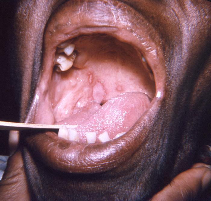
Lesões no palato mole e na língua causadas pelo vírus herpes simplex
Imagem : “This photograph depicts a close view of an elderly African American female patient’s oral cavity” por CDC/Robert E. Sumpter. Licença: Domínio Público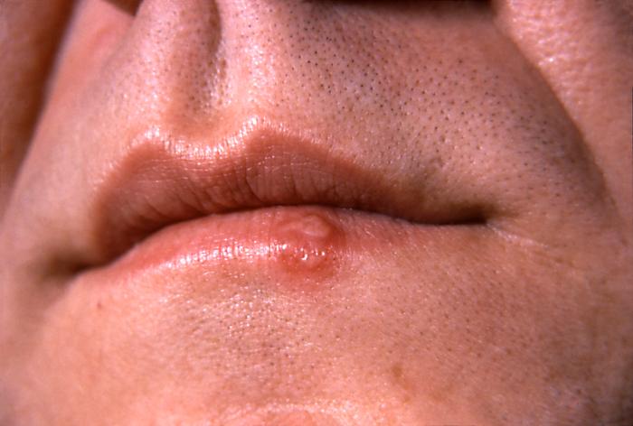
Lesão de herpes simplex no bordo do lábio inferior, no 2º dia após o início
Imagem: “This photograph depicts a close-up of the lips of a patient with a herpes simplex lesion on the lower lip” por CDC/ Dr. Hermann. Licença: Domínio PúblicoPanarício herpético:
Herpes gladiatorum Herpes gladiatorum Skin infection of the face, neck, and arms of wrestlers and rugby players Herpes Simplex Virus 1 and 2:
Eritema multiforme:
Eczema Eczema Atopic dermatitis, also known as eczema, is a chronic, relapsing, pruritic, inflammatory skin disease that occurs more frequently in children, although adults can also be affected. The condition is often associated with elevated serum levels of IgE and a personal or family history of atopy. Skin dryness, erythema, oozing, crusting, and lichenification are present. Atopic Dermatitis (Eczema) herpético:
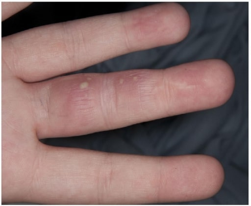
Paciente com vesículas no dedo devido a panarício herpético
Imagem: “Herpetic Whitlow” por Salford Royal Foundation Trust, Manchester, Reino Unido. Licença: CC BY 3.0
Lesões vesiculares difusas na face de um paciente com eczema herpético durante a terapêutica com UV
Imagem: “Multiple pustules on the face surface” por AUTOR. Licença: CC BY 2.0Infeções oculares ocorrem em < 5% dos pacientes com infeções por VHS-1, levando à perda de visão e/ou cegueira.
Queratite:
Necrose aguda da retina Retina The ten-layered nervous tissue membrane of the eye. It is continuous with the optic nerve and receives images of external objects and transmits visual impulses to the brain. Its outer surface is in contact with the choroid and the inner surface with the vitreous body. The outermost layer is pigmented, whereas the inner nine layers are transparent. Eye: Anatomy:
Conjuntivite e blefarite:
Coriorretinite (uveíte posterior):
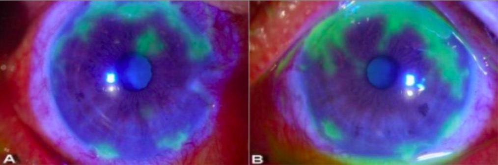
Exame com lâmpada de fenda a demonstrar lesões dendríticas na queratite herpética:
É possível observar padrões geográficos irregulares de úlceras, destacados a verde após a aplicação de corante amarelo-alaranjado de fluoresceína. O corante é absorvido pela córnea lesada (onde a superfície foi interrompida) e a área aparece verde sob luz azul cobalto.
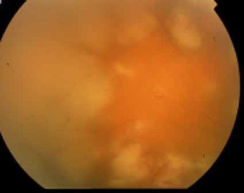
Imagem de fundoscopia de um paciente com necrose retiniana aguda:
Podem ser observadas áreas necróticas confluentes características. A retinite caracteriza-se pelas manchas profundas, multifocais, amarelo-esbranquiçadas, que geralmente começam à periferia e depois se tornam concêntricas e confluentes e propagam-se em direção ao pólo posterior.
Encefalite:
Meningite assética:
Outras manifestações:
Epiglotite ou laringite (crupe herpética):
Pneumonite por VHS:
Esofagite por VHS:
Hepatite fulminante:
As condições descritas abaixo estão frequentemente associadas a infeções por VHS-2 e algumas estão também associadas ao VHS-1.
Infeções genitais primárias por VHS-2:
Infeção não primária do 1º episódio (reativação):
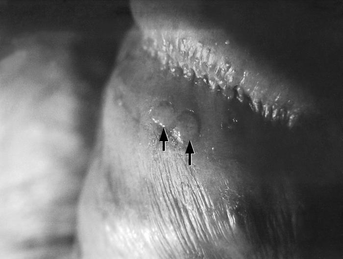
Bolhas penianas (setas) devido a uma infeção recorrente pelo vírus herpes simplex-2 (VHS-2)
Imagem: “Penile blisters (arrows), due to a recurring herpes simplex-2 (HSV-2) virus infection” por CDC/ Susan Lindsley. Licença: Domínio Público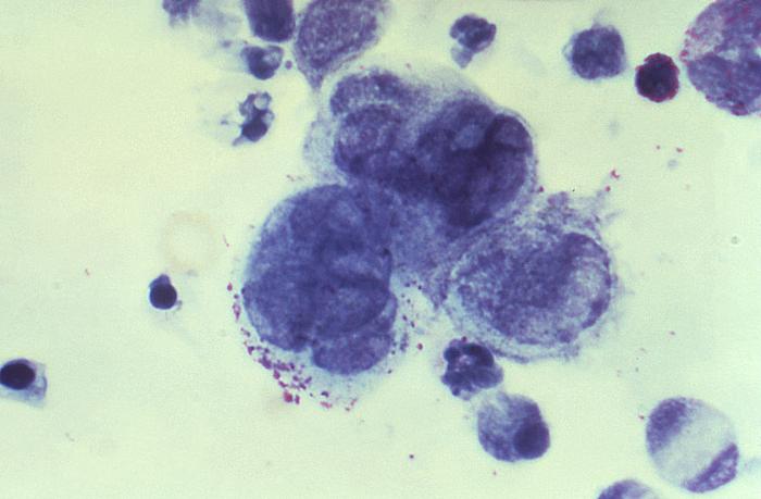
Imagem de um esfregaço de Tzanck obtido de uma lesão peniana:
São observadas células gigantes multinucleadas, sugestivas de uma infeção por herpes.
Antivirais:
Analgésicos:
A tabela exibida abaixo compara e diferencia as espécies VHS-1 e VHS-2:
| Serotipo | VHS-1 | VHS-2 |
|---|---|---|
| Transmissão |
|
|
| Infeção lítica | Células mucoepiteliais | Células mucoepiteliais |
| Latência | Gânglios do trigémeo | Gânglios sacrais |
| Doenças |
|
|
A tabela abaixo compara os 9 herpesvírus considerados endémicos em humanos. Existem 115 diferentes espécies conhecidas de herpesvírus, agrupadas em 3 famílias:
| HHV | Nome comum | Principais células-alvo | Local de latência | Apresentação clínica* |
|---|---|---|---|---|
|
1 (grupo alfa) |
HSV-1 | Células mucoepiteliais | Gânglios da raiz dorsal |
|
|
2 (grupo alfa) |
HSV-2 |
|
||
|
3 (grupo alfa) |
VZV |
|
||
|
4 (grupo gama) |
EBV EBV Epstein-barr virus (EBV) is a linear, double-stranded DNA virus belonging to the herpesviridae family. This highly prevalent virus is mostly transmitted through contact with oropharyngeal secretions from an infected individual. The virus can infect epithelial cells and B lymphocytes, where it can undergo lytic replication or latency. Epstein-Barr Virus |
|
Células B de memória |
|
|
5 (grupo beta) |
CMV |
|
Células progenitoras hematopoiéticas da medula óssea |
|
|
6A, 6B (grupo beta) |
HHV-6 HHV-6 Human herpesvirus (HHV)-6 and HHV-7 are similar double-stranded DNA viruses belonging to the Herpesviridae family. Human herpesviruses are ubiquitous and infections are commonly contracted during childhood. Human Herpesvirus 6 and 7 | células T | Monócitos | Roséola |
|
7 (grupo beta) |
HHV-7 HHV-7 Human herpesvirus (HHV)-6 and HHV-7 are similar double-stranded DNA viruses belonging to the Herpesviridae family. Human herpesviruses are ubiquitous and infections are commonly contracted during childhood. Human Herpesvirus 6 and 7 | células T | ||
|
8 (grupo gama) |
Herpesvírus associado ao sarcoma de Kaposi Kaposi A multicentric, malignant neoplastic vascular proliferation characterized by the development of bluish-red cutaneous nodules, usually on the lower extremities, most often on the toes or feet, and slowly increasing in size and number and spreading to more proximal areas. The tumors have endothelium-lined channels and vascular spaces admixed with variably sized aggregates of spindle-shaped cells, and often remain confined to the skin and subcutaneous tissue, but widespread visceral involvement may occur. Hhv-8 is the suspected cause. There is also a high incidence in AIDS patients. AIDS-defining Conditions |
|
células B | Sarcoma de Kaposi Kaposi A multicentric, malignant neoplastic vascular proliferation characterized by the development of bluish-red cutaneous nodules, usually on the lower extremities, most often on the toes or feet, and slowly increasing in size and number and spreading to more proximal areas. The tumors have endothelium-lined channels and vascular spaces admixed with variably sized aggregates of spindle-shaped cells, and often remain confined to the skin and subcutaneous tissue, but widespread visceral involvement may occur. Hhv-8 is the suspected cause. There is also a high incidence in AIDS patients. AIDS-defining Conditions |