Domina Conceptos Médicos

Estudia para la escuela de medicina y tus examenes con Lecturio.

Eritema Multiforme

El eritema multiforme es una reacción de hipersensibilidad aguda caracterizada por lesiones cutáneas en EN Erythema nodosum is an immune-mediated panniculitis (inflammation of the subcutaneous fat) caused by a type IV (delayed-type) hypersensitivity reaction. It commonly manifests in young women as tender, erythematous nodules on the shins. Erythema Nodosum diana con múltiples anillos y centros oscuros. Las lesiones pueden ir acompañadas de síntomas sistémicos (e.g., fiebre) y lesiones mucosas (e.g., bullas). La mayoría de los LOS Neisseria pacientes con eritema multiforme tienen un antecedente de infección viral reciente (especialmente el virus Virus Viruses are infectious, obligate intracellular parasites composed of a nucleic acid core surrounded by a protein capsid. Viruses can be either naked (non-enveloped) or enveloped. The classification of viruses is complex and based on many factors, including type and structure of the nucleoid and capsid, the presence of an envelope, the replication cycle, and the host range. Virology herpes simple) o bacteriana (generalmente Mycoplasma pneumoniae Mycoplasma pneumoniae Short filamentous organism of the genus mycoplasma, which binds firmly to the cells of the respiratory epithelium. It is one of the etiologic agents of non-viral primary atypical pneumonia in man. Mycoplasma). El eritema multiforme se diagnostica clínicamente y el tratamiento se dirige a remover el agente causal y al AL Amyloidosis alivio de los LOS Neisseria síntomas.

Last updated: Dec 15, 2025

Editorial responsibility: Stanley Oiseth, Lindsay Jones, Evelin Maza

Descripción General

Epidemiología

  • Incidencia a nivel mundial de 1,6–2 millones de casos al AL Amyloidosis año
  • Más frecuente en EN Erythema nodosum is an immune-mediated panniculitis (inflammation of the subcutaneous fat) caused by a type IV (delayed-type) hypersensitivity reaction. It commonly manifests in young women as tender, erythematous nodules on the shins. Erythema Nodosum adultos jóvenes (< 30 años)
  • Hombres > mujeres

Etiología

El eritema multiforme es una reacción inmunitaria mediada por células (reacción de tipo IV) dirigida contra los LOS Neisseria antígenos del agente causante, los LOS Neisseria cuales se depositan en EN Erythema nodosum is an immune-mediated panniculitis (inflammation of the subcutaneous fat) caused by a type IV (delayed-type) hypersensitivity reaction. It commonly manifests in young women as tender, erythematous nodules on the shins. Erythema Nodosum la piel.

  • Clasificación Etiologías Ejemplos
    Causas infecciosas (más comunes, 90% de los LOS Neisseria casos) Bacteriana
    • Mycoplasma pneumoniae Mycoplasma pneumoniae Short filamentous organism of the genus mycoplasma, which binds firmly to the cells of the respiratory epithelium. It is one of the etiologic agents of non-viral primary atypical pneumonia in man. Mycoplasma
    • Rickettsia Rickettsia Rickettsiae are a diverse collection of obligate intracellular, gram-negative bacteria that have a tropism for vascular endothelial cells. The vectors for transmission vary by species but include ticks, fleas, mites, and lice. Rickettsia
    • Chlamydophila psittaci
    • Salmonella typhi Salmonella typhi A serotype of Salmonella enterica which is the etiologic agent of typhoid fever. Enteric Fever (Typhoid Fever)
    • Mycobacterium tuberculosis Mycobacterium tuberculosis Tuberculosis (TB) is an infectious disease caused by Mycobacterium tuberculosis complex bacteria. The bacteria usually attack the lungs but can also damage other parts of the body. Approximately 30% of people around the world are infected with this pathogen, with the majority harboring a latent infection. Tuberculosis spreads through the air when a person with active pulmonary infection coughs or sneezes. Tuberculosis
    • Histoplasma capsulatum Histoplasma capsulatum Histoplasma/Histoplasmosis
    Viral
    • Virus Virus Viruses are infectious, obligate intracellular parasites composed of a nucleic acid core surrounded by a protein capsid. Viruses can be either naked (non-enveloped) or enveloped. The classification of viruses is complex and based on many factors, including type and structure of the nucleoid and capsid, the presence of an envelope, the replication cycle, and the host range. Virology herpes simple ( HSV HSV Herpes simplex virus (HSV) is a double-stranded DNA virus belonging to the family Herpesviridae. Herpes simplex virus commonly causes recurrent infections involving the skin and mucosal surfaces, including the mouth, lips, eyes, and genitals. Herpes Simplex Virus 1 and 2, por sus siglas en EN Erythema nodosum is an immune-mediated panniculitis (inflammation of the subcutaneous fat) caused by a type IV (delayed-type) hypersensitivity reaction. It commonly manifests in young women as tender, erythematous nodules on the shins. Erythema Nodosum inglés) tipos 1 y 2
      • Causa más común
      • Aproximadamente el 50% de los LOS Neisseria casos
    • Mononucleosis Mononucleosis Infectious mononucleosis (IM), also known as “the kissing disease,” is a highly contagious viral infection caused by the Epstein-Barr virus. Its common name is derived from its main method of transmission: the spread of infected saliva via kissing. Clinical manifestations of IM include fever, tonsillar pharyngitis, and lymphadenopathy. Mononucleosis infecciosa ( Virus Virus Viruses are infectious, obligate intracellular parasites composed of a nucleic acid core surrounded by a protein capsid. Viruses can be either naked (non-enveloped) or enveloped. The classification of viruses is complex and based on many factors, including type and structure of the nucleoid and capsid, the presence of an envelope, the replication cycle, and the host range. Virology de Epstein Barr)
    Fúngica Histoplasmosis Histoplasmosis Histoplasmosis is an infection caused by Histoplasma capsulatum, a dimorphic fungus. Transmission is through inhalation, and exposure to soils containing bird or bat droppings increases the risk of infection. Most infections are asymptomatic; however, immunocompromised individuals generally develop acute pulmonary infection, chronic infection, or even disseminated disease. Histoplasma/Histoplasmosis, dermatofitos
    Causas no infecciosas Medicamentos
    • Penicilinas
    • Sulfonamida
    • Medicamentos anticonvulsivantes
    • Antiinflamatorios no esteroideos (AINE)
    • Barbitúricos
    Malignidad
    • Leucemia
    • Linfoma
    Misceláneas
    • Sarcoidosis Sarcoidosis Sarcoidosis is a multisystem inflammatory disease that causes noncaseating granulomas. The exact etiology is unknown. Sarcoidosis usually affects the lungs and thoracic lymph nodes, but it can also affect almost every system in the body, including the skin, heart, and eyes, most commonly. Sarcoidosis
    • Lupus eritematoso sistémico
    • Poliarteritis nodosa

    Clasificación

    El eritema multiforme se clasifica en EN Erythema nodosum is an immune-mediated panniculitis (inflammation of the subcutaneous fat) caused by a type IV (delayed-type) hypersensitivity reaction. It commonly manifests in young women as tender, erythematous nodules on the shins. Erythema Nodosum función de la presencia de lesiones en EN Erythema nodosum is an immune-mediated panniculitis (inflammation of the subcutaneous fat) caused by a type IV (delayed-type) hypersensitivity reaction. It commonly manifests in young women as tender, erythematous nodules on the shins. Erythema Nodosum las mucosas y de los LOS Neisseria síntomas sistémicos en EN Erythema nodosum is an immune-mediated panniculitis (inflammation of the subcutaneous fat) caused by a type IV (delayed-type) hypersensitivity reaction. It commonly manifests in young women as tender, erythematous nodules on the shins. Erythema Nodosum eritema multiforme menor y mayor.

    Lesiones cutáneas Afectación de la mucosa Síntomas sistémicos
    Eritema multiforme menor
    Eritema multiforme mayor

    Presentación Clínica

    El eritema multiforme generalmente se resuelve espontáneamente en EN Erythema nodosum is an immune-mediated panniculitis (inflammation of the subcutaneous fat) caused by a type IV (delayed-type) hypersensitivity reaction. It commonly manifests in young women as tender, erythematous nodules on the shins. Erythema Nodosum el plazo de 1 mes, sin secuelas a largo plazo. En EN Erythema nodosum is an immune-mediated panniculitis (inflammation of the subcutaneous fat) caused by a type IV (delayed-type) hypersensitivity reaction. It commonly manifests in young women as tender, erythematous nodules on the shins. Erythema Nodosum raras ocasiones, el eritema multiforme puede reaparecer, hasta 6 veces/año durante 10 años (generalmente asociada a una infección por HSV HSV Herpes simplex virus (HSV) is a double-stranded DNA virus belonging to the family Herpesviridae. Herpes simplex virus commonly causes recurrent infections involving the skin and mucosal surfaces, including the mouth, lips, eyes, and genitals. Herpes Simplex Virus 1 and 2).

    Lesiones cutáneas

    • Comienzan como máculas eritematosas que evolucionan a lo largo de 3–5 días hasta convertirse en EN Erythema nodosum is an immune-mediated panniculitis (inflammation of the subcutaneous fat) caused by a type IV (delayed-type) hypersensitivity reaction. It commonly manifests in young women as tender, erythematous nodules on the shins. Erythema Nodosum pápulaslesiones en EN Erythema nodosum is an immune-mediated panniculitis (inflammation of the subcutaneous fat) caused by a type IV (delayed-type) hypersensitivity reaction. It commonly manifests in young women as tender, erythematous nodules on the shins. Erythema Nodosum diana con 3 zonas diferentes
      • Zona central: oscura y eritematosa
      • Zona media: anillo circundante pálido
      • Zona periférica: anillo exterior eritematoso con márgenes bien definidos
    • Distribución acral simétrica (i.e., manos, pies y cara)
    • Superficies extensoras
    • Propagación centrípeta (distal a proximal)
    • Redondeadas, < 3 cm de diámetro
    • Puede haber prurito o sensación urente.

    Lesiones de la mucosa

    • Bullosa → erosiones/úlceras dolorosas
    • 3 lugares:
      • Mucosa oral (más común)
      • Mucosa ocular
      • Mucosa genital

    Síntomas sistémicos

    • Fiebre
    • Malestar
    • Mialgia
    • Artralgia/artritis
    Presentación clínica del eritema multiforme

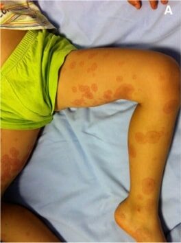
    Lesiones en diana con una forma regular y redonda y 3 zonas concéntricas: una zona central de color rojo más oscuro, una zona rosa más pálida y un anillo rojo periférico

    Imagen: “Erythema multiforme” por Pediatric Unit, Maternal & Infant Department, S. Chiara University-Hospital, Via Roma 67, Pisa 56126, Italy. Licencia: CC BY 2.0, editada por Lecturio.

    Diagnóstico y Tratamiento

    Diagnóstico

    • Generalmente se diagnostica clínicamente
    • Pruebas serológicas para identificar posibles causas:
      • M. pneumoniae en EN Erythema nodosum is an immune-mediated panniculitis (inflammation of the subcutaneous fat) caused by a type IV (delayed-type) hypersensitivity reaction. It commonly manifests in young women as tender, erythematous nodules on the shins. Erythema Nodosum pacientes con síntomas respiratorios
      • HSV HSV Herpes simplex virus (HSV) is a double-stranded DNA virus belonging to the family Herpesviridae. Herpes simplex virus commonly causes recurrent infections involving the skin and mucosal surfaces, including the mouth, lips, eyes, and genitals. Herpes Simplex Virus 1 and 2 en EN Erythema nodosum is an immune-mediated panniculitis (inflammation of the subcutaneous fat) caused by a type IV (delayed-type) hypersensitivity reaction. It commonly manifests in young women as tender, erythematous nodules on the shins. Erythema Nodosum pacientes con eritema multiforme recurrente
    • En EN Erythema nodosum is an immune-mediated panniculitis (inflammation of the subcutaneous fat) caused by a type IV (delayed-type) hypersensitivity reaction. It commonly manifests in young women as tender, erythematous nodules on the shins. Erythema Nodosum casos inciertos, la biopsia de piel puede mostrar:
      • Degeneración vacuolada de las células basales
      • Necrosis Necrosis The death of cells in an organ or tissue due to disease, injury or failure of the blood supply. Ischemic Cell Damage dispersa de queratinocitos
      • Inflamación y edema Edema Edema is a condition in which excess serous fluid accumulates in the body cavity or interstitial space of connective tissues. Edema is a symptom observed in several medical conditions. It can be categorized into 2 types, namely, peripheral (in the extremities) and internal (in an organ or body cavity). Edema

    Tratamiento

    • El eritema multiforme suele ser autolimitado y no requiere tratamiento.
    • El tratamiento se dirige al AL Amyloidosis agente causante y al AL Amyloidosis alivio de los LOS Neisseria síntomas.
      • Eliminación del agente causante (e.g., medicamentos, alérgeno)
      • Tratamiento sintomático: corticoides tópicos/antihistamínicos orales para el prurito y enjuague bucal anestésico para las lesiones orales dolorosas
    • Tratamiento específico
      • Depende de la ubicación:
        • Lesiones oculares: referencia a un oftalmólogo
        • Lesiones orales graves: ingreso para el tratamiento de dolor Dolor Inflammation e hidratación
      • Eritema multiforme recurrente: aciclovir continuo durante 6 meses (terapia supresiva)
    Cambios epidérmicos en el eritema multiforme

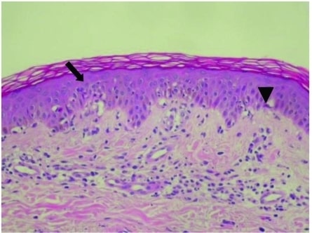
    Cambios epidérmicos en el eritema multiforme con degeneración vacuolada de las células basales (flecha) y necrosis de queratinocitos individuales dispersos (cabeza de flecha)

    Imagen: “Epidermal Changes in Erythema Multiforme” por Division of Gastroenterology and Hepatology, Department of Internal Medicine, Taipei Medical University Hospital, Taipei, Taiwan. Licencia: CC BY 2.5

    Diagnóstico Diferencial

    • Urticaria Urticaria Urticaria is raised, well-circumscribed areas (wheals) of edema (swelling) and erythema (redness) involving the dermis and epidermis with associated pruritus (itch). Urticaria is not a single disease but rather is a reaction pattern representing cutaneous mast cell degranulation. Urticaria (Hives): enfermedad autolimitada que consiste en EN Erythema nodosum is an immune-mediated panniculitis (inflammation of the subcutaneous fat) caused by a type IV (delayed-type) hypersensitivity reaction. It commonly manifests in young women as tender, erythematous nodules on the shins. Erythema Nodosum lesiones superficiales, eritematosas y pruriginosas resultantes de la activación de los LOS Neisseria mastocitos y la liberación de histamina en EN Erythema nodosum is an immune-mediated panniculitis (inflammation of the subcutaneous fat) caused by a type IV (delayed-type) hypersensitivity reaction. It commonly manifests in young women as tender, erythematous nodules on the shins. Erythema Nodosum la dermis Dermis A layer of vascularized connective tissue underneath the epidermis. The surface of the dermis contains innervated papillae. Embedded in or beneath the dermis are sweat glands; hair follicles; and sebaceous glands. Skin: Structure and Functions. Las lesiones pueden durar desde < 24 horas hasta > 6 semanas. Causada comúnmente por infecciones virales o por reacciones alérgicas a medicamentos o alimentos, otras infecciones, estrés, frío/ calor Calor Inflammation y trastornos autoinmunes. El tratamiento, además de abordar la afección subyacente, incluye antihistamínicos y esteroides.
    • Penfigoide ampolloso: afección inmunomediada caracterizada por la destrucción de proteínas adhesivas extracelulares (e.g., hemidesmosomas) por autoanticuerpos de inmunoglobulina G. Los LOS Neisseria pacientes presentan urticaria Urticaria Urticaria is raised, well-circumscribed areas (wheals) of edema (swelling) and erythema (redness) involving the dermis and epidermis with associated pruritus (itch). Urticaria is not a single disease but rather is a reaction pattern representing cutaneous mast cell degranulation. Urticaria (Hives) y pápulas eccematosas durante el estadio prodrómico. Después, se desarrollan bullas cutáneas y tensas. Esto puede durar de meses a años, y el tratamiento se centra en EN Erythema nodosum is an immune-mediated panniculitis (inflammation of the subcutaneous fat) caused by a type IV (delayed-type) hypersensitivity reaction. It commonly manifests in young women as tender, erythematous nodules on the shins. Erythema Nodosum la atención sintomática de soporte con esteroides y antihistamínicos.
    • Síndrome de Stevens-Johnson: reacción de hipersensibilidad inmunomediada potencialmente mortal que afecta a las membranas mucosas y a la piel (< 10% de la superficie corporal). Por lo general, los LOS Neisseria pacientes tienen antecedentes de haber tomado ciertos medicamentos (e.g., sulfamidas) o una infección reciente. La presentación clínica implica lesiones cutáneas maculares con 2 zonas de cambio de color, vesículas, bullas y síntomas sistémicos (e.g., fiebre). El tratamiento comprende cuidados de soporte para la piel y los LOS Neisseria ojos y el control de síntomas.
    • Necrólisis epidérmica tóxica: un proceso de enfermedad de hipersensibilidad inmunomediada potencialmente mortal y que es una extensión del síndrome de Stevens-Johnson. La necrólisis epidérmica tóxica es una reacción de hipersensibilidad inmunomediada con afectación mucocutánea (> 30% de la superficie corporal). La tasa de mortalidad depende de la gravedad tanto en EN Erythema nodosum is an immune-mediated panniculitis (inflammation of the subcutaneous fat) caused by a type IV (delayed-type) hypersensitivity reaction. It commonly manifests in young women as tender, erythematous nodules on the shins. Erythema Nodosum el síndrome de Stevens-Johnson como en EN Erythema nodosum is an immune-mediated panniculitis (inflammation of the subcutaneous fat) caused by a type IV (delayed-type) hypersensitivity reaction. It commonly manifests in young women as tender, erythematous nodules on the shins. Erythema Nodosum la necrólisis epidérmica tóxica, siendo aproximadamente del 10% en EN Erythema nodosum is an immune-mediated panniculitis (inflammation of the subcutaneous fat) caused by a type IV (delayed-type) hypersensitivity reaction. It commonly manifests in young women as tender, erythematous nodules on the shins. Erythema Nodosum el síndrome de Stevens-Johnson y de hasta el 50% en EN Erythema nodosum is an immune-mediated panniculitis (inflammation of the subcutaneous fat) caused by a type IV (delayed-type) hypersensitivity reaction. It commonly manifests in young women as tender, erythematous nodules on the shins. Erythema Nodosum la necrólisis epidérmica tóxica. Ambas afecciones requieren el ingreso en EN Erythema nodosum is an immune-mediated panniculitis (inflammation of the subcutaneous fat) caused by a type IV (delayed-type) hypersensitivity reaction. It commonly manifests in young women as tender, erythematous nodules on the shins. Erythema Nodosum una unidad de quemados para el cuidado de la piel.

    References

    1. Weston, W. L. (2005). Herpes-associated erythema multiforme. Journal of Investigative Dermatology. https://linkinghub.elsevier.com/retrieve/pii/S0022-202X(15)32327-7
    2. Zoghaib, S., & Kechichian, E. et al. (2019). Triggers, clinical manifestations, and management of pediatric erythema multiforme: A systematic review. Journal of the American Academy of Dermatology, 81(3), 813–822. https://doi.org/10.1016/j.jaad.2019.04.035
    3. de Risi-Pugliese, T., & Sbidian, E. et al. (2019). Interventions for erythema multiforme: A systematic review. Journal of the European Academy of Dermatology and Venereology. https://doi.org/10.1111/jdv.15447

    ¡Crea tu cuenta gratis o inicia una sesión para seguir leyendo!

    Regístrate ahora y obtén acceso gratuito a Lecturio con páginas de concepto, videos médicos y cuestionarios para tu educación médica.

    User Reviews

    Que tengas una sesión de estudio alegre y navideña 🎁 Ahorra 50% en todos los planes >>

    Details