Domina Conceptos Médicos

Estudia para la escuela de medicina y tus examenes con Lecturio.

Derrame Pericárdico y Taponamiento Cardíaco

El derrame pericárdico es la acumulación de líquido en EN Erythema nodosum is an immune-mediated panniculitis (inflammation of the subcutaneous fat) caused by a type IV (delayed-type) hypersensitivity reaction. It commonly manifests in young women as tender, erythematous nodules on the shins. Erythema Nodosum el espacio pericárdico que rodea del corazón. El pericardio no se expande fácilmente, por lo que la acumulación rápida de líquido provoca un aumento de la presión alrededor del corazón. El aumento de la presión restringe el llenado cardíaco, lo que provoca una disminución del gasto cardíaco y taponamiento cardíaco. Los LOS Neisseria signos y síntomas suelen aparecer en EN Erythema nodosum is an immune-mediated panniculitis (inflammation of the subcutaneous fat) caused by a type IV (delayed-type) hypersensitivity reaction. It commonly manifests in young women as tender, erythematous nodules on the shins. Erythema Nodosum el contexto del taponamiento cardíaco e incluyen disnea, hipotensión, ruidos cardíacos disminuidos, ingurgitación yugular y pulso paradójico. El diagnóstico de derrame pericárdico se confirma con ultrasonido cardíaco. Los LOS Neisseria derrames pequeños en EN Erythema nodosum is an immune-mediated panniculitis (inflammation of the subcutaneous fat) caused by a type IV (delayed-type) hypersensitivity reaction. It commonly manifests in young women as tender, erythematous nodules on the shins. Erythema Nodosum pacientes hemodinámicamente estables se tratan médicamente. Los LOS Neisseria derrames más grandes y el taponamiento cardíaco pueden requerir una pericardiocentesis Pericardiocentesis Puncture and aspiration of fluid from the pericardium. Cardiac Surgery o una pericardiotomía.

Last updated: Dec 15, 2025

Editorial responsibility: Stanley Oiseth, Lindsay Jones, Evelin Maza

Epidemiología y Etiología

Definición

El derrame pericárdico es la acumulación de líquido en EN Erythema nodosum is an immune-mediated panniculitis (inflammation of the subcutaneous fat) caused by a type IV (delayed-type) hypersensitivity reaction. It commonly manifests in young women as tender, erythematous nodules on the shins. Erythema Nodosum el espacio pericárdico.

El taponamiento cardíaco es la acumulación de líquido pericárdico suficiente para impedir el llenado cardíaco y causar compromiso hemodinámico. Lo más importante es la velocidad de acumulación de líquido, y no necesariamente la cantidad.

Epidemiología

Derrame pericárdico:

  • La incidencia es desconocida.
  • Se ha HA Hemolytic anemia (HA) is the term given to a large group of anemias that are caused by the premature destruction/hemolysis of circulating red blood cells (RBCs). Hemolysis can occur within (intravascular hemolysis) or outside the blood vessels (extravascular hemolysis). Hemolytic Anemia observado en EN Erythema nodosum is an immune-mediated panniculitis (inflammation of the subcutaneous fat) caused by a type IV (delayed-type) hypersensitivity reaction. It commonly manifests in young women as tender, erythematous nodules on the shins. Erythema Nodosum aproximadamente el 3% de las autopsias en EN Erythema nodosum is an immune-mediated panniculitis (inflammation of the subcutaneous fat) caused by a type IV (delayed-type) hypersensitivity reaction. It commonly manifests in young women as tender, erythematous nodules on the shins. Erythema Nodosum estudios
  • Edad:
    • Puede ocurrir en EN Erythema nodosum is an immune-mediated panniculitis (inflammation of the subcutaneous fat) caused by a type IV (delayed-type) hypersensitivity reaction. It commonly manifests in young women as tender, erythematous nodules on the shins. Erythema Nodosum todos los LOS Neisseria grupos etarios
    • Media: 50–60 años

Taponamiento cardíaco:

  • Incidencia: 2 casos por cada 10 000 personas en EN Erythema nodosum is an immune-mediated panniculitis (inflammation of the subcutaneous fat) caused by a type IV (delayed-type) hypersensitivity reaction. It commonly manifests in young women as tender, erythematous nodules on the shins. Erythema Nodosum Estados Unidos
  • Ocurre en EN Erythema nodosum is an immune-mediated panniculitis (inflammation of the subcutaneous fat) caused by a type IV (delayed-type) hypersensitivity reaction. It commonly manifests in young women as tender, erythematous nodules on the shins. Erythema Nodosum aproximadamente el 2% de las lesiones penetrantes
  • Más común en EN Erythema nodosum is an immune-mediated panniculitis (inflammation of the subcutaneous fat) caused by a type IV (delayed-type) hypersensitivity reaction. It commonly manifests in young women as tender, erythematous nodules on the shins. Erythema Nodosum niños y hombres

Etiología

Múltiples trastornos están asociados al AL Amyloidosis derrame pericárdico, incluyendo:

  • Infección:
    • Viral (más común):
      • Coxsackievirus Coxsackievirus Coxsackievirus is a member of a family of viruses called Picornaviridae and the genus Enterovirus. Coxsackieviruses are single-stranded, positive-sense RNA viruses, and are divided into coxsackie group A and B viruses. Both groups of viruses cause upper respiratory infections, rashes, aseptic meningitis, or encephalitis. Coxsackievirus grupo B
      • Influenza Influenza Influenza viruses are members of the Orthomyxoviridae family and the causative organisms of influenza, a highly contagious febrile respiratory disease. There are 3 primary influenza viruses (A, B, and C) and various subtypes, which are classified based on their virulent surface antigens, hemagglutinin (HA) and neuraminidase (NA). Influenza typically presents with a fever, myalgia, headache, and symptoms of an upper respiratory infection. Influenza Viruses/Influenza
      • Echovirus Echovirus Echoviruses are single-stranded positive-sense RNA viruses belonging to the genus Enterovirus. Transmission is most commonly through the fecal-oral route. The majority of patients are asymptomatic. Patients who are symptomatic can exhibit a wide range of illnesses ranging from nonspecific URIs and exanthems to severe and life-threatening illnesses. Echovirus
      • VIH
      • EBV EBV Epstein-barr virus (EBV) is a linear, double-stranded DNA virus belonging to the herpesviridae family. This highly prevalent virus is mostly transmitted through contact with oropharyngeal secretions from an infected individual. The virus can infect epithelial cells and B lymphocytes, where it can undergo lytic replication or latency. Epstein-Barr Virus
      • CMV
      • Parvovirus B19 Parvovirus B19 Primate erythroparvovirus 1 (generally referred to as parvovirus B19, B19 virus, or sometimes erythrovirus B19) ranks among the smallest DNA viruses. Parvovirus B19 is of the family Parvoviridae and genus Erythrovirus. In immunocompetent humans, parvovirus B19 classically results in erythema infectiosum (5th disease) or “slapped cheek syndrome.” Parvovirus B19
      • Varicela
    • Bacteriana:
      • Staphylococcus aureus Staphylococcus aureus Potentially pathogenic bacteria found in nasal membranes, skin, hair follicles, and perineum of warm-blooded animals. They may cause a wide range of infections and intoxications. Brain Abscess
      • Streptococcus Streptococcus Streptococcus is one of the two medically important genera of gram-positive cocci, the other being Staphylococcus. Streptococci are identified as different species on blood agar on the basis of their hemolytic pattern and sensitivity to optochin and bacitracin. There are many pathogenic species of streptococci, including S. pyogenes, S. agalactiae, S. pneumoniae, and the viridans streptococci. Streptococcus
      • Neisseria Neisseria Neisseria is a genus of bacteria commonly present on mucosal surfaces. Several species exist, but only 2 are pathogenic to humans: N. gonorrhoeae and N. meningitidis. Neisseria species are non-motile, gram-negative diplococci most commonly isolated on modified Thayer-Martin (MTM) agar. Neisseria
      • Legionella Legionella Legionella is a facultative intracellular, gram-negative bacilli. Legionella does not grow on common culture media because it requires certain supplementation (cysteine and iron). Legionella pneumophila (L. pneumophila) accounts for the majority of human infections. Legionella/Legionellosis
      • Treponema pallidum Treponema pallidum The causative agent of venereal and non-venereal syphilis as well as yaws. Treponema
      • Mycobacterium tuberculosis Mycobacterium tuberculosis Tuberculosis (TB) is an infectious disease caused by Mycobacterium tuberculosis complex bacteria. The bacteria usually attack the lungs but can also damage other parts of the body. Approximately 30% of people around the world are infected with this pathogen, with the majority harboring a latent infection. Tuberculosis spreads through the air when a person with active pulmonary infection coughs or sneezes. Tuberculosis
    • Micótica:
      • Candida Candida Candida is a genus of dimorphic, opportunistic fungi. Candida albicans is part of the normal human flora and is the most common cause of candidiasis. The clinical presentation varies and can include localized mucocutaneous infections (e.g., oropharyngeal, esophageal, intertriginous, and vulvovaginal candidiasis) and invasive disease (e.g., candidemia, intraabdominal abscess, pericarditis, and meningitis). Candida/Candidiasis
      • Histoplasmosis Histoplasmosis Histoplasmosis is an infection caused by Histoplasma capsulatum, a dimorphic fungus. Transmission is through inhalation, and exposure to soils containing bird or bat droppings increases the risk of infection. Most infections are asymptomatic; however, immunocompromised individuals generally develop acute pulmonary infection, chronic infection, or even disseminated disease. Histoplasma/Histoplasmosis
      • Coccidioidomicosis
  • Malignidad:
    • Tumores cardíacos primarios
    • Enfermedad metastásica
  • Traumatismo:
    • Traumatismo penetrante de tórax
    • Post reanimación cardíaca
  • Ocurrencia post-procedimiento:
    • Cirugía cardíaca (síndrome postpericardiotomía)
    • Radiación
  • Enfermedades autoinmunes y del tejido conectivo:
    • Lupus eritematoso sistémico
    • Artritis reumatoide
    • Espondilitis anquilosante
    • Escleroderma
    • Sarcoidosis Sarcoidosis Sarcoidosis is a multisystem inflammatory disease that causes noncaseating granulomas. The exact etiology is unknown. Sarcoidosis usually affects the lungs and thoracic lymph nodes, but it can also affect almost every system in the body, including the skin, heart, and eyes, most commonly. Sarcoidosis
    • Síndrome de Sjögren
    • Vasculitis Vasculitis Inflammation of any one of the blood vessels, including the arteries; veins; and rest of the vasculature system in the body. Systemic Lupus Erythematosus
  • Otras condiciones médicas:
    • Post-infarto de miocardio (síndrome de Dressler)
    • Insuficiencia cardíaca
    • Disección aórtica (tipo A)
    • Uremia Uremia A clinical syndrome associated with the retention of renal waste products or uremic toxins in the blood. It is usually the result of renal insufficiency. Most uremic toxins are end products of protein or nitrogen catabolism, such as urea or creatinine. Severe uremia can lead to multiple organ dysfunctions with a constellation of symptoms. Acute Kidney Injury (insuficiencia renal crónica)
    • Mixedema
    • Amiloidosis
  • Puede ser inducido por medicamentos:
    • Procainamida
    • Hidralazina
    • Isoniazida
    • Minoxidil
    • Fenitoína
    • Anticoagulantes
  • Idiopática

Fisiopatología

Fisiología normal

  • El espacio pericárdico contiene normalmente un pequeño volumen de líquido seroso.
  • En EN Erythema nodosum is an immune-mediated panniculitis (inflammation of the subcutaneous fat) caused by a type IV (delayed-type) hypersensitivity reaction. It commonly manifests in young women as tender, erythematous nodules on the shins. Erythema Nodosum circunstancias normales, el líquido pericárdico:
    • Amortigua el corazón
    • Proporciona un entorno de baja fricción
    • Permite que el corazón se mueva con facilidad.

Derrame pericárdico y taponamiento cardíaco

  • El pericardio tiene una elasticidad limitada.
  • Acumulación de líquido pericárdico → ↑ presión en EN Erythema nodosum is an immune-mediated panniculitis (inflammation of the subcutaneous fat) caused by a type IV (delayed-type) hypersensitivity reaction. It commonly manifests in young women as tender, erythematous nodules on the shins. Erythema Nodosum el saco pericárdico
  • A medida que el derrame pericárdico sigue aumentando → ↑ compresión del corazón:
    • ↓ Llenado diastólico → congestión venosa
    • ↓ Volumen sistólico
    • ↓ Gasto cardíaco → hipotensión y shock Shock Shock is a life-threatening condition associated with impaired circulation that results in tissue hypoxia. The different types of shock are based on the underlying cause: distributive (↑ cardiac output (CO), ↓ systemic vascular resistance (SVR)), cardiogenic (↓ CO, ↑ SVR), hypovolemic (↓ CO, ↑ SVR), obstructive (↓ CO), and mixed. Types of Shock obstructivo (cardiogénico)
    • FC Fc Crystallizable fragments composed of the carboxy-terminal halves of both immunoglobulin heavy chains linked to each other by disulfide bonds. Fc fragments contain the carboxy-terminal parts of the heavy chain constant regions that are responsible for the effector functions of an immunoglobulin (complement fixation, binding to the cell membrane via fc receptors, and placental transport). This fragment can be obtained by digestion of immunoglobulins with the proteolytic enzyme papain. Immunoglobulins: Types and Functions para mantener el gasto cardíaco como mecanismo compensatorio
  • La tasa de acumulación de líquido es importante:
    • Si el líquido llenara el espacio pericárdico rápidamente (e.g., traumatismo torácico), tan solo 150 ml podrían provocar un taponamiento.
    • Si el líquido se acumula lentamente, el saco pericárdico puede estirarse hasta acomodar aproximadamente 2 L de líquido.

Presentación Clínica

Síntomas

Sin taponamiento cardíaco:

  • Por lo general, no hay síntomas específicos del derrame
  • Los LOS Neisseria síntomas pueden estar relacionados con la enfermedad subyacente (e.g., infección, uremia Uremia A clinical syndrome associated with the retention of renal waste products or uremic toxins in the blood. It is usually the result of renal insufficiency. Most uremic toxins are end products of protein or nitrogen catabolism, such as urea or creatinine. Severe uremia can lead to multiple organ dysfunctions with a constellation of symptoms. Acute Kidney Injury, enfermedad autoinmune).

Taponamiento cardíaco:

  • Disnea
  • Dolor Dolor Inflammation torácico ( pericarditis Pericarditis Pericarditis is an inflammation of the pericardium, often with fluid accumulation. It can be caused by infection (often viral), myocardial infarction, drugs, malignancies, metabolic disorders, autoimmune disorders, or trauma. Acute, subacute, and chronic forms exist. Pericarditis):
    • Peor en EN Erythema nodosum is an immune-mediated panniculitis (inflammation of the subcutaneous fat) caused by a type IV (delayed-type) hypersensitivity reaction. It commonly manifests in young women as tender, erythematous nodules on the shins. Erythema Nodosum posición supina
    • Mejora al AL Amyloidosis incorporarse
  • Mareo
  • Síncope
  • Palpitaciones
  • Disfonía
  • Ansiedad o confusión
  • Fatiga
  • Hipo

Examen físico

En EN Erythema nodosum is an immune-mediated panniculitis (inflammation of the subcutaneous fat) caused by a type IV (delayed-type) hypersensitivity reaction. It commonly manifests in young women as tender, erythematous nodules on the shins. Erythema Nodosum los LOS Neisseria derrames pericárdicos de gran tamaño y en EN Erythema nodosum is an immune-mediated panniculitis (inflammation of the subcutaneous fat) caused by a type IV (delayed-type) hypersensitivity reaction. It commonly manifests in young women as tender, erythematous nodules on the shins. Erythema Nodosum el taponamiento cardíaco pueden observarse lo siguiente:

Signos vitales:

  • Hipotensión
  • Taquicardia

Hallazgos cardiovasculares:

  • Roce pericárdico ( pericarditis Pericarditis Pericarditis is an inflammation of the pericardium, often with fluid accumulation. It can be caused by infection (often viral), myocardial infarction, drugs, malignancies, metabolic disorders, autoimmune disorders, or trauma. Acute, subacute, and chronic forms exist. Pericarditis)
  • Ruidos cardíacos disminuidos
  • Ingurgitación yugular
  • Reflujo hepatoyugular
  • Pulso periférico disminuido
  • Pulso paradójico: caída de la presión arterial sistólica > 10 mm Hg durante la inspiración

Hallazgos respiratorios:

  • Signo de Ewart:
    • Matidez a la percusión bajo el ángulo de la escápula izquierda
    • Ruidos respiratorios tubulares
    • Egofonía
  • Disminución de los LOS Neisseria ruidos respiratorios (si hay derrame pleural)

Hallazgos periféricos:

  • Edema Edema Edema is a condition in which excess serous fluid accumulates in the body cavity or interstitial space of connective tissues. Edema is a symptom observed in several medical conditions. It can be categorized into 2 types, namely, peripheral (in the extremities) and internal (in an organ or body cavity). Edema
  • Cianosis

Tríada de Beck

La tríada describe los LOS Neisseria hallazgos clásicos en EN Erythema nodosum is an immune-mediated panniculitis (inflammation of the subcutaneous fat) caused by a type IV (delayed-type) hypersensitivity reaction. It commonly manifests in young women as tender, erythematous nodules on the shins. Erythema Nodosum el taponamiento cardíaco:

  • Hipotensión
  • Ingurgitación yugular
  • Ruidos cardíacos disminuidos a la auscultación

Diagnóstico

Imagenología

ECG ECG An electrocardiogram (ECG) is a graphic representation of the electrical activity of the heart plotted against time. Adhesive electrodes are affixed to the skin surface allowing measurement of cardiac impulses from many angles. The ECG provides 3-dimensional information about the conduction system of the heart, the myocardium, and other cardiac structures. Electrocardiogram (ECG):

  • Taquicardia sinusal
  • Disminución del voltaje de los LOS Neisseria complejos QRS
  • Elevación difusa del ST con depresión del PR ( pericarditis Pericarditis Pericarditis is an inflammation of the pericardium, often with fluid accumulation. It can be caused by infection (often viral), myocardial infarction, drugs, malignancies, metabolic disorders, autoimmune disorders, or trauma. Acute, subacute, and chronic forms exist. Pericarditis)
  • Alternancia eléctrica:
    • Complejos QRS consecutivos que se alternan en EN Erythema nodosum is an immune-mediated panniculitis (inflammation of the subcutaneous fat) caused by a type IV (delayed-type) hypersensitivity reaction. It commonly manifests in young women as tender, erythematous nodules on the shins. Erythema Nodosum elevación
    • Un artefacto de movimiento debido a la oscilación pendular del corazón dentro del espacio pericárdico
    • Se ve VE Ventilation: Mechanics of Breathing en EN Erythema nodosum is an immune-mediated panniculitis (inflammation of the subcutaneous fat) caused by a type IV (delayed-type) hypersensitivity reaction. It commonly manifests in young women as tender, erythematous nodules on the shins. Erythema Nodosum el derrame pericárdico grande o en EN Erythema nodosum is an immune-mediated panniculitis (inflammation of the subcutaneous fat) caused by a type IV (delayed-type) hypersensitivity reaction. It commonly manifests in young women as tender, erythematous nodules on the shins. Erythema Nodosum el taponamiento cardíaco
Alternancia eléctrica derrame pericárdico

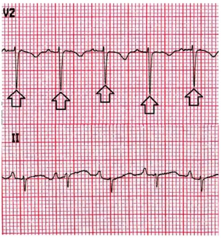
Alternancias eléctricas en un ECG en un paciente con un gran derrame pericárdico:
Las flechas señalan la amplitud alternante del complejo QRS.

Imagen: “Electrical alternans” por Eric Williams Medical Sciences Complex, The University of the West Indies, Champs Fleurs, Trinidad and Tobago. Licencia: CC BY 4.0

Radiografía de tórax:

  • Puede parecer normal en EN Erythema nodosum is an immune-mediated panniculitis (inflammation of the subcutaneous fat) caused by a type IV (delayed-type) hypersensitivity reaction. It commonly manifests in young women as tender, erythematous nodules on the shins. Erythema Nodosum condiciones con poca acumulación de líquido
  • Agrandamiento de la silueta cardíaca:
    • Se produce cuando se han acumulado > 250 mL de líquido
    • Tiene forma de “botella de agua”.
  • Los LOS Neisseria campos pulmonares suelen estar libres.
Cardiomegalia por derrame pericárdico antes y después del drenaje

Cardiomegalia por derrame pericárdico antes y después del drenaje:
(a) Radiografía de tórax que muestra una cardiomegalia debido a la acumulación de un derrame pericárdico
(b) Resolución de la cardiomegalia tras el drenaje del líquido.

Imagen: “CXR” por Division of Cardiology, Saint Luke’s University Health Network, Bethlehem, PA 18015, USA. Licencia: CC BY 3.0

Ultrasonido cardíaco:

  • Prueba diagnóstica de elección
  • Alta sensibilidad y especificidad
  • Proporciona información hemodinámica
  • El derrame pericárdico luce como un espacio ecolúcido en EN Erythema nodosum is an immune-mediated panniculitis (inflammation of the subcutaneous fat) caused by a type IV (delayed-type) hypersensitivity reaction. It commonly manifests in young women as tender, erythematous nodules on the shins. Erythema Nodosum el saco pericárdico.
  • Hallazgos de taponamiento cardíaco:
    • Colapso de la pared libre de la aurícula derecha durante la sístole
    • Colapso del ventrículo derecho durante la diástole
    • Arqueo septal
    • Dilatación de la vena cava inferior sin variación durante la respiración
Ecocardiograma con derrame pericárdico

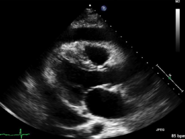
Ultrasonido cardíaco transtorácico que muestra un derrame pericárdico (región ecolúcida alrededor del corazón)

Imagen: “Transthoracic echocardiography” por Department of Internal Medicine, The University of New Mexico, Albuquerque, NM 87106, USA. Licencia: CC BY 4.0

TC y RM:

  • No son las modalidades diagnósticas de elección
  • Puede utilizarse si el ultrasonido no es diagnóstico
  • Puede evaluar la patología pericárdica
  • Puede ser más sensible para identificar derrames loculados
Émbolo pulmonar en TAC que muestra derrame pericárdico

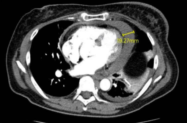
Una TC que muestra un derrame pericárdico de 19,27 mm.

Imagen: “CT pulmonary embolus” por Stanford Hospital and Clinics, Stanford, California. Licencia: CC BY 2.0

Análisis del líquido pericárdico y biopsia pericárdica

Se puede realizar un análisis del líquido pericárdico y una biopsia pericárdica para determinar la causa del derrame. Se pueden realizar las siguientes pruebas en EN Erythema nodosum is an immune-mediated panniculitis (inflammation of the subcutaneous fat) caused by a type IV (delayed-type) hypersensitivity reaction. It commonly manifests in young women as tender, erythematous nodules on the shins. Erythema Nodosum el líquido pericárdico:

  • Tinción de Gram y cultivos (incluyendo para hongos)
  • Recuento de células con diferencial
  • Citología
  • Tinción y cultivo de bacilos ácido-alcohol resistentes
  • Pruebas de PCR PCR Polymerase chain reaction (PCR) is a technique that amplifies DNA fragments exponentially for analysis. The process is highly specific, allowing for the targeting of specific genomic sequences, even with minuscule sample amounts. The PCR cycles multiple times through 3 phases: denaturation of the template DNA, annealing of a specific primer to the individual DNA strands, and synthesis/elongation of new DNA molecules. Polymerase Chain Reaction (PCR) viral

Evaluación de laboratorio

Para determinar la etiología de un derrame pericárdico se pueden realizar las siguientes pruebas:

  • Hemograma con diferencial
  • BUN y creatinina
  • Velocidad de eritrosedimentación y proteína C reactiva
  • Troponina
  • Hormona estimulante de la tiroides (TSH, por sus siglas en EN Erythema nodosum is an immune-mediated panniculitis (inflammation of the subcutaneous fat) caused by a type IV (delayed-type) hypersensitivity reaction. It commonly manifests in young women as tender, erythematous nodules on the shins. Erythema Nodosum inglés)
  • Niveles de factor reumatoide
  • ANA
  • Niveles de complemento
  • Ensayo Quantiferon-TB
  • Serología para VIH

Tratamiento

Tratamiento del derrame pericárdico

  • Depende de la estabilidad hemodinámica del paciente y de la causa subyacente del derrame
  • Identificar y tratar los LOS Neisseria trastornos subyacentes.
  • Tratamiento médico de los LOS Neisseria derrames inflamatorios o de la pericarditis Pericarditis Pericarditis is an inflammation of the pericardium, often with fluid accumulation. It can be caused by infection (often viral), myocardial infarction, drugs, malignancies, metabolic disorders, autoimmune disorders, or trauma. Acute, subacute, and chronic forms exist. Pericarditis asociada:
    • AINE
    • Colchicina
  • Los LOS Neisseria pequeños derrames en EN Erythema nodosum is an immune-mediated panniculitis (inflammation of the subcutaneous fat) caused by a type IV (delayed-type) hypersensitivity reaction. It commonly manifests in young women as tender, erythematous nodules on the shins. Erythema Nodosum un paciente estable suelen resolverse de manera espontánea → no es necesaria ninguna intervención
  • El drenaje pericárdico puede considerarse en EN Erythema nodosum is an immune-mediated panniculitis (inflammation of the subcutaneous fat) caused by a type IV (delayed-type) hypersensitivity reaction. It commonly manifests in young women as tender, erythematous nodules on the shins. Erythema Nodosum:
    • Derrames grandes y sintomáticos
    • Etiología incierta

Tratamiento del taponamiento cardíaco

Consideraciones generales:

  • Administrar oxígeno suplementario
  • Medidas para ↑ el gasto cardíaco:
    • Reanimación con líquidos IV
    • Apoyo inotrópico (e.g., dobutamina)

Pericardiocentesis Pericardiocentesis Puncture and aspiration of fluid from the pericardium. Cardiac Surgery:

  • Se introduce una aguja en EN Erythema nodosum is an immune-mediated panniculitis (inflammation of the subcutaneous fat) caused by a type IV (delayed-type) hypersensitivity reaction. It commonly manifests in young women as tender, erythematous nodules on the shins. Erythema Nodosum el espacio pericárdico.
  • Se evacúa el líquido para aliviar la presión en EN Erythema nodosum is an immune-mediated panniculitis (inflammation of the subcutaneous fat) caused by a type IV (delayed-type) hypersensitivity reaction. It commonly manifests in young women as tender, erythematous nodules on the shins. Erythema Nodosum el corazón.
  • Se puede colocar un catéter para el drenaje periódico.
Abordaje subxifoide para la pericardiocentesis

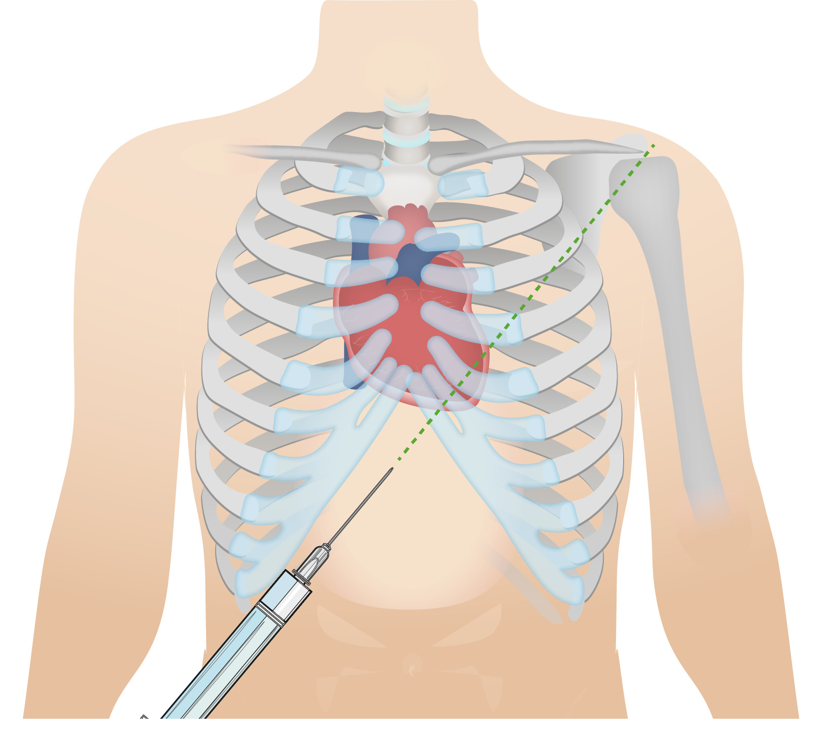
Abordaje subxifoideo para la pericardiocentesis:
Este abordaje permite el drenaje del líquido pericárdico.

Imagen por Lecturio.

Tratamiento quirúrgico:

  • Permite realizar una biopsia pericárdica
  • Preferible en EN Erythema nodosum is an immune-mediated panniculitis (inflammation of the subcutaneous fat) caused by a type IV (delayed-type) hypersensitivity reaction. It commonly manifests in young women as tender, erythematous nodules on the shins. Erythema Nodosum derrames pericárdicos traumáticos
  • Opciones:
    • Pericardiotomía
    • Ventana pericárdica

Diagnóstico Diferencial

  • Pericarditis Pericarditis Pericarditis is an inflammation of the pericardium, often with fluid accumulation. It can be caused by infection (often viral), myocardial infarction, drugs, malignancies, metabolic disorders, autoimmune disorders, or trauma. Acute, subacute, and chronic forms exist. Pericarditis: inflamación del pericardio que resulta de una infección, enfermedad autoinmune, radiación, intervención quirúrgica, infarto de miocardio o una cirugía cardíaca. Los LOS Neisseria pacientes pueden tener fiebre, dolor Dolor Inflammation torácico pleurítico y un roce pericárdico en EN Erythema nodosum is an immune-mediated panniculitis (inflammation of the subcutaneous fat) caused by a type IV (delayed-type) hypersensitivity reaction. It commonly manifests in young women as tender, erythematous nodules on the shins. Erythema Nodosum la auscultación cardíaca. El diagnóstico se confirma con la elevación difusa del ST en EN Erythema nodosum is an immune-mediated panniculitis (inflammation of the subcutaneous fat) caused by a type IV (delayed-type) hypersensitivity reaction. It commonly manifests in young women as tender, erythematous nodules on the shins. Erythema Nodosum el ECG ECG An electrocardiogram (ECG) is a graphic representation of the electrical activity of the heart plotted against time. Adhesive electrodes are affixed to the skin surface allowing measurement of cardiac impulses from many angles. The ECG provides 3-dimensional information about the conduction system of the heart, the myocardium, and other cardiac structures. Electrocardiogram (ECG), y engrosamiento y derrame pericárdico en EN Erythema nodosum is an immune-mediated panniculitis (inflammation of the subcutaneous fat) caused by a type IV (delayed-type) hypersensitivity reaction. It commonly manifests in young women as tender, erythematous nodules on the shins. Erythema Nodosum el ultrasonido cardíaco. El tratamiento puede incluir AINE, colchicina y esteroides.
  • Miocarditis: enfermedad inflamatoria del miocardio. La miocarditis suele provocar signos y síntomas de insuficiencia cardíaca. La evolución de la miocarditis puede variar en EN Erythema nodosum is an immune-mediated panniculitis (inflammation of the subcutaneous fat) caused by a type IV (delayed-type) hypersensitivity reaction. It commonly manifests in young women as tender, erythematous nodules on the shins. Erythema Nodosum función de la etiología y la progresión de los LOS Neisseria síntomas. El diagnóstico se apoya en EN Erythema nodosum is an immune-mediated panniculitis (inflammation of the subcutaneous fat) caused by a type IV (delayed-type) hypersensitivity reaction. It commonly manifests in young women as tender, erythematous nodules on the shins. Erythema Nodosum los LOS Neisseria hallazgos clínicos, las pruebas de laboratorio y la imagenología cardíaca. Rara vez se requiere un diagnóstico definitivo mediante una biopsia endomiocárdica. El tratamiento es de soporte y está dirigido a tratar las complicaciones.
  • Embolia pulmonar: obstrucción de las arterias pulmonares, casi siempre debida a la migración de un trombo desde el sistema venoso profundo. Los LOS Neisseria signos y síntomas incluyen dolor Dolor Inflammation torácico pleurítico, disnea, taquipnea y taquicardia. Los LOS Neisseria casos severos pueden resultar en EN Erythema nodosum is an immune-mediated panniculitis (inflammation of the subcutaneous fat) caused by a type IV (delayed-type) hypersensitivity reaction. It commonly manifests in young women as tender, erythematous nodules on the shins. Erythema Nodosum inestabilidad hemodinámica o paro cardiorespiratorio. Una TC de tórax con angiografía es el método principal de diagnóstico. El tratamiento incluye oxigenación, anticoagulación y terapia trombolítica para pacientes inestables.
  • Neumotórax: enfermedad potencialmente mortal en EN Erythema nodosum is an immune-mediated panniculitis (inflammation of the subcutaneous fat) caused by a type IV (delayed-type) hypersensitivity reaction. It commonly manifests in young women as tender, erythematous nodules on the shins. Erythema Nodosum la que se acumula aire en EN Erythema nodosum is an immune-mediated panniculitis (inflammation of the subcutaneous fat) caused by a type IV (delayed-type) hypersensitivity reaction. It commonly manifests in young women as tender, erythematous nodules on the shins. Erythema Nodosum el espacio pleural, provocando un colapso parcial o total del pulmón. Un neumotórax puede ser traumático o espontáneo. Los LOS Neisseria pacientes se presentan con una aparición repentina de dolor Dolor Inflammation torácico agudo, disnea y disminución de los LOS Neisseria sonidos respiratorios en EN Erythema nodosum is an immune-mediated panniculitis (inflammation of the subcutaneous fat) caused by a type IV (delayed-type) hypersensitivity reaction. It commonly manifests in young women as tender, erythematous nodules on the shins. Erythema Nodosum el examen. Un neumotórax grande o a tensión puede provocar un colapso cardiopulmonar. El diagnóstico se realiza con base en EN Erythema nodosum is an immune-mediated panniculitis (inflammation of the subcutaneous fat) caused by a type IV (delayed-type) hypersensitivity reaction. It commonly manifests in young women as tender, erythematous nodules on the shins. Erythema Nodosum los LOS Neisseria hallazgos imagenológicos. El tratamiento incluye la descompresión con aguja y la colocación de un tubo de tórax (drenaje pleural).

Referencias

  1. Hoit, B.D. (2025). Etiology of pericardial disease. In Yeon, S.B. (Ed.), UpToDate. Retrieved April 26, 2025, from https://www.uptodate.com/contents/etiology-of-pericardial-disease
  2. Hoit, B. D. (2024). Pericardial effusion: Approach to diagnosis. In M. M. LeWinter & D. J. Sexton (Eds.), UpToDate. Wolters Kluwer. Retrieved April 26, 2025, from https://www.uptodate.com/contents/pericardial-effusion-approach-to-diagnosis
  3. Hoit, B.D. (2025). Cardiac tamponade. In Yeon, S.B. (Ed.), UpToDate. Retrieved April 26, 2025, from https://www.uptodate.com/contents/cardiac-tamponade
  4. Sagristà-Sauleda, J., Angel, J., Sambola, A., Permanyer-Miralda, G. (2008). Hemodynamic effects of volume expansion in patients with cardiac tamponade. Circulation. 117:1545–1549. https://doi.org/10.1161/CIRCULATIONAHA.107.737841
  5. Strimel, W.J., Ayub, B., Contractor, T. (2018). Pericardial effusion. In O’Brien, T.X. (Ed.), Medscape. Retrieved April 26, 2025, from https://emedicine.medscape.com/article/157325-overview
  6. Yarlagadda, C. (2021). Cardiac tamponade. In O’Brien, T.X. (Ed.), Medscape. Retrieved April 26, 2025, from https://emedicine.medscape.com/article/152083-overview
  7. Weiser, T.G. (2024). Cardiac tamponade. [Online] MSD Manual Professional Version. Retrieved April 26, 2025, from https://www.msdmanuals.com/professional/injuries-poisoning/thoracic-trauma/cardiac-tamponade
  8. Willner, D.A., Goyal, A., Grigorova, Y., Kiel, J. (2024). Pericardial effusion. [Online] StatPearls. Retrieved April 26, 2025, from https://www.ncbi.nlm.nih.gov/books/NBK431089/
  9. Stashko, E., Meer, J.M. (2023). Cardiac tamponade. [Online] StatPearls. Retrieved April 26, 2025, from https://www.ncbi.nlm.nih.gov/books/NBK431090

¡Crea tu cuenta gratis o inicia una sesión para seguir leyendo!

Regístrate ahora y obtén acceso gratuito a Lecturio con páginas de concepto, videos médicos y cuestionarios para tu educación médica.

User Reviews

Que tengas una sesión de estudio alegre y navideña 🎁 Ahorra 50% en todos los planes >>

Details