El síndrome de Stevens-Johnson (SSJ) es una reacción de hipersensibilidad cutánea, inmunomediada, que suele ser desencadenada por medicamentos, como los LOS Neisseria antiepilépticos y los LOS Neisseria antibióticos. La afección se sitúa en EN Erythema nodosum is an immune-mediated panniculitis (inflammation of the subcutaneous fat) caused by a type IV (delayed-type) hypersensitivity reaction. It commonly manifests in young women as tender, erythematous nodules on the shins. Erythema Nodosum un espectro con la necrólisis epidérmica tóxica (NET) basado en EN Erythema nodosum is an immune-mediated panniculitis (inflammation of the subcutaneous fat) caused by a type IV (delayed-type) hypersensitivity reaction. It commonly manifests in young women as tender, erythematous nodules on the shins. Erythema Nodosum la cantidad de superficie corporal afectada. El síndrome de Stevens-Johnson se caracteriza por la necrosis Necrosis The death of cells in an organ or tissue due to disease, injury or failure of the blood supply. Ischemic Cell Damage de los LOS Neisseria queratinocitos y la separación de la epidermis Epidermis The external, nonvascular layer of the skin. It is made up, from within outward, of five layers of epithelium: (1) basal layer (stratum basale epidermidis); (2) spinous layer (stratum spinosum epidermidis); (3) granular layer (stratum granulosum epidermidis); (4) clear layer (stratum lucidum epidermidis); and (5) horny layer (stratum corneum epidermidis). Skin: Structure and Functions de la dermis Dermis A layer of vascularized connective tissue underneath the epidermis. The surface of the dermis contains innervated papillae. Embedded in or beneath the dermis are sweat glands; hair follicles; and sebaceous glands. Skin: Structure and Functions. Los LOS Neisseria pacientes presentan un pródromo gripal, seguido de ampollas cutáneas y descamación en EN Erythema nodosum is an immune-mediated panniculitis (inflammation of the subcutaneous fat) caused by a type IV (delayed-type) hypersensitivity reaction. It commonly manifests in young women as tender, erythematous nodules on the shins. Erythema Nodosum la cara, tórax y mucosas. El síndrome de Stevens-Johnson se considera una emergencia médica, y el tratamiento es principalmente de soporte. Se requiere la retirada del agente causal. La vigilancia y el tratamiento de la sobreinfección son esenciales debido al AL Amyloidosis alto riesgo de muerte asociado a estos casos.
Last updated: Apr 13, 2022
| Subtipo | Área de superficie corporal implicada |
|---|---|
|
Síndrome de Stevens-Johnson |
<10% de superficie corporal afectada |
|
Superposición de SSJ/TEN |
10%–30% de superficie corporal |
|
Necrólisis epidérmica tóxica |
>30% de área de superficie corporal |
En EN Erythema nodosum is an immune-mediated panniculitis (inflammation of the subcutaneous fat) caused by a type IV (delayed-type) hypersensitivity reaction. It commonly manifests in young women as tender, erythematous nodules on the shins. Erythema Nodosum la siguiente tabla se enumeran las principales causas infecciosas y por medicamentos más comunes del SSJ/NET:
| Tipos | Ejemplos | |
|---|---|---|
|
Medicamentos |
Antiepilépticos |
Lamotrigina, fenobarbital, carbamazepina, fenitoína |
|
Sulfa |
Cotrimoxazol, sulfasalazina |
|
|
Otros antibióticos |
Aminopenicilinas, fluoroquinolonas, cefalosporinas |
|
|
AINE |
Meloxicam Meloxicam A benzothiazine and thiazole derivative that acts as a nsaid and cyclooxygenase-2 (cox-2) inhibitor. It is used in the treatment of rheumatoid arthritis; osteoarthritis; and ankylosing spondylitis. Nonsteroidal Antiinflammatory Drugs (NSAIDs), piroxicam Piroxicam A cyclooxygenase inhibiting, non-steroidal anti-inflammatory agent (nsaid) that is well established in treating rheumatoid arthritis and osteoarthritis and used for musculoskeletal disorders, dysmenorrhea, and postoperative pain. Its long half-life enables it to be administered once daily. Nonsteroidal Antiinflammatory Drugs (NSAIDs) |
|
|
Antirretrovirales |
Nevirapina |
|
|
Misceláneos |
Alopurinol, clormezanona |
|
|
Infecciosa |
Viral |
Virus Virus Viruses are infectious, obligate intracellular parasites composed of a nucleic acid core surrounded by a protein capsid. Viruses can be either naked (non-enveloped) or enveloped. The classification of viruses is complex and based on many factors, including type and structure of the nucleoid and capsid, the presence of an envelope, the replication cycle, and the host range. Virology del herpes simple, VIH, coxsackievirus Coxsackievirus Coxsackievirus is a member of a family of viruses called Picornaviridae and the genus Enterovirus. Coxsackieviruses are single-stranded, positive-sense RNA viruses, and are divided into coxsackie group A and B viruses. Both groups of viruses cause upper respiratory infections, rashes, aseptic meningitis, or encephalitis. Coxsackievirus, hepatitis, gripe, paperas, virus Virus Viruses are infectious, obligate intracellular parasites composed of a nucleic acid core surrounded by a protein capsid. Viruses can be either naked (non-enveloped) or enveloped. The classification of viruses is complex and based on many factors, including type and structure of the nucleoid and capsid, the presence of an envelope, the replication cycle, and the host range. Virology de Epstein-Barr, enterovirus Enterovirus A genus of the family picornaviridae whose members preferentially inhabit the intestinal tract of a variety of hosts. The genus contains many species. Newly described members of human enteroviruses are assigned continuous numbers with the species designated ‘human enterovirus’. Coxsackievirus |
|
Bacteriana |
Estreptococos beta-hemolíticos del grupo A, brucelosis, micobacterias, Mycoplasma pneumoniae Mycoplasma pneumoniae Short filamentous organism of the genus mycoplasma, which binds firmly to the cells of the respiratory epithelium. It is one of the etiologic agents of non-viral primary atypical pneumonia in man. Mycoplasma, rickettsias, tularemia | |
Se desconoce el mecanismo exacto, pero hay varias teorías:
Esta alteración de la integridad de la piel puede provocar:
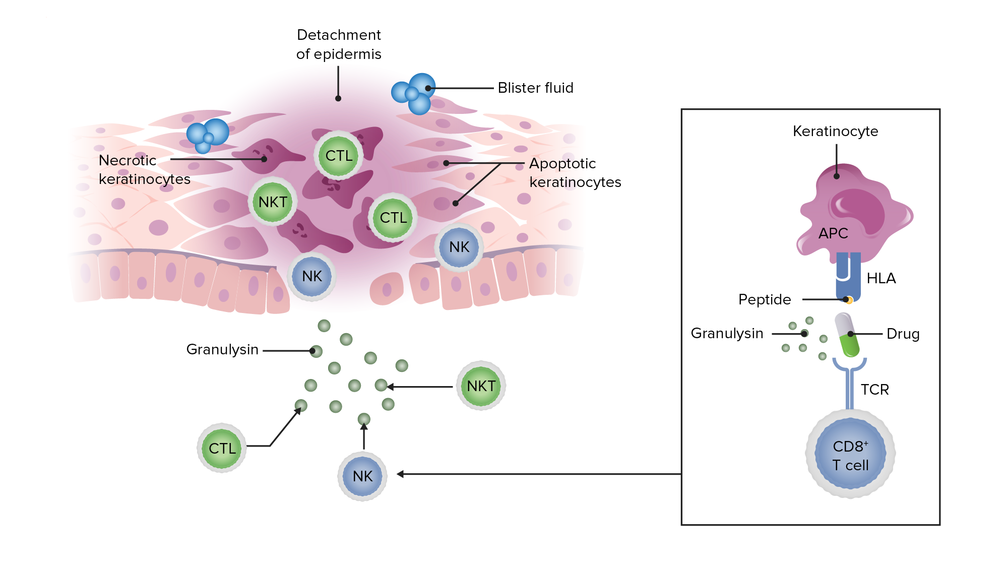
Esquema que muestra cómo un antígeno peptídico (en este caso, de un medicamento) presentado en los queratinocitos puede provocar una respuesta inflamatoria citotóxica que da lugar a la liberación de granulisina, a la apoptosis y necrosis de los queratinocitos, al desprendimiento de la epidermis y a la formación de ampollas en el SSJ y el NET.
Imagen por Lecturio.Pródromo:
Fase aguda:
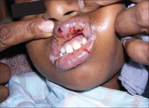
Lesiones orales por SSJ observadas en un paciente que toma un medicamento antirretroviral (nevirapina).
Imagen: “Oral lesions associated with nevirapine-related Stevens Johnson syndrome: A report of four cases.” por lasundaram S, Ranganathan K, Umadevi K, Gunaseelan R, Kumaraswamy N, Solomon S, Devaleenol B, Ambrose P – Journal of oral and maxillofacial pathology : JOMFP (2011). Licencia:CC BY 2.0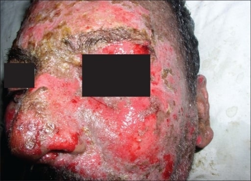
Lesiones características del SSJ/NET en un paciente con eritema y descamación.
Imagen: “Oral lesions associated with nevirapine-related Stevens Johnson syndrome: A report of four cases” por Balasundaram S, Ranganathan K, Umadevi K, Gunaseelan R, Kumaraswamy N, Solomon S, Devaleenol B, Ambrose P – Journal of oral and maxillofacial pathology : JOMFP (2011) . Licencia:CC BY 2.0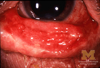
Queratoconjuntivitis en un paciente con SSJ/NET-
Imagen: “Conjunctivitis in SJS” por Jonathan Trobe, M.D., University of Michigan Kellogg Eye Center. Licencia: CC BY 3.0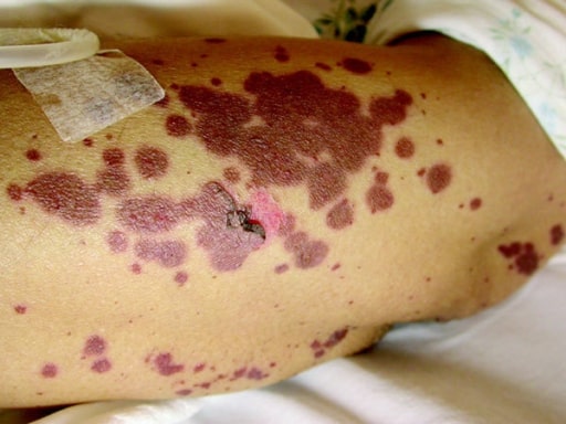
Demostración del signo de Nikolsky provocando la descamación de la piel con la aplicación de presión.
Imagen: “Curcumin in stevens-johnsons syndrome: culprit or bystander?” por Irani C, Haddad F, Maalouly G, Nemnoum R. Licencia: CC BY 2.0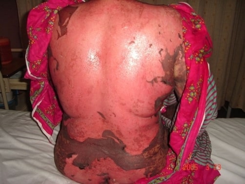
Necrólisis epidérmica tóxica inducida por medicamentos con descamación de la piel en la espalda y nalgas.
Imagen: “Drug induced toxic epidermal necrolysis: two case reports” por Qadir SN, Raza N, Qadir F. License: CC BY 3.0
Fotografía de un paciente con denudación generalizada de la epidermis en láminas, consistente con necrólisis epidérmica tóxica.
Imagen: “Review of Toxic Epidermal Necrolysis” por International Journal of Molecular Sciences. Licencia: CC BY 4.0El diagnóstico es clínico, basado en EN Erythema nodosum is an immune-mediated panniculitis (inflammation of the subcutaneous fat) caused by a type IV (delayed-type) hypersensitivity reaction. It commonly manifests in young women as tender, erythematous nodules on the shins. Erythema Nodosum los LOS Neisseria antecedentes y el examen físico.
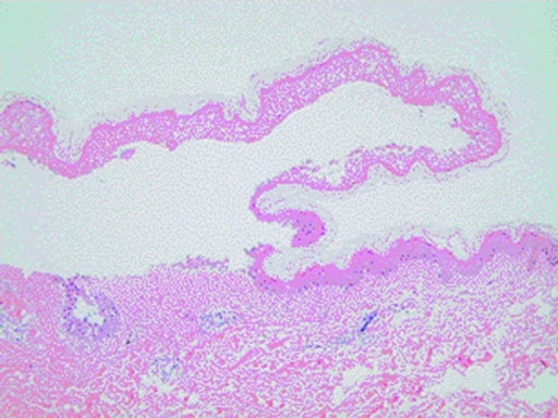
Histopatología de una biopsia de piel tomada a un paciente con SSJ/NET que muestra el desprendimiento epidérmico característico y la infiltración de linfocitos en la dermis
Imagen: “Curcumin in stevens-johnsons syndrome: culprit or bystander?” por Irani C, Haddad F, Maalouly G, Nemnoum R. Licencia: CC BY 2.0| Factores pronósticos | Puntuación |
|---|---|
|
Edad ≥ 40 años |
1 |
|
Malignidad presente |
1 |
|
Superficie corporal desprendida ≥ 10%. |
1 |
|
Taquicardia ≥ 120/min. |
1 |
|
Urea Urea A compound formed in the liver from ammonia produced by the deamination of amino acids. It is the principal end product of protein catabolism and constitutes about one half of the total urinary solids. Urea Cycle sérica > 10 mmol/L |
1 |
|
Glucosa sérica > 14 mmol/L |
1 |
|
Bicarbonato sérico <20 mmol/L |
1 |
La puntuación SCORTEN se utiliza para ayudar a determinar la gravedad, el pronóstico y el entorno adecuado para el tratamiento de los LOS Neisseria pacientes con SSJ/NET.