Domina Conceptos Médicos

Estudia para la escuela de medicina y tus examenes con Lecturio.

Cáncer de Pene

Las lesiones malignas del pene surgen del epitelio escamoso del glande, el prepucio o el cuerpo del pene. El cáncer de pene es poco frecuente en EN Erythema nodosum is an immune-mediated panniculitis (inflammation of the subcutaneous fat) caused by a type IV (delayed-type) hypersensitivity reaction. It commonly manifests in young women as tender, erythematous nodules on the shins. Erythema Nodosum los LOS Neisseria Estados Unidos, pero hay una mayor prevalencia en EN Erythema nodosum is an immune-mediated panniculitis (inflammation of the subcutaneous fat) caused by a type IV (delayed-type) hypersensitivity reaction. It commonly manifests in young women as tender, erythematous nodules on the shins. Erythema Nodosum regiones socioeconómicas más bajas. El subtipo histológico más común es el carcinoma de células escamosas. Los LOS Neisseria hombres no circuncidados y los LOS Neisseria infectados por el VPH son los LOS Neisseria que tienen mayor riesgo de padecer neoplasias penianas. El diagnóstico se hace HACE Altitude Sickness mediante una combinación del examen físico, los LOS Neisseria antecedentes, los LOS Neisseria estudios imagenológicos y la biopsia de tejido. Es necesaria una correcta estadificación TNM para determinar el tratamiento correcto, que va VA Ventilation: Mechanics of Breathing desde la terapia tópica local hasta un abordaje multimodal de cirugía/radiación/quimioterapia.

Last updated: Dec 15, 2025

Editorial responsibility: Stanley Oiseth, Lindsay Jones, Evelin Maza

Descripción General

Epidemiología

  • En EN Erythema nodosum is an immune-mediated panniculitis (inflammation of the subcutaneous fat) caused by a type IV (delayed-type) hypersensitivity reaction. It commonly manifests in young women as tender, erythematous nodules on the shins. Erythema Nodosum general, las neoplasias penianas son poco frecuentes.
  • Mayor incidencia: grupo de edad de 50–70 años
  • En EN Erythema nodosum is an immune-mediated panniculitis (inflammation of the subcutaneous fat) caused by a type IV (delayed-type) hypersensitivity reaction. It commonly manifests in young women as tender, erythematous nodules on the shins. Erythema Nodosum Estados Unidos:
    • Incidencia anual estimada de aproximadamente 2000 nuevos casos
    • Incidencia promedio: 1 de cada 100 000 hombres al AL Amyloidosis año
  • En EN Erythema nodosum is an immune-mediated panniculitis (inflammation of the subcutaneous fat) caused by a type IV (delayed-type) hypersensitivity reaction. It commonly manifests in young women as tender, erythematous nodules on the shins. Erythema Nodosum los LOS Neisseria países altamente industrializados (Estados Unidos, Europa, Canadá), las tasas de incidencia anual son menores.
  • Las tasas de prevalencia son 5–10 veces mayores en EN Erythema nodosum is an immune-mediated panniculitis (inflammation of the subcutaneous fat) caused by a type IV (delayed-type) hypersensitivity reaction. It commonly manifests in young women as tender, erythematous nodules on the shins. Erythema Nodosum los LOS Neisseria países no industrializados.

Factores de riesgo

  • Hombres no circuncidados:
    • El mayor riesgo contribuyente a las lesiones malignas del pene
    • Riesgo 22 veces superior al AL Amyloidosis de los LOS Neisseria varones circuncidados
    • Reducción máxima del riesgo de cáncer de pene con la circuncisión realizada en EN Erythema nodosum is an immune-mediated panniculitis (inflammation of the subcutaneous fat) caused by a type IV (delayed-type) hypersensitivity reaction. It commonly manifests in young women as tender, erythematous nodules on the shins. Erythema Nodosum el periodo neonatal
  • Antecedentes de infección por VPH o VIH:
    • Aumento de 8 veces de lesiones penianas
    • Hasta el 50% de los LOS Neisseria cánceres de pene están asociados a los LOS Neisseria serotipos 16 (el más común), 6 y 18 del VPH.
    • Riesgo especialmente elevado de carcinomas basaloides y condilomatosos del pene
  • VIH
  • Bajo estatus socioeconómico
  • Mala higiene genital (acumulación de esmegma)
  • Tabaquismo
  • Psoralenos y fotoquimioterapia ultravioleta A
  • Afecciones inflamatorias crónicas:

Fisiopatología

Anatomía

  • La mayoría de los LOS Neisseria cánceres de pene surgen del epitelio escamoso de la epidermis Epidermis The external, nonvascular layer of the skin. It is made up, from within outward, of five layers of epithelium: (1) basal layer (stratum basale epidermidis); (2) spinous layer (stratum spinosum epidermidis); (3) granular layer (stratum granulosum epidermidis); (4) clear layer (stratum lucidum epidermidis); and (5) horny layer (stratum corneum epidermidis). Skin: Structure and Functions y la dermis Dermis A layer of vascularized connective tissue underneath the epidermis. The surface of the dermis contains innervated papillae. Embedded in or beneath the dermis are sweat glands; hair follicles; and sebaceous glands. Skin: Structure and Functions.
  • Debajo de la piel del cuerpo del pene se originan dos capas fasciales distintas:
    • Fascia Fascia Layers of connective tissue of variable thickness. The superficial fascia is found immediately below the skin; the deep fascia invests muscles, nerves, and other organs. Cellulitis superficial o dartos
    • Fascia Fascia Layers of connective tissue of variable thickness. The superficial fascia is found immediately below the skin; the deep fascia invests muscles, nerves, and other organs. Cellulitis profunda o de Buck
  • Dentro de la fascia Fascia Layers of connective tissue of variable thickness. The superficial fascia is found immediately below the skin; the deep fascia invests muscles, nerves, and other organs. Cellulitis de Buck se encuentran los LOS Neisseria dos cuerpos cavernosos dorsalmente y un único cuerpo esponjoso ventralmente.
  • El drenaje linfático del pene se realiza principalmente a través de los LOS Neisseria ganglios linfáticos inguinales de la ingle:
Anatomía reproductiva externa masculina

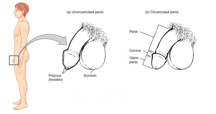
Anatomía reproductiva masculina (externa)

Imagen: “Male Reproductive System” por Phil Schatz. Licencia: CC BY 4.0
Sección transversal del pene

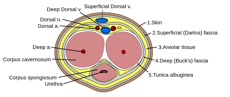
Sección transversal del pene:
La imagen muestra la forma que adoptan los tejidos eréctiles dentro del cuerpo del pene. Aquí, observe la túnica albugínea que encierra ambos tipos de tejido eréctil, así como la uretra, que está completamente dentro del cuerpo esponjoso.

Imagen: “Penis cross section” por Mcstrother. Licencia: CC BY 3.0

Patogénesis y patología

  • Crecimiento del cáncer:
    • Lesiones pequeñas al AL Amyloidosis principio, a menudo observadas en EN Erythema nodosum is an immune-mediated panniculitis (inflammation of the subcutaneous fat) caused by a type IV (delayed-type) hypersensitivity reaction. It commonly manifests in young women as tender, erythematous nodules on the shins. Erythema Nodosum el glande o el prepucio
    • Carcinomas de pene mediados por el VPH:
      • Las oncoproteínas E6 y E7 producidas por las infecciones por VPH pueden interferir con la función de los LOS Neisseria genes Genes A category of nucleic acid sequences that function as units of heredity and which code for the basic instructions for the development, reproduction, and maintenance of organisms. DNA Types and Structure supresores de tumores.
      • Se producen alteraciones de las vías genéticas RB1 y p53 y de los LOS Neisseria reguladores del ciclo celular p21 y p16.
  • Carcinoma in situ Carcinoma in situ A lesion with cytological characteristics associated with invasive carcinoma but the tumor cells are confined to the epithelium of origin, without invasion of the basement membrane. Leukoplakia ( CIS CIS Multiple Sclerosis) o neoplasia intraepitelial del pene:
    • La enfermedad de Bowen se presenta como lesiones en EN Erythema nodosum is an immune-mediated panniculitis (inflammation of the subcutaneous fat) caused by a type IV (delayed-type) hypersensitivity reaction. It commonly manifests in young women as tender, erythematous nodules on the shins. Erythema Nodosum el cuerpo del pene.
    • La eritroplasia de Queyrat se presenta con lesiones en EN Erythema nodosum is an immune-mediated panniculitis (inflammation of the subcutaneous fat) caused by a type IV (delayed-type) hypersensitivity reaction. It commonly manifests in young women as tender, erythematous nodules on the shins. Erythema Nodosum el glande.
  • Carcinoma de células escamosas:
    • Variante común:
      • Comprende hasta dos tercios de los LOS Neisseria casos del carcinoma de células escamosas
      • Suele invadir el cuerpo esponjoso
      • Las metástasis en EN Erythema nodosum is an immune-mediated panniculitis (inflammation of the subcutaneous fat) caused by a type IV (delayed-type) hypersensitivity reaction. It commonly manifests in young women as tender, erythematous nodules on the shins. Erythema Nodosum los LOS Neisseria ganglios inguinales están presentes en EN Erythema nodosum is an immune-mediated panniculitis (inflammation of the subcutaneous fat) caused by a type IV (delayed-type) hypersensitivity reaction. It commonly manifests in young women as tender, erythematous nodules on the shins. Erythema Nodosum 25%–40% de los LOS Neisseria casos.
    • Carcinoma papilar: 2%–15% de los LOS Neisseria casos
      • Generalmente de bajo grado o bien diferenciado, pero es superficialmente invasivo
      • Evidencia microscópica de hiperqueratosis y papilomatosis
      • No está relacionado con la infección por el VPH
      • No suele asociarse a invasión vascular o perineural
    • Condilomatoso: 7%–10% de los LOS Neisseria casos
      • Lesiones similares a una coliflor
      • Núcleo fibrovascular prominente con formaciones papilomatosas
      • No suele alcanzar a las estructuras vasculares y neurales
      • Vinculado a la infección por el VPH
      • Metástasis en EN Erythema nodosum is an immune-mediated panniculitis (inflammation of the subcutaneous fat) caused by a type IV (delayed-type) hypersensitivity reaction. It commonly manifests in young women as tender, erythematous nodules on the shins. Erythema Nodosum los LOS Neisseria ganglios inguinales en EN Erythema nodosum is an immune-mediated panniculitis (inflammation of the subcutaneous fat) caused by a type IV (delayed-type) hypersensitivity reaction. It commonly manifests in young women as tender, erythematous nodules on the shins. Erythema Nodosum el 17%–25% de los LOS Neisseria casos
    • Carcinoma basaloide: 4%–10% de los LOS Neisseria casos
      • Masas irregulares y ulceradas relacionadas con el VPH
      • Histológicamente, se presentan como células hipercromáticas uniformes y pequeñas con necrosis Necrosis The death of cells in an organ or tissue due to disease, injury or failure of the blood supply. Ischemic Cell Damage central.
      • Suele ser profundamente invasivo en EN Erythema nodosum is an immune-mediated panniculitis (inflammation of the subcutaneous fat) caused by a type IV (delayed-type) hypersensitivity reaction. It commonly manifests in young women as tender, erythematous nodules on the shins. Erythema Nodosum el cuerpo cavernoso o esponjoso
    • Carcinoma verrucoso: 3%–7% de los LOS Neisseria casos
      • Suele ser de bajo grado y exofítico
      • Papilas histológicamente rectas revestidas por células neoplásicas extremadamente bien diferenciadas
      • Superficie hiperqueratósica con queratina interpapilar
    • Carcinoma sarcomatoide (de células fusiformes): 1%–6% de los LOS Neisseria casos
      • Variante más agresiva
      • Masas polipoides mal diferenciadas, ulceradas o redondeadas.
      • Suele presentar invasión vascular y perineural
  • Carcinoma de células basales: bajo riesgo de metástasis
  • Sarcoma (incluido el sarcoma de Kaposi Kaposi A multicentric, malignant neoplastic vascular proliferation characterized by the development of bluish-red cutaneous nodules, usually on the lower extremities, most often on the toes or feet, and slowly increasing in size and number and spreading to more proximal areas. The tumors have endothelium-lined channels and vascular spaces admixed with variably sized aggregates of spindle-shaped cells, and often remain confined to the skin and subcutaneous tissue, but widespread visceral involvement may occur. Hhv-8 is the suspected cause. There is also a high incidence in AIDS patients. AIDS-defining Conditions, que se asocia con el HHV-8)
  • Melanoma Melanoma Melanoma is a malignant tumor arising from melanocytes, the melanin-producing cells of the epidermis. These tumors are most common in fair-skinned individuals with a history of excessive sun exposure and sunburns. Melanoma: cáncer de piel más agresivo
  • La metástasis en EN Erythema nodosum is an immune-mediated panniculitis (inflammation of the subcutaneous fat) caused by a type IV (delayed-type) hypersensitivity reaction. It commonly manifests in young women as tender, erythematous nodules on the shins. Erythema Nodosum el pene es rara.

Presentación Clínica y Diagnóstico

Presentación

  • Inicialmente, se presenta como una anomalía cutánea o una lesión palpable en EN Erythema nodosum is an immune-mediated panniculitis (inflammation of the subcutaneous fat) caused by a type IV (delayed-type) hypersensitivity reaction. It commonly manifests in young women as tender, erythematous nodules on the shins. Erythema Nodosum el pene:
  • Las lesiones pueden variar en EN Erythema nodosum is an immune-mediated panniculitis (inflammation of the subcutaneous fat) caused by a type IV (delayed-type) hypersensitivity reaction. It commonly manifests in young women as tender, erythematous nodules on the shins. Erythema Nodosum tamaño y características:
    • Lesiones ulceradas
    • Infiltrativas con crecimiento progresivo
  • La adenopatía inguinal se presenta en EN Erythema nodosum is an immune-mediated panniculitis (inflammation of the subcutaneous fat) caused by a type IV (delayed-type) hypersensitivity reaction. It commonly manifests in young women as tender, erythematous nodules on the shins. Erythema Nodosum un 30%–60% de los LOS Neisseria casos.
  • Metástasis a distancia:
    • Suele significar una presentación tardía de la enfermedad
    • Afecta a órganos genitourinarios adyacentes o a órganos distantes
    • Presente en EN Erythema nodosum is an immune-mediated panniculitis (inflammation of the subcutaneous fat) caused by a type IV (delayed-type) hypersensitivity reaction. It commonly manifests in young women as tender, erythematous nodules on the shins. Erythema Nodosum alrededor del 1%–10% de los LOS Neisseria casos

Examen físico

  • Examen constitucional generalizado (evaluar el estado de funcionamiento general)
  • Examen genitourinario específico:
    • Examen del pene:
      • Determinar estado de circuncisión.
      • Caracterizar el tamaño, la profundidad, la localización y las características de la lesión.
      • Evaluar si hay secreción, eritema y aumento de volumen.
    • Examen inguinal:
      • Evaluar la presencia de linfadenopatía inguinal.
      • Número de ganglios inguinales palpables
      • Nódulos fijos o móviles
Cáncer de pene

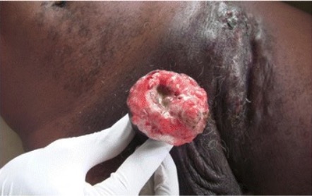
Carcinoma de células escamosas del pene

Imagen: “Cancer of the penis” por Service d’Urologie, CHU de Cocody, Abidjan, Ivory Coast, Africa. Licencia: CC BY 4.0

Diagnóstico

  • Biopsia de tejido confirmatoria:
    • En EN Erythema nodosum is an immune-mediated panniculitis (inflammation of the subcutaneous fat) caused by a type IV (delayed-type) hypersensitivity reaction. It commonly manifests in young women as tender, erythematous nodules on the shins. Erythema Nodosum sacabocados (punch)
    • Incisional
    • Escisional
  • Imagenología:
    • Opciones:
      • Ultrasonido
      • RM/TC
    • Funciones:
      • Evaluar el estadio del tumor Tumor Inflammation primario y el enfoque quirúrgico
      • Identifica la invasión en EN Erythema nodosum is an immune-mediated panniculitis (inflammation of the subcutaneous fat) caused by a type IV (delayed-type) hypersensitivity reaction. It commonly manifests in young women as tender, erythematous nodules on the shins. Erythema Nodosum el cuerpo cavernoso o esponjoso
      • Determina la afectación de los LOS Neisseria ganglios linfáticos regionales
      • Realizar estudios imagenológicos de otras localizaciones en EN Erythema nodosum is an immune-mediated panniculitis (inflammation of the subcutaneous fat) caused by a type IV (delayed-type) hypersensitivity reaction. It commonly manifests in young women as tender, erythematous nodules on the shins. Erythema Nodosum caso de metástasis
  • Estadificación de los LOS Neisseria ganglios linfáticos inguinales:
    • Aspiración con aguja fina:
      • Se realiza bajo anestesia local con una aguja de calibre 23
      • Para tomar muestras de ganglios clínicamente palpables
    • Biopsia dinámica del ganglio centinela:
      • Implica la inyección de un tinte trazador radiactivo
      • Examina el drenaje de los LOS Neisseria ganglios linfáticos inguinales
      • Permite el mapeo linfático y la biopsia del ganglio centinela
    • Disección de ganglios linfáticos inguinales superficiales:
      • Proporciona información directa sobre los LOS Neisseria nódulos sospechosos
      • Realizado por cirujanos pélvicos experimentados
      • Mayores tasas de complicaciones en EN Erythema nodosum is an immune-mediated panniculitis (inflammation of the subcutaneous fat) caused by a type IV (delayed-type) hypersensitivity reaction. It commonly manifests in young women as tender, erythematous nodules on the shins. Erythema Nodosum general

Estadificación

Tabla: Sistema de estadificación Tumor Tumor Inflammation, Nódulo y Metástasis (TNM) para el cáncer de pene (American Joint Committee on Cancer)
T: tumor Tumor Inflammation
Tumor Tumor Inflammation primario (T) Descripción
Tx El tumor Tumor Inflammation primario no puede ser evaluado.
T0 No hay evidencia de tumor Tumor Inflammation
Tis Carcinoma in situ Carcinoma in situ A lesion with cytological characteristics associated with invasive carcinoma but the tumor cells are confined to the epithelium of origin, without invasion of the basement membrane. Leukoplakia
T1
  • Glande: El tumor Tumor Inflammation invade la lámina propia.
  • Prepucio: El tumor Tumor Inflammation invade la dermis Dermis A layer of vascularized connective tissue underneath the epidermis. The surface of the dermis contains innervated papillae. Embedded in or beneath the dermis are sweat glands; hair follicles; and sebaceous glands. Skin: Structure and Functions, la lámina propia o la fascia Fascia Layers of connective tissue of variable thickness. The superficial fascia is found immediately below the skin; the deep fascia invests muscles, nerves, and other organs. Cellulitis de dartos.
  • Cuerpo del pene: El tumor Tumor Inflammation invade el tejido conectivo entre la epidermis Epidermis The external, nonvascular layer of the skin. It is made up, from within outward, of five layers of epithelium: (1) basal layer (stratum basale epidermidis); (2) spinous layer (stratum spinosum epidermidis); (3) granular layer (stratum granulosum epidermidis); (4) clear layer (stratum lucidum epidermidis); and (5) horny layer (stratum corneum epidermidis). Skin: Structure and Functions y los LOS Neisseria cuerpos cavernosos/esponjoso.
T1a Tumor Tumor Inflammation sin invasión linfovascular o perineural y no de alto grado
T1b Tumor Tumor Inflammation con invasión linfovascular y/o invasión perineural o el tumor Tumor Inflammation es de alto grado
T2 El tumor Tumor Inflammation invade el cuerpo esponjoso con o sin invasión uretral.
T3 T3 A T3 thyroid hormone normally synthesized and secreted by the thyroid gland in much smaller quantities than thyroxine (T4). Most T3 is derived from peripheral monodeiodination of T4 at the 5′ position of the outer ring of the iodothyronine nucleus. The hormone finally delivered and used by the tissues is mainly t3. Thyroid Hormones El tumor Tumor Inflammation invade los LOS Neisseria cuerpos cavernosos con o sin invasión uretral.
T4 T4 The major hormone derived from the thyroid gland. Thyroxine is synthesized via the iodination of tyrosines (monoiodotyrosine) and the coupling of iodotyrosines (diiodotyrosine) in the thyroglobulin. Thyroxine is released from thyroglobulin by proteolysis and secreted into the blood. Thyroxine is peripherally deiodinated to form triiodothyronine which exerts a broad spectrum of stimulatory effects on cell metabolism. Thyroid Hormones El tumor Tumor Inflammation invade las estructuras adyacentes.
Tabla: Sistema de estadificación Tumor Tumor Inflammation, Nódulo y Metástasis (TNM) para el cáncer de pene (American Joint Committee on Cancer)
N: nódulo
Ganglios (nódulos) linfáticos regionales (N) Descripción
cNx Los LOS Neisseria ganglios linfáticos regionales no pueden ser evaluados.
cN0 No hay ganglios linfáticos inguinales palpables o visiblemente agrandados
cN1 Ganglio linfático inguinal unilateral palpable y móvil
cN2 ≥ 2 ganglios inguinales unilaterales palpables y móviles, o ganglios inguinales bilaterales
cN3 Masa ganglionar inguinal fija palpable, o linfadenopatía pélvica unilateral o bilateral
Tabla: Sistema de estadificación Tumor Tumor Inflammation, Nódulo y Metástasis (TNM) para el cáncer de pene (American Joint Committee on Cancer)
M: metástasis
Metástasis a distancia (M) Descripción
M0 No hay metástasis a distancia
M1 Metástasis a distancia

Tratamiento

Opciones de tratamiento

Tabla: Opciones de tratamiento para lesiones cancerosas del pene
Estrategia de tratamiento Explicación
Intervenciones para preservar el pene
  • Escisión local con reconstrucción
  • Glandectomía
  • Terapia láser
  • Radioterapia
  • Braquiterapia
Amputación parcial/total La decisión de la escisión parcial debe realizarse con gran precaución en EN Erythema nodosum is an immune-mediated panniculitis (inflammation of the subcutaneous fat) caused by a type IV (delayed-type) hypersensitivity reaction. It commonly manifests in young women as tender, erythematous nodules on the shins. Erythema Nodosum pacientes selectos con tumores localmente invasivos.
Vigilancia de las metástasis hacia los LOS Neisseria ganglios linfáticos Paciente de bajo riesgo sin invasión vascular
Linfadenectomía temprana Para pacientes de alto riesgo o con invasión vascular

Abordaje terapéutico

Carcinoma in situ Carcinoma in situ A lesion with cytological characteristics associated with invasive carcinoma but the tumor cells are confined to the epithelium of origin, without invasion of the basement membrane. Leukoplakia o Tis–T1:

  • Terapia tópica:
    • Fluorouracilo en EN Erythema nodosum is an immune-mediated panniculitis (inflammation of the subcutaneous fat) caused by a type IV (delayed-type) hypersensitivity reaction. It commonly manifests in young women as tender, erythematous nodules on the shins. Erythema Nodosum crema indicado para el CIS CIS Multiple Sclerosis
    • Imiquimod Imiquimod A topically-applied aminoquinoline immune modulator that induces interferon production. It is used in the treatment of external genital and perianal warts, superficial carcinoma, basal cell; and actinic keratosis. Hypertrophic and Keloid Scars crema para el CIS CIS Multiple Sclerosis
  • Ablación láser:
    • Se suele utilizar el láser de CO2
    • Para la ablación local de lesiones superficiales
    • Suele ser exitosa para el CIS CIS Multiple Sclerosis
  • Reconstrucción total del glande:
    • Elimina las capas de la piel y la lámina propia del glande del pene
    • Seguido de la colocación de un injerto de piel
    • Método eficaz para el CIS CIS Multiple Sclerosis
  • Cirugía micrográfica de Mohs:
    • Escisión por capas de la lesión del pene
    • Requiere múltiples procedimientos y coordinación
  • Radioterapia:
    • Mediante haz externo o braquiterapia intersticial
    • Requiere múltiples sesiones y altas dosis de radiación
    • Efectos secundarios significativos: mucositis Mucositis Stomatitis is a general term referring to inflammation of the mucous membranes of the mouth, which may include sores. Stomatitis can be caused by infections, autoimmune disorders, allergic reactions, or exposure to irritants. The typical presentation may be either solitary or a group of painful oral lesions. Stomatitis uretral, edema Edema Edema is a condition in which excess serous fluid accumulates in the body cavity or interstitial space of connective tissues. Edema is a symptom observed in several medical conditions. It can be categorized into 2 types, namely, peripheral (in the extremities) and internal (in an organ or body cavity). Edema, riesgo de infección secundaria

Tumores voluminosos T2– T4 T4 The major hormone derived from the thyroid gland. Thyroxine is synthesized via the iodination of tyrosines (monoiodotyrosine) and the coupling of iodotyrosines (diiodotyrosine) in the thyroglobulin. Thyroxine is released from thyroglobulin by proteolysis and secreted into the blood. Thyroxine is peripherally deiodinated to form triiodothyronine which exerts a broad spectrum of stimulatory effects on cell metabolism. Thyroid Hormones:

  • Escisiones limitadas para tumores T2 distales y más pequeños
  • Amputación del pene:
    • Determinado por la extensión y localización del tumor Tumor Inflammation primario
    • Amputación parcial para tumores invasivos distales
    • Amputación total en EN Erythema nodosum is an immune-mediated panniculitis (inflammation of the subcutaneous fat) caused by a type IV (delayed-type) hypersensitivity reaction. It commonly manifests in young women as tender, erythematous nodules on the shins. Erythema Nodosum caso de tumores extensamente invasivos
  • Quimioterapia ± radioterapia también utilizada para reducir el tumor Tumor Inflammation o para los LOS Neisseria ganglios linfáticos grandes

Ganglios linfáticos:

  • No palpable:
    • Bajo riesgo (hasta T1): vigilancia de las metástasis en EN Erythema nodosum is an immune-mediated panniculitis (inflammation of the subcutaneous fat) caused by a type IV (delayed-type) hypersensitivity reaction. It commonly manifests in young women as tender, erythematous nodules on the shins. Erythema Nodosum ganglios linfáticos
    • Alto riesgo: confirmar con biopsia ganglionar y proceder a la linfadenectomía si es positiva
  • Ganglios linfáticos palpables: linfadenectomía inguinal

Diagnóstico Diferencial

  • Angioqueratomas: pápulas violáceas benignas poco frecuentes, con una escama superpuesta: Los LOS Neisseria angioqueratomas suelen aparecer en EN Erythema nodosum is an immune-mediated panniculitis (inflammation of the subcutaneous fat) caused by a type IV (delayed-type) hypersensitivity reaction. It commonly manifests in young women as tender, erythematous nodules on the shins. Erythema Nodosum el glande del pene, pero también pueden encontrarse en EN Erythema nodosum is an immune-mediated panniculitis (inflammation of the subcutaneous fat) caused by a type IV (delayed-type) hypersensitivity reaction. It commonly manifests in young women as tender, erythematous nodules on the shins. Erythema Nodosum el escroto. En EN Erythema nodosum is an immune-mediated panniculitis (inflammation of the subcutaneous fat) caused by a type IV (delayed-type) hypersensitivity reaction. It commonly manifests in young women as tender, erythematous nodules on the shins. Erythema Nodosum la mayoría de los LOS Neisseria casos, las lesiones son asintomáticas y no requieren tratamiento.
  • Liquen plano (pene): se caracteriza por pápulas violáceas en EN Erythema nodosum is an immune-mediated panniculitis (inflammation of the subcutaneous fat) caused by a type IV (delayed-type) hypersensitivity reaction. It commonly manifests in young women as tender, erythematous nodules on the shins. Erythema Nodosum el glande: El liquen plano rara vez es sintomático, aunque algunos pacientes pueden experimentar prurito. El tratamiento será para el control sintomático si es necesario.
  • Condilomas acuminados: crecimientos papilomatosos blandos muy asociados a la infección por el VPH: La mayoría de los LOS Neisseria condilomas acuminados son benignos y están asociados a los LOS Neisseria tipos 6 y 11 del VPH de bajo riesgo. El tratamiento consiste en EN Erythema nodosum is an immune-mediated panniculitis (inflammation of the subcutaneous fat) caused by a type IV (delayed-type) hypersensitivity reaction. It commonly manifests in young women as tender, erythematous nodules on the shins. Erythema Nodosum la eliminación de las lesiones con tratamiento láser, agentes tópicos o escisión quirúrgica.
  • Psoriasis Psoriasis Psoriasis is a common T-cell-mediated inflammatory skin condition. The etiology is unknown, but is thought to be due to genetic inheritance and environmental triggers. There are 4 major subtypes, with the most common form being chronic plaque psoriasis. Psoriasis genital: placas eritematosas con márgenes bien definidos en EN Erythema nodosum is an immune-mediated panniculitis (inflammation of the subcutaneous fat) caused by a type IV (delayed-type) hypersensitivity reaction. It commonly manifests in young women as tender, erythematous nodules on the shins. Erythema Nodosum la zona genital: Estas lesiones suelen ser asintomáticas, pero pueden ser pruriginosas. La presencia de otras placas psoriásicas en EN Erythema nodosum is an immune-mediated panniculitis (inflammation of the subcutaneous fat) caused by a type IV (delayed-type) hypersensitivity reaction. It commonly manifests in young women as tender, erythematous nodules on the shins. Erythema Nodosum otras partes del cuerpo distingue esta enfermedad del cáncer de pene.

Referencias

  1. Chipollni, J. (2021). Penile neoplasms. AUA Urology Core Curriculum. Retrieved April 3, 2021, from https://auau.auanet.org/core
  2. Engelsgjerd J. S., LaGrange, C. A. (2020). Penile cancer. StatPearls. https://www.ncbi.nlm.nih.gov/books/NBK499930/
  3. Pagliaro L. C. (2016). Penile cancer. Kantarjian H. M., Wolff R. A. (Eds.), MD Anderson Manual of Medical Oncology, 3ª ed. McGraw-Hill. https://accessmedicine.mhmedical.com/content.aspx?bookid=1772&sectionid=121901369
  4. Pettaway, C. (2020). Carcinoma of the penis: Clinical presentation, diagnosis, and staging. UpToDate. Retrieved April 3, 2021, from https://www.uptodate.com/contents/carcinoma-of-the-penis-clinical-presentation-diagnosis-and-staging
  5. Pettaway, C. (2021). Carcinoma of the penis: epidemiology, risk factors, and pathology. UpToDate. Retrieved April 3, 2021, from https://www.uptodate.com/contents/carcinoma-of-the-penis-epidemiology-risk-factors-and-pathology
  6. Pettaway, C. (2021). Carcinoma of the penis: Surgical and medical treatment. UpToDate. Retrieved April 3, 2021, from https://www.uptodate.com/contents/carcinoma-of-the-penis-surgical-and-medical-treatment

¡Crea tu cuenta gratis o inicia una sesión para seguir leyendo!

Regístrate ahora y obtén acceso gratuito a Lecturio con páginas de concepto, videos médicos y cuestionarios para tu educación médica.

User Reviews

Que tengas una sesión de estudio alegre y navideña 🎁 Ahorra 50% en todos los planes >>

Details