El sistema inmunológico está dotado de un variado repertorio de mecanismos de defensa frente a patógenos. Funcionalmente, el sistema inmunológico se diferencia en EN Erythema nodosum is an immune-mediated panniculitis (inflammation of the subcutaneous fat) caused by a type IV (delayed-type) hypersensitivity reaction. It commonly manifests in young women as tender, erythematous nodules on the shins. Erythema Nodosum componentes innatos y adaptativos. La inmunidad innata, la 1era capa protectora de defensa, es un sistema que reconoce los LOS Neisseria microbios amenazantes, distingue los LOS Neisseria tejidos propios de los LOS Neisseria patógenos y, posteriormente, elimina a los LOS Neisseria invasores extraños. La respuesta es inespecífica y utiliza diferentes capas de protección: barreras como la piel, receptores de reconocimiento de patrones (PRR, por sus siglas en EN Erythema nodosum is an immune-mediated panniculitis (inflammation of the subcutaneous fat) caused by a type IV (delayed-type) hypersensitivity reaction. It commonly manifests in young women as tender, erythematous nodules on the shins. Erythema Nodosum inglés), así como proteínas circulantes (e.g., complemento) que transmiten señales de una amenaza, y células inmunológicas que ayudan a eliminar el microbio. Se identifican patrones moleculares asociados a patógenos (PAMP, por sus siglas en EN Erythema nodosum is an immune-mediated panniculitis (inflammation of the subcutaneous fat) caused by a type IV (delayed-type) hypersensitivity reaction. It commonly manifests in young women as tender, erythematous nodules on the shins. Erythema Nodosum inglés) en EN Erythema nodosum is an immune-mediated panniculitis (inflammation of the subcutaneous fat) caused by a type IV (delayed-type) hypersensitivity reaction. It commonly manifests in young women as tender, erythematous nodules on the shins. Erythema Nodosum microorganismos y patrones moleculares asociados a daños (DAMP, por sus siglas en EN Erythema nodosum is an immune-mediated panniculitis (inflammation of the subcutaneous fat) caused by a type IV (delayed-type) hypersensitivity reaction. It commonly manifests in young women as tender, erythematous nodules on the shins. Erythema Nodosum inglés) de tejidos lesionados, y se reclutan las células apropiadas. Las células involucradas incluyen fagocitos y células accesorias. Los LOS Neisseria patógenos agresores son engullidos por los LOS Neisseria fagocitos para su destrucción. En EN Erythema nodosum is an immune-mediated panniculitis (inflammation of the subcutaneous fat) caused by a type IV (delayed-type) hypersensitivity reaction. It commonly manifests in young women as tender, erythematous nodules on the shins. Erythema Nodosum las células presentadoras de antígenos (la más potente de las cuales es la célula dendrítica), partes del material patógeno o péptidos se transportan a la superficie celular. A través de un mecanismo único de carga de antígeno específico para CMH I o II, los LOS Neisseria péptidos de antígeno procesados se presentan luego a los LOS Neisseria linfocitos T apropiados, lo que lleva a la activación de los LOS Neisseria linfocitos T. Esta interacción vincula la inmunidad innata con la inmunidad adaptativa.
Last updated: Jan 20, 2026
El sistema inmunológico brinda defensa (inmunidad) contra patógenos invasores que van desde virus Virus Viruses are infectious, obligate intracellular parasites composed of a nucleic acid core surrounded by a protein capsid. Viruses can be either naked (non-enveloped) or enveloped. The classification of viruses is complex and based on many factors, including type and structure of the nucleoid and capsid, the presence of an envelope, the replication cycle, and the host range. Virology hasta parásitos, y los LOS Neisseria componentes están interconectados por la circulación sanguínea y linfática.
Hay 2 líneas de defensa (que se superponen):
| Inmunidad innata | Inmunidad adaptativa | |
|---|---|---|
| Genética | Línea germinal codificada | Reordenamientos genéticos implicados en EN Erythema nodosum is an immune-mediated panniculitis (inflammation of the subcutaneous fat) caused by a type IV (delayed-type) hypersensitivity reaction. It commonly manifests in young women as tender, erythematous nodules on the shins. Erythema Nodosum el desarrollo de linfocitos |
| Respuesta inmune | No específica | Altamente específica |
| Tiempo de respuesta | Inmediato (minutos a horas) | Se desarrolla durante un período de tiempo más largo |
| Respuesta de memoria | Ninguna | Responde rápidamente al AL Amyloidosis reconocimiento del antígeno con respuesta de memoria |
| Reconocimiento del patógeno | Los LOS Neisseria receptores de reconocimiento de patrones (PRR), como los LOS Neisseria receptores tipo toll, reconocen patrones moleculares asociados a patógenos (PAMP) |
|
| Componentes |
|
|
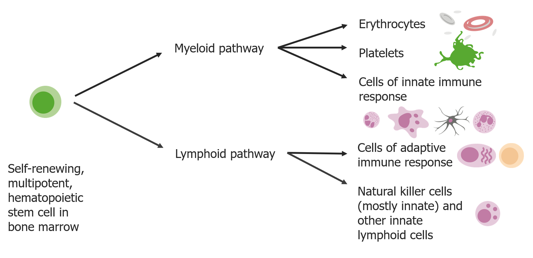
Las células madre se diferencian en 2 vías:
Las vías mieloides producen eritrocitos, plaquetas y células de la respuesta inmune innata. Las vías linfoides producen las células de respuesta adaptativa y los linfocitos asesinos naturales.
Los LOS Neisseria fagocitos “comen” el material extraño y ayudan a detectar, limpiar y reparar el tejido dañado, reconociendo patógenos a través de PRR u opsonización (por complemento o inmunoglobulinas).
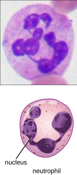
Neutrófilo: Granulocitos con núcleo multilobulado y gránulos finos ligeramente rosados
Imagen: “Granulocytes can be distinguished by the number of lobes in their nuclei and the staining properties of their granuless”. por Parker N et al. Licencia: CC BY 4.0, recortado por Lecturio.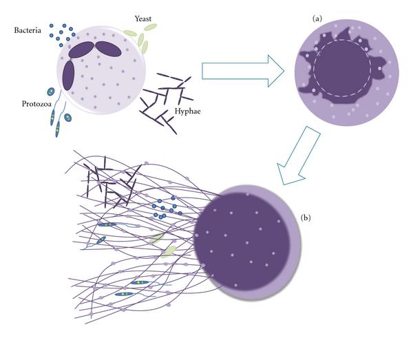
Mecanismo de liberación de la trampa extracelular de neutrófilos:
Los neutrófilos son estimulados por el contacto con bacterias, protozoos, hongos (formas de levaduras e hifas), lo que conduce a: (a) Alteraciones ultraestructurales de la forma nuclear con descondensación de la cromatina y membrana nuclear inflamada y fragmentada, que permite la asociación de gránulos y proteínas citoplasmáticas con la cromatina, y (b) liberación de estructuras extracelulares que consisten en un esqueleto de ADN, decorado con histonas, neutrófilos, proteínas granulares y citoplasmáticas, que atrapan y matan a los microorganismos.
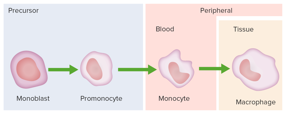
El desarrollo de monocitos comienza a partir de células madre hematopoyéticas y progresa a través de etapas hasta la unidad formadora de colonias de granulocitos-macrófagos:
El 1er precursor de monocitos es el monoblasto, que tiene un núcleo redondo u ovalado.
Sigue el promonocito y tiene un núcleo enrevesado.
El monocito surge con un núcleo dentado y se libera de la médula ósea para convertirse en un macrófago en los tejidos.
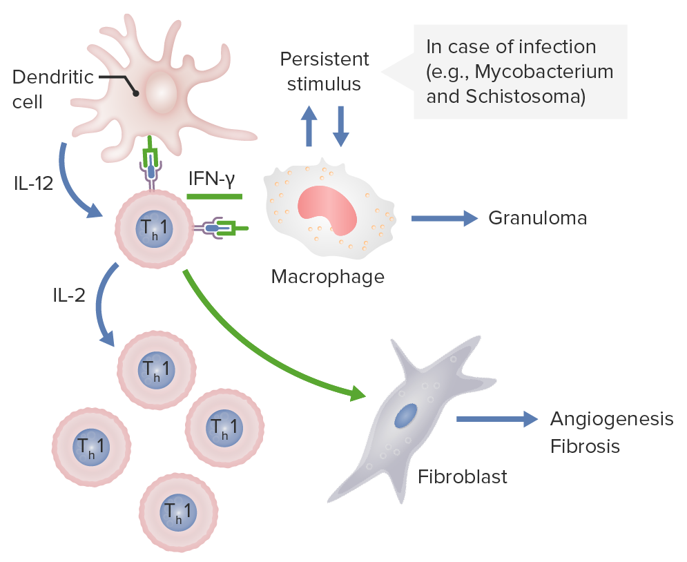
Las células dendríticas liberan IL-12, que activa las células CD4 Th1. Estas células Th1 producen IL-2, estimulando la producción de más subconjuntos de células T Th1. Las células Th1 también liberan IFN-γ, que activa los macrófagos y activa los fibroblastos para causar angiogénesis y fibrosis. Si estos macrófagos son estimulados persistentemente por patógenos como Mycobacterium y Schistosoma, se forman granulomas.
Imagen por Lecturio.Es importante tener en EN Erythema nodosum is an immune-mediated panniculitis (inflammation of the subcutaneous fat) caused by a type IV (delayed-type) hypersensitivity reaction. It commonly manifests in young women as tender, erythematous nodules on the shins. Erythema Nodosum cuenta que las células dendríticas foliculares no tienen ninguna relación con las células dendríticas en EN Erythema nodosum is an immune-mediated panniculitis (inflammation of the subcutaneous fat) caused by a type IV (delayed-type) hypersensitivity reaction. It commonly manifests in young women as tender, erythematous nodules on the shins. Erythema Nodosum linaje y función.
Células dendríticas foliculares:
| Células dendríticas | Células dendríticas foliculares | |
|---|---|---|
| Origen | Derivadas de células madre hematopoyéticas | Derivadas de células madre mesenquimatosas |
| Sitios | Presentes en EN Erythema nodosum is an immune-mediated panniculitis (inflammation of the subcutaneous fat) caused by a type IV (delayed-type) hypersensitivity reaction. It commonly manifests in young women as tender, erythematous nodules on the shins. Erythema Nodosum todo el cuerpo | Presentes solo en EN Erythema nodosum is an immune-mediated panniculitis (inflammation of the subcutaneous fat) caused by a type IV (delayed-type) hypersensitivity reaction. It commonly manifests in young women as tender, erythematous nodules on the shins. Erythema Nodosum centros germinales de tejidos linfoides secundarios |
| Clase de CMH y moléculas coestimuladoras | Poseen CMH II y moléculas coestimuladoras (e.g., B7) | Carecen de CMH II y moléculas coestimuladoras |
| Funciones |
|
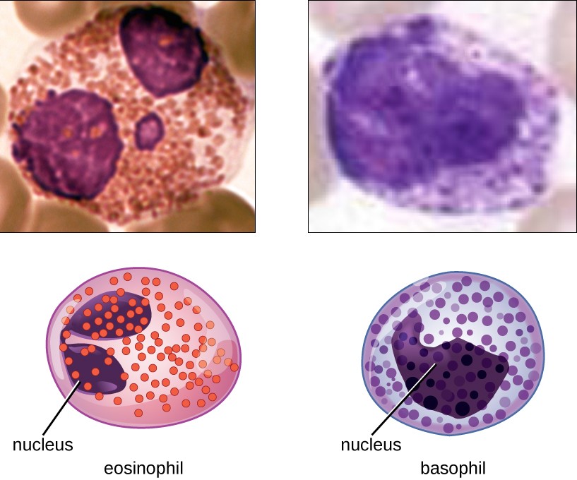
Eosinófilos y basófilos
Ambos son granulocitos, con eosinófilos que poseen un núcleo bilobulado y gránulos de color rosa oscuro y basófilos que tienen un núcleo bilobulado o trilobulado y gránulos de color azul oscuro.
Las células presentadoras de antígenos (como las células dendríticas y los LOS Neisseria macrófagos) detectan, procesan y presentan los LOS Neisseria antígenos a los LOS Neisseria linfocitos T, lo que permite que la inmunidad adaptativa reconozca y genere una respuesta cada vez que se encuentra el patógeno (memoria inmunológica).
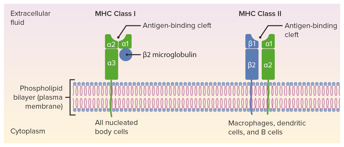
Estructuras de CMH I y CMH II:
El CMH I tiene 1 cadena corta y 1 cadena larga (cadena ɑ con 3 dominios: ɑ1, ɑ2 y ɑ3), asociada con la β₂-microglobulina. El CMH II tiene 2 cadenas ɑ y 2 β. El antígeno peptídico va a la hendidura de unión al antígeno.
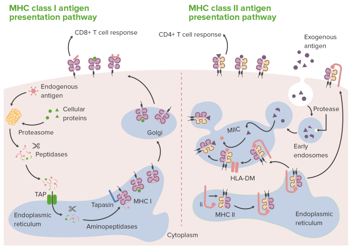
Vías de presentación de antígenos por moléculas CMH de clase I y II:
En la presentación de antígeno de clase I (izquierda), los proteosomas degradan antígenos proteçínas endógenos (dentro de la célula) en péptidos. Los fragmentos de péptidos se transportan (a través del transportador asociado con el procesamiento de antígenos) al retículo endoplasmático, donde las aminopeptidasas los recortan aún más y los cargan en la molécula CMH de clase I. Los complejos cargados con CMH de clase I van al aparato de Golgi para su modificación postraduccional. Luego, los complejos se transportan a la superficie celular, donde se presentan a los linfocitos T CD8+. En la presentación de antígenos de clase II (derecha), las células presentadoras de antígenos captan antígenos extracelulares/exógenos dentro de los fagosomas. A continuación, los fagosomas se fusionan con los lisosomas llenos de enzimas proteolíticas. Esto da lugar a la descomposición de las proteínas fagocitadas en pequeños péptidos. Mientras tanto, en el retículo endoplasmático se sintetizan nuevas moléculas de CMH de clase II. Estas moléculas tienen la cadena invariante (estructura rosa en la imagen de la derecha, marcada como Ii), que se une a la hendidura de unión al antígeno. Con la hendidura ocluida (por la cadena invariante), los péptidos residentes en el retículo endoplasmático no pueden unirse. La cadena invariante dirige el complejo del CMH II hacia el endosoma acidificado (donde están los péptidos del antígeno) al salir del retículo endoplasmático. Cuando los complejos de CMH II llegan al endosoma, la cadena invariante se libera, lo que permite la carga de los péptidos antigénicos (acompañados por una proteína, HLA-DM) en las moléculas CMH de clase II. Una vez cargados, los complejos antígeno-péptido-CMH de clase II son llevados a la superficie celular, listos para presentar el antígeno a los linfocitos T CD4+.
Ii: cadena invariante asociada a CMH de clase II
MIIC: compartimento CMH de clase II
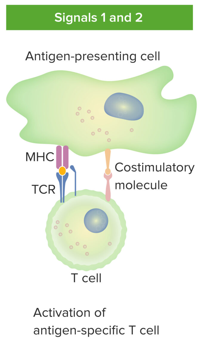
Interacción de células presentadoras de antígenos y linfocitos T:
La célula presentadora de antígenos interactúa con el linfocito T a través de la señal 1 (el receptor del linfocito T se une al antígeno afín presentado por la molécula CMH en la célula presentadora de antígeno) y la señal 2 (interacción de las moléculas coestimuladoras entre la célula presentadora de antígenos y el linfocito T). Con la presentación adecuada del antígeno, el linfocito T maduro se activa.
| CMH I | CMH II | |
|---|---|---|
| Loci | HLA-A HLA-A Polymorphic class I human histocompatibility (HLA) surface antigens present on almost all nucleated cells. At least 20 antigens have been identified which are encoded by the a locus of multiple alleles on chromosome 6. They serve as targets for t-cell cytolytic responses and are involved with acceptance or rejection of tissue/organ grafts. Organ Transplantation, HLA-B HLA-B Class I human histocompatibility (HLA) surface antigens encoded by more than 30 detectable alleles on locus B of the HLA complex, the most polymorphic of all the HLA specificities. Several of these antigens (e.g., hla-b27, -b7, -b8) are strongly associated with predisposition to rheumatoid and other autoimmune disorders. Like other class I HLA determinants, they are involved in the cellular immune reactivity of cytolytic T lymphocytes. Organ Transplantation, HLA-C HLA-C Class I human histocompatibility (HLA) antigens encoded by a small cluster of structural genes at the c locus on chromosome 6. They have significantly lower immunogenicity than the hla-a and -b determinants and are therefore of minor importance in donor/recipient crossmatching. Their primary role is their high-risk association with certain disease manifestations (e.g., spondyloarthritis, psoriasis, multiple myeloma). Organ Transplantation | HLA-DP, HLA-DQ, HLA-DR |
| Unión | Linfocito T CD8 | Linfocito T CD4 |
| Distribución | Todas las células nucleadas (ninguno en EN Erythema nodosum is an immune-mediated panniculitis (inflammation of the subcutaneous fat) caused by a type IV (delayed-type) hypersensitivity reaction. It commonly manifests in young women as tender, erythematous nodules on the shins. Erythema Nodosum los LOS Neisseria eritrocitos) | Células presentadoras de antígenos |
| Rol | Presentar antígenos endógenos a los LOS Neisseria linfocitos T CD8+ (citolíticos) | Presentar antígenos exógenos a los LOS Neisseria linfocitos T CD4+ |
| Estructura |
|
2 cadenas de igual longitud (2 ɑ, 2 β) |
| Proteína asociada | β₂-microglobulina | Cadena invariante |
| Carga de antígeno | Carga de péptido antigénico en EN Erythema nodosum is an immune-mediated panniculitis (inflammation of the subcutaneous fat) caused by a type IV (delayed-type) hypersensitivity reaction. It commonly manifests in young women as tender, erythematous nodules on the shins. Erythema Nodosum CMH I en EN Erythema nodosum is an immune-mediated panniculitis (inflammation of the subcutaneous fat) caused by a type IV (delayed-type) hypersensitivity reaction. It commonly manifests in young women as tender, erythematous nodules on the shins. Erythema Nodosum el retículo endoplasmático (administrado a través de TAP) | Carga de péptido antigénico en EN Erythema nodosum is an immune-mediated panniculitis (inflammation of the subcutaneous fat) caused by a type IV (delayed-type) hypersensitivity reaction. It commonly manifests in young women as tender, erythematous nodules on the shins. Erythema Nodosum CMH II en EN Erythema nodosum is an immune-mediated panniculitis (inflammation of the subcutaneous fat) caused by a type IV (delayed-type) hypersensitivity reaction. It commonly manifests in young women as tender, erythematous nodules on the shins. Erythema Nodosum el fagolisosoma acidificado después de la liberación de la cadena invariante |
La región HLA codifica varias moléculas que realizan funciones clave en EN Erythema nodosum is an immune-mediated panniculitis (inflammation of the subcutaneous fat) caused by a type IV (delayed-type) hypersensitivity reaction. It commonly manifests in young women as tender, erythematous nodules on the shins. Erythema Nodosum el sistema inmunológico. Existe una fuerte asociación entre la región HLA y varias enfermedades.
| Subtipo HLA | Afecciones |
|---|---|
| A3 | Hemocromatosis |
| B8 |
|
| B27 |
|
| C | Psoriasis Psoriasis Psoriasis is a common T-cell-mediated inflammatory skin condition. The etiology is unknown, but is thought to be due to genetic inheritance and environmental triggers. There are 4 major subtypes, with the most common form being chronic plaque psoriasis. Psoriasis |
| DQ2/DQ8 | Enfermedad celíaca |
| DR2 |
|
| DR3 |
|
| DR4 |
|
| DR5 | Tiroiditis de Hashimoto |
| DR7 | Síndrome nefrótico sensible a esteroides |