El hígado es la glándula más grande del cuerpo humano. El hígado se encuentra en EN Erythema nodosum is an immune-mediated panniculitis (inflammation of the subcutaneous fat) caused by a type IV (delayed-type) hypersensitivity reaction. It commonly manifests in young women as tender, erythematous nodules on the shins. Erythema Nodosum el cuadrante superior derecho del abdomen y pesa aproximadamente 1,5 kilogramos. Sus funciones principales son la desintoxicación, metabolismo (carbohidratos, proteínas, lípidos, hormonas), almacenamiento de nutrientes (e.g., hierro y vitaminas), síntesis de factores de coagulación, formación de bilis, filtración y almacenamiento de sangre. El hígado se puede dividir en EN Erythema nodosum is an immune-mediated panniculitis (inflammation of the subcutaneous fat) caused by a type IV (delayed-type) hypersensitivity reaction. It commonly manifests in young women as tender, erythematous nodules on the shins. Erythema Nodosum 4 lóbulos u 8 segmentos. Microscópicamente, se divide en EN Erythema nodosum is an immune-mediated panniculitis (inflammation of the subcutaneous fat) caused by a type IV (delayed-type) hypersensitivity reaction. It commonly manifests in young women as tender, erythematous nodules on the shins. Erythema Nodosum lobulillos hepáticos. Su haz neurovascular principal se encuentra dentro de la fisura transversa del hígado, también llamada porta hepatis Porta hepatis Liver: Anatomy.
Last updated: Dec 15, 2025
El hígado es la glándula más grande del cuerpo. Se extiende desde la región hipocondríaca derecha a la izquierda (¾ del hígado está en EN Erythema nodosum is an immune-mediated panniculitis (inflammation of the subcutaneous fat) caused by a type IV (delayed-type) hypersensitivity reaction. It commonly manifests in young women as tender, erythematous nodules on the shins. Erythema Nodosum el cuadrante superior derecho).
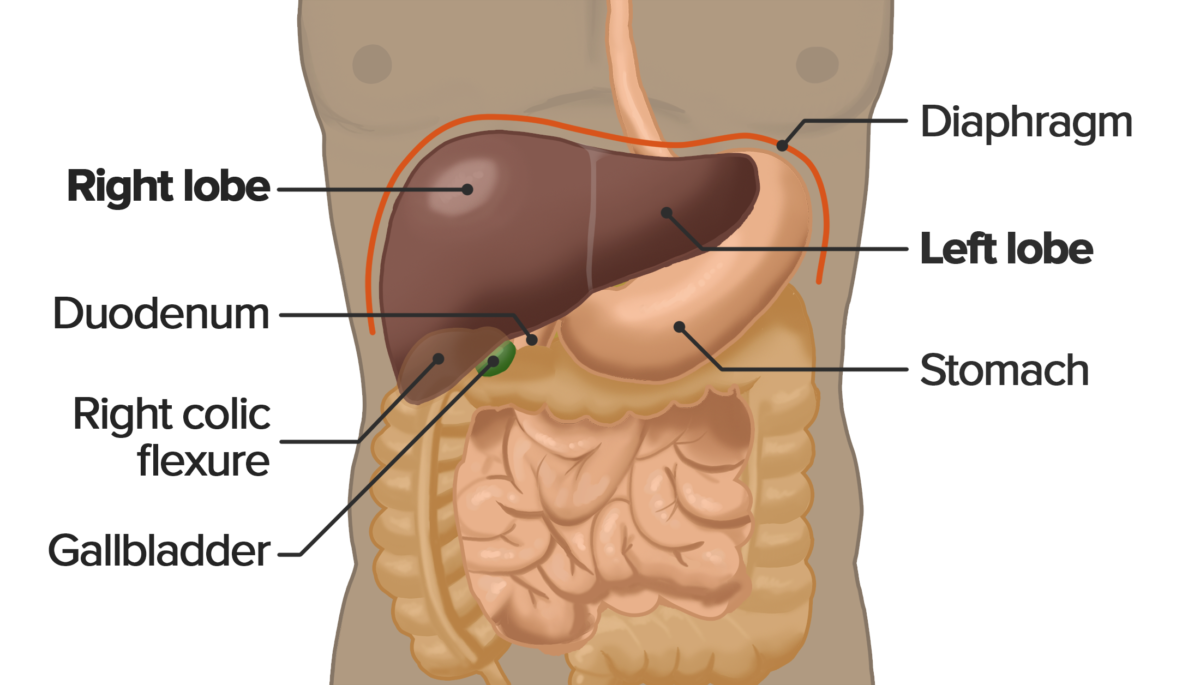
Localización del hígado dentro de la cavidad abdominal
Imagen por Lecturio.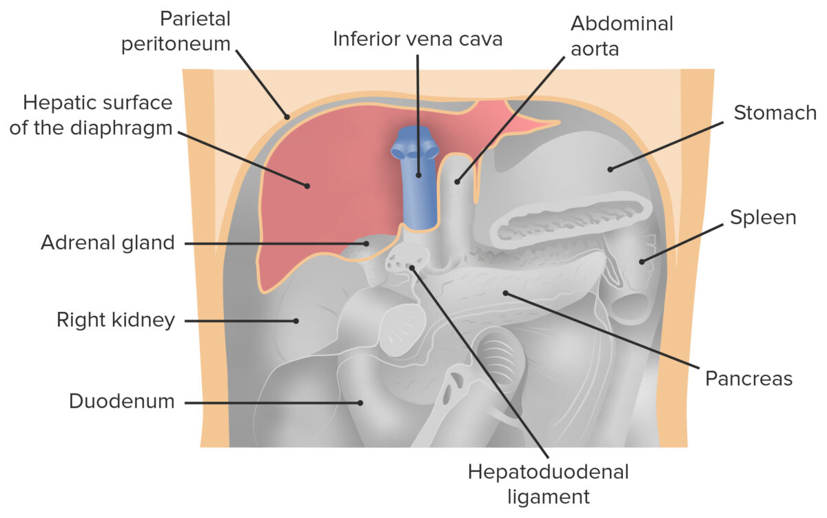
Vista frontal de la cavidad abdominal con el hígado removido. La imagen muestra la posición del hígado en la cavidad peritoneal en relación con los órganos vecinos.
Imagen por Lecturio.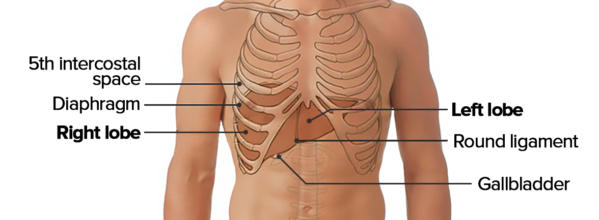
Topografía del hígado
Imagen por Lecturio.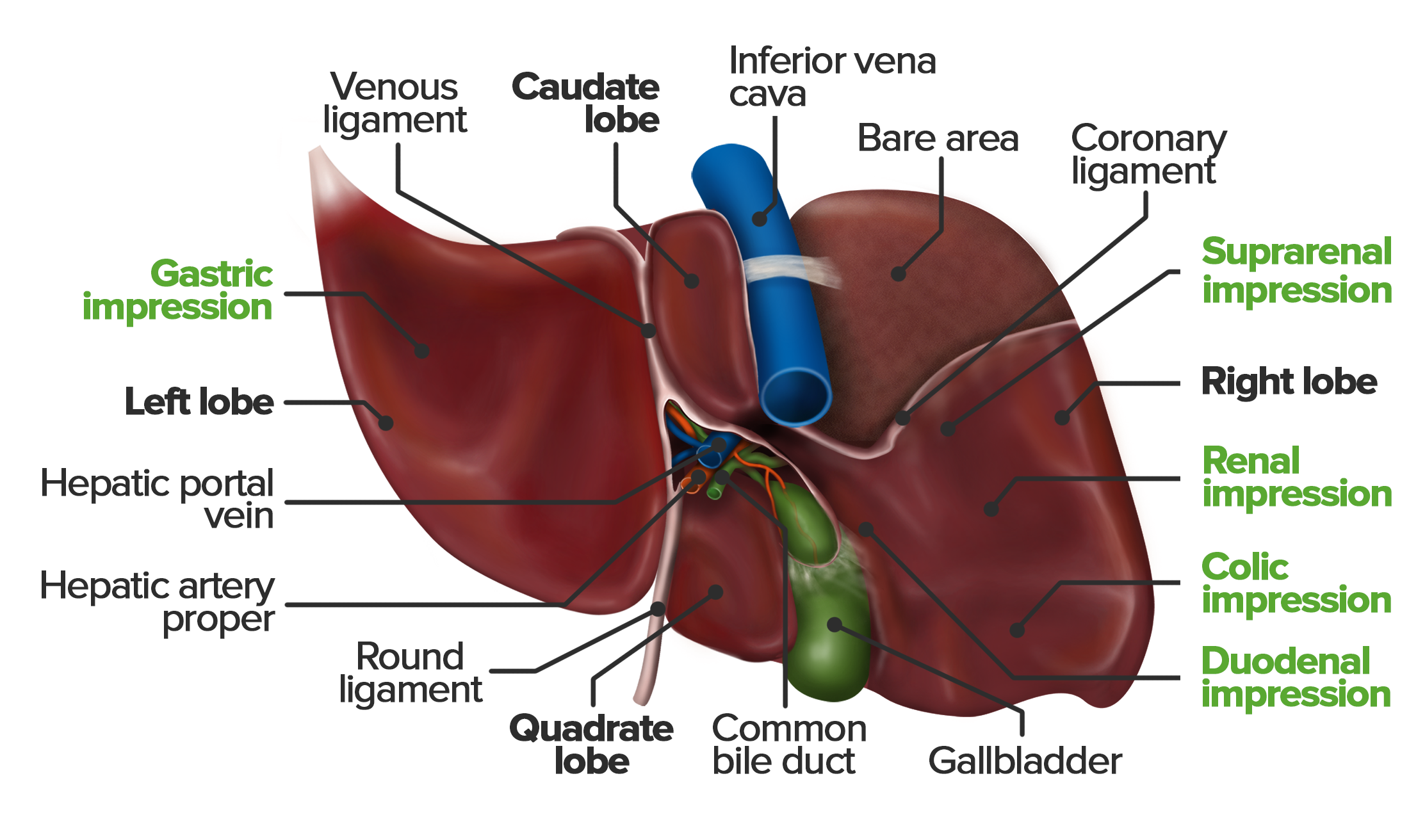
Vista inferior de la superficie visceral del hígado. Obsérvese la estructura desigual que resulta de las impresiones de los órganos vecinos. La impresión cólica es causada por la flexura hepática del colon; la porción descendente del duodeno forma la impresión duodenal.
Imagen por Lecturio.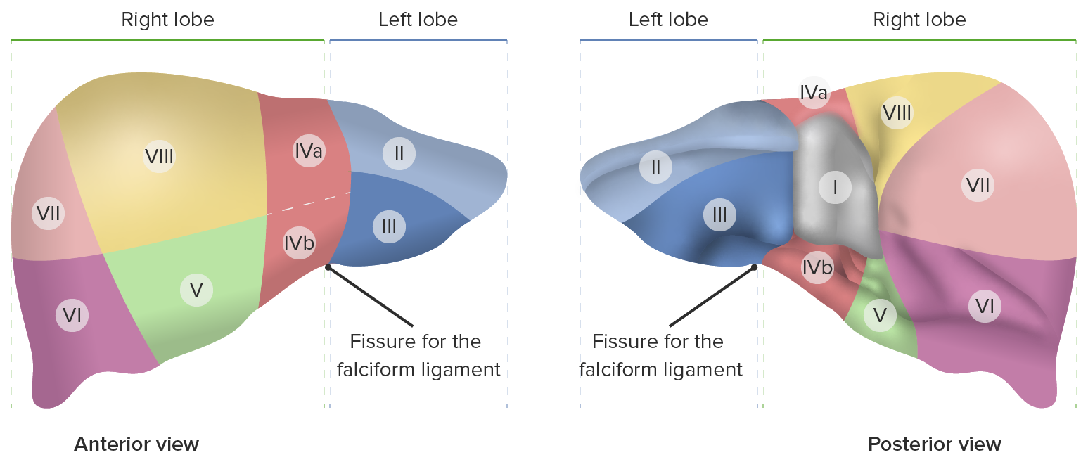
Ocho segmentos hepáticos según el sistema de clasificación de Couinaud.
Imagen por Lecturio.El porta hepatis Porta hepatis Liver: Anatomy (también llamado portal hepático) es una fisura transversal que separa los LOS Neisseria lóbulos caudado y cuadrado y sirve como vía de paso para lo siguiente:
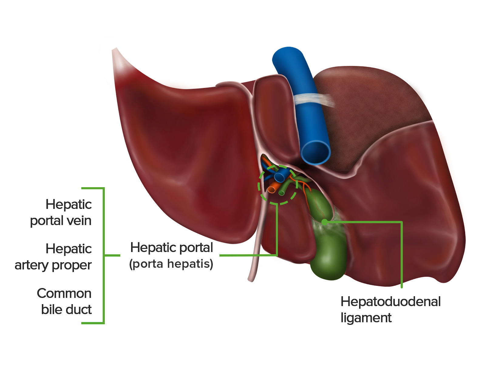
Vista inferior de la superficie visceral del hígado, mostrando el portal hepático y el ligamento hepatoduodenal circundante.
Imagen por Lecturio.Definición: Los LOS Neisseria ligamentos del hígado son capas dobles de peritoneo visceral que fijan la posición del hígado al AL Amyloidosis unirlo a las estructuras circundantes.
|
Ligamentos coronarios (anterior y posterior) |
Reflexión peritoneal desde el diafragma hasta el hígado. Demarca el área desnuda (superficie del hígado sin cubierta peritoneal) |
| Ligamento falciforme |
Reflexión peritoneal desde el ombligo hasta el hígado Remanente del mesenterio ventral embrionario Su borde libre contiene el ligamento redondo del hígado. |
| Ligamento hepatoduodenal |
Porción del epiplón menor Se extiende desde la porta hepatis Porta hepatis Liver: Anatomy hasta la parte superior del duodeno Contenido: arteria hepática propiamente dicha, vena porta, conducto hepático común |
| Ligamento hepatogástrico |
Se extiende desde el hígado hasta la curvatura menor del estómago Contenido: arterias gástricas |
|
Ligamento redondo (también conocido como ligamentum teres Ligamentum teres A cord-like remnant structure formed from the closed left fetal umbilical vein. It is located along the lower edge of the falciform ligament. Liver: Anatomy) |
Remanente de la porción intraabdominal de la vena umbilical Se extiende desde el ombligo hasta el hígado en EN Erythema nodosum is an immune-mediated panniculitis (inflammation of the subcutaneous fat) caused by a type IV (delayed-type) hypersensitivity reaction. It commonly manifests in young women as tender, erythematous nodules on the shins. Erythema Nodosum el borde libre del ligamento falciforme |
| Ligamentos triangulares |
Formado por la fusión de
los
LOS
Neisseria pliegues anterior y posterior del ligamento coronario 1 izquierdo y 1 derecho; ambos se extienden desde el hígado hasta el diafragma |
|
Ligamento venoso (también conocido como ligamentum venosum Ligamentum Venosum Liver: Anatomy) |
Remanente del conducto venoso Se extiende desde el remanente de la porción intraabdominal de la vena umbilical hasta la vena cava inferior |
| Fisura izquierda | Impresiones de los LOS Neisseria ligamentos redondo y venoso |
| Fisura derecha | Impresiones de la vesícula biliar y la vena cava inferior |
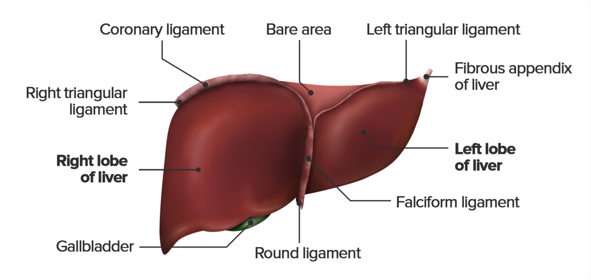
Vista anterior de la superficie diafragmática del hígado, mostrando los ligamentos falciforme, triangular, redondo y coronario. Observe que el ligamento redondo se extiende desde el borde libre del ligamento falciforme.
Imagen por Lecturio.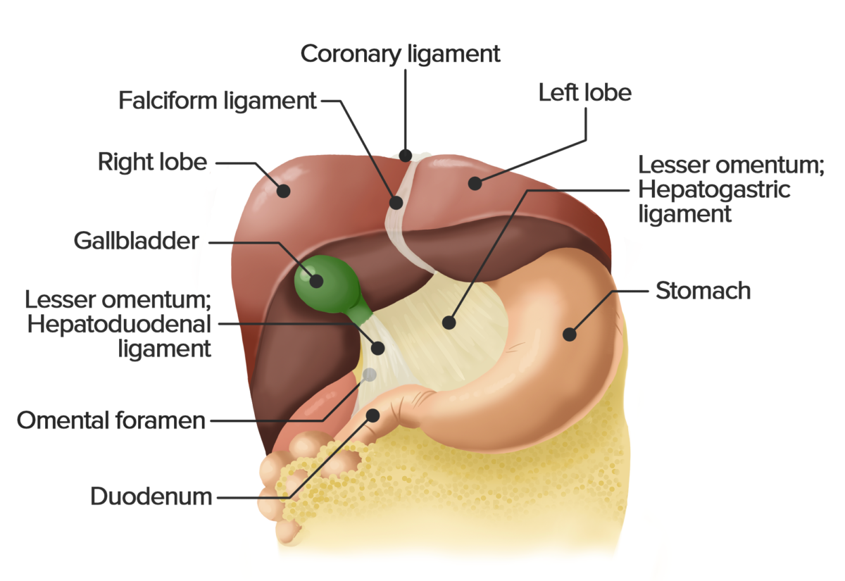
Vista anterior del hígado. Se levantó el hígado para mostrar el epiplón menor, que consiste en el ligamento hepatogástrico y hepatoduodenal. Esta doble capa de peritoneo conecta el hígado con la curvatura menor del estómago y el duodeno.
Imagen por Lecturio.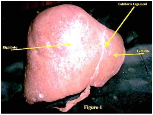
Vista anterosuperior de un hígado humano
Imagen: “Hypoplastic left lobe of liver with accessory caudate lobe.” por Department of Anatomy, AIIMS, Rishikesh, Uttarakhand 249201, India. Licencia: CC BY 3.0
Vista posteroinferior de un hígado
Imagen: “Hypoplastic left lobe of liver with accessory caudate lobe.” por Department of Anatomy, AIIMS, Rishikesh, Uttarakhand 249201, India. Licencia: CC BY 3.0
Diagrama esquemático de la arquitectura del hígado, mostrando el lobulillo hepático. Las tríadas portales en las esquinas consisten en ramas de la vena porta, la arteria hepática propiamente dicha y un conducto biliar. La rama de la vena porta transporta sangre rica en nutrientes, pero desoxigenada del intestino delgado, la rama de la arteria hepática irriga sangre oxigenada a los hepatocitos. El conducto biliar drena la bilis de los hepatocitos hacia los conductos más grandes y la vesícula biliar.
Imagen por Lecturio.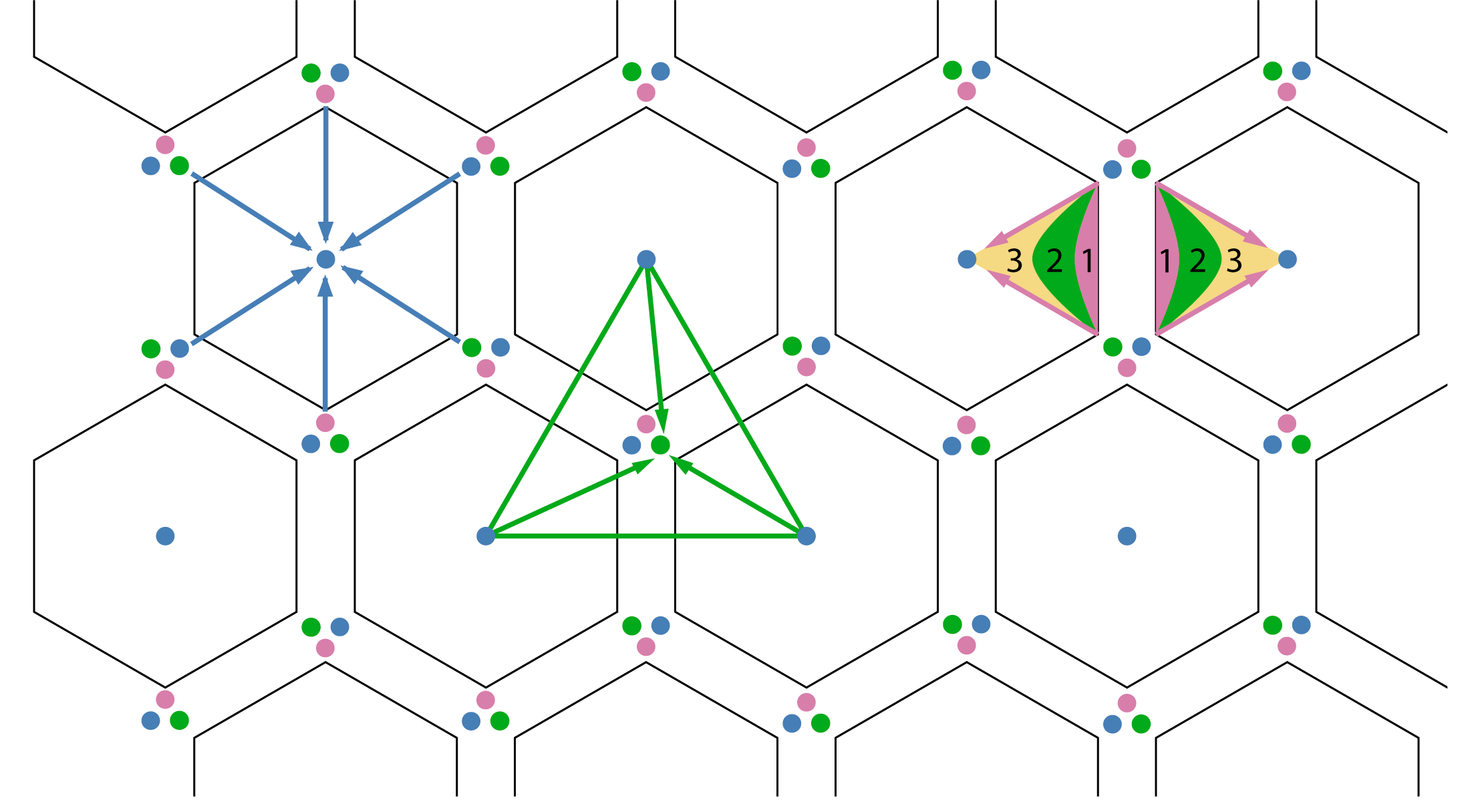
Diagrama esquemático de los 3 tipos de unidades hepáticas. Observe las tríadas portales ubicadas en los vértices de las unidades hexagonales y las 3 zonas histológicas dentro del acino hepático.
Imagen por Lecturio.
Representación esquemática de un sinusoide y placa de hepatocitos separados por el espacio de Disse. Observe las células especializadas del hígado: células de Kupffer, Pit y Stellate.
Imagen por Lecturio.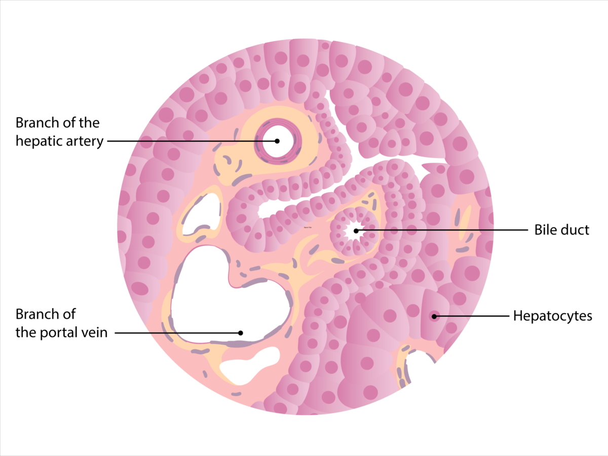
Diagrama esquemático de la histología del hígado. Observe que la rama de la vena porta se puede identificar por la luz ancha y la pared delgada de células de músculo liso. La rama de la arteria hepática tiene un calibre más pequeño rodeada por una capa más gruesa de músculo liso. Las paredes del conducto biliar contienen células epiteliales en forma cuboide (colangiocitos), que lo distinguen de la arteria hepática.
Imagen por Lecturio.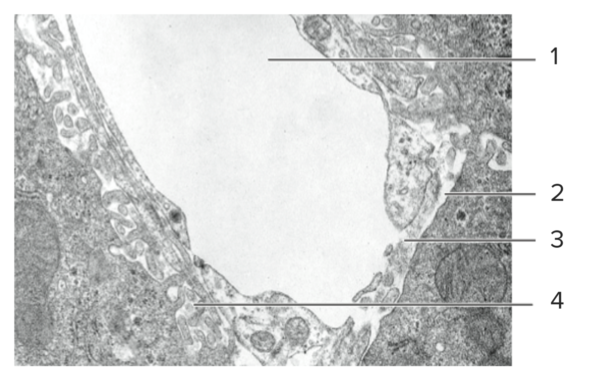
Corte histológico de tejido hepático mostrando un sinusoide (1), su lámina basal discontinua (2), endotelio fenestrado (3) y el espacio perisinusoidal de Disse (4).
Imagen por Lecturio.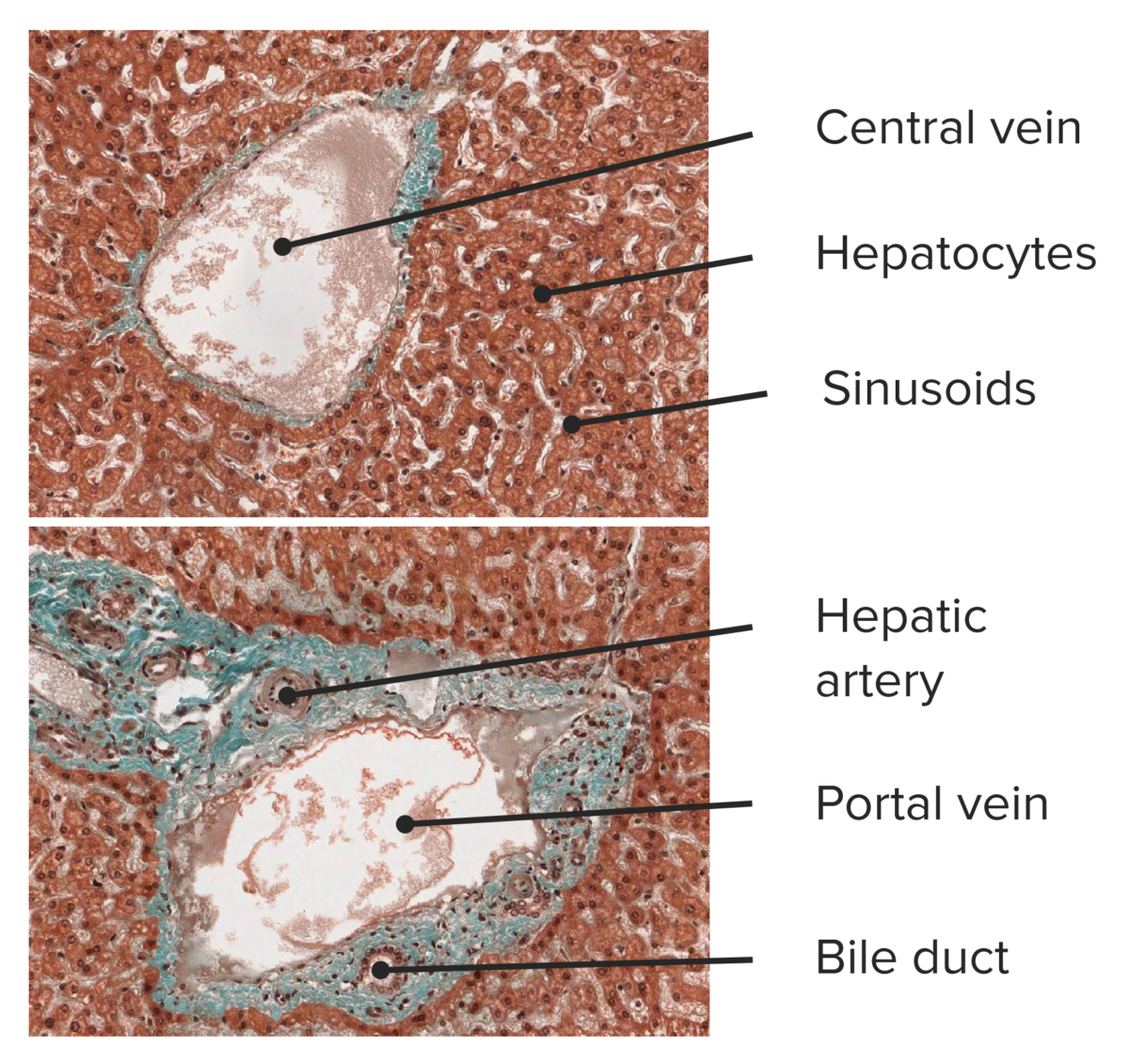
Corte histológico de tejido hepático, mostrando una vena central (arriba) y una tríada portal (abajo).
Imagen por Lecturio.El hígado tiene un suministro de sangre dual especial que proporciona una mezcla de sangre oxigenada, desoxigenada y rica en EN Erythema nodosum is an immune-mediated panniculitis (inflammation of the subcutaneous fat) caused by a type IV (delayed-type) hypersensitivity reaction. It commonly manifests in young women as tender, erythematous nodules on the shins. Erythema Nodosum nutrientes.
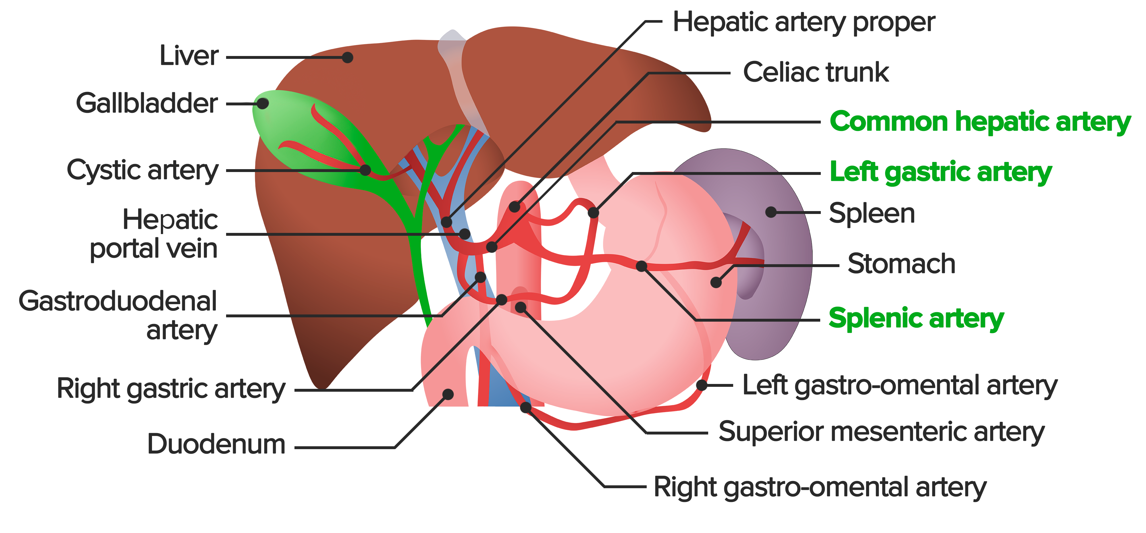
Descripción general de la irrigación arterial abdominal. El tronco celíaco es la primera rama principal de la aorta abdominal. Irriga sangre oxigenada al hígado, estómago, bazo, páncreas y partes del esófago y duodeno. El tronco celíaco da origen a la arteria gástrica izquierda, arteria esplénica y arteria hepática común. La arteria hepática común se divide en la arteria hepática propiamente dicha, arteria gastroduodenal y arteria gástrica derecha, todas las cuales se pueden ver aquí.
Imagen por Lecturio.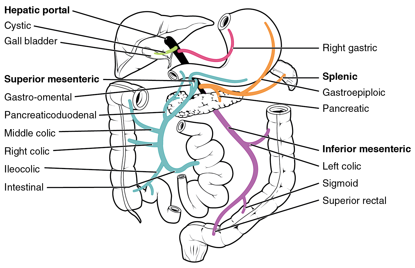
Diagrama del sistema portal venoso. La vena porta hepática se forma más comúnmente por la unión de la vena esplénica y las venas mesentéricas superiores. Otras tributarias incluyen las venas mesentérica inferior, cística y gástrica izquierda y derecha. En su conjunto, el sistema portal recoge el drenaje venoso del bazo, estómago, vesícula biliar, intestino delgado y grueso y el páncreas.
Imagen: “Hepatic Portal Vein System” por OpenStax College. Licencia: CC BY 3.0Ganglios linfáticos hepáticos: localizados alrededor de la porta hepatis Porta hepatis Liver: Anatomy → grupo celíaco de ganglios linfáticos → cisterna chyli (saco dilatado que recibe la linfa del tronco gastrointestinal y 2 troncos linfáticos lumbares) → conducto torácico
Canalículos biliares → conductos biliares intrahepáticos → conductos hepáticos izquierdo y derecho → conducto hepático común → conducto biliar común → duodeno
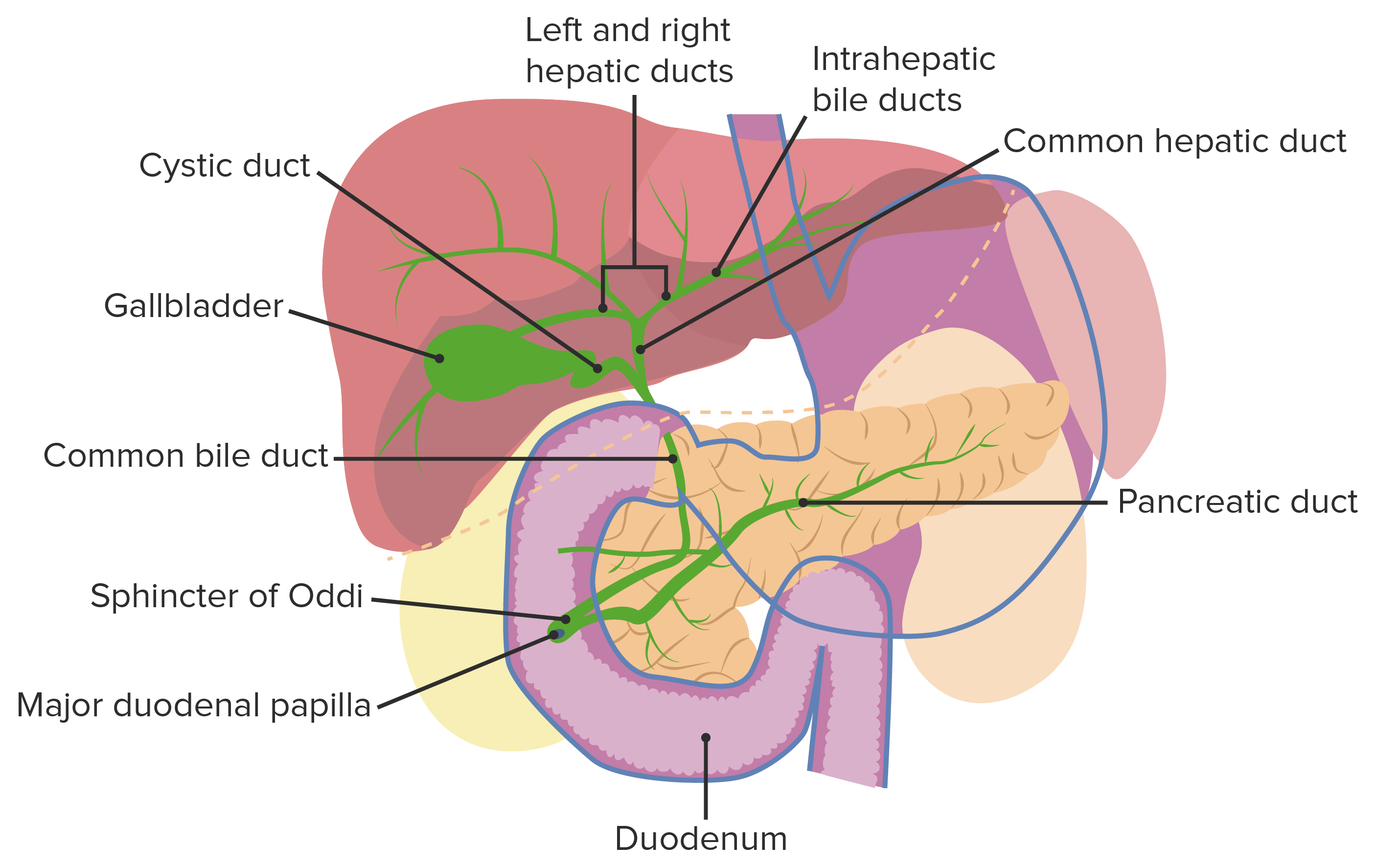
Vesícula biliar y vías biliares
Imagen por Lecturio.El hígado elimina los LOS Neisseria productos de degradación obtenidos a través de la resorción del tracto gastrointestinal. Hace HACE Altitude Sickness que las sustancias liposolubles sean solubles en EN Erythema nodosum is an immune-mediated panniculitis (inflammation of the subcutaneous fat) caused by a type IV (delayed-type) hypersensitivity reaction. It commonly manifests in young women as tender, erythematous nodules on the shins. Erythema Nodosum agua a través de la modificación enzimática. Esto permite la excreción a través de las vías biliares o a través de la orina.

Diagrama esquemático que muestra las diversas vías metabólicas en las que participa el hígado
Imagen por Lecturio.Neoplasias
Infecciones
Trastornos inflamatorios
Trastornos hereditarios
Trastornos varios