El derrame pleural se refiere a la acumulación de líquido entre las capas de la pleuraPleuraThe pleura is a serous membrane that lines the walls of the thoracic cavity and the surface of the lungs. This structure of mesodermal origin covers both lungs, the mediastinum, the thoracic surface of the diaphragm, and the inner part of the thoracic cage. The pleura is divided into a visceral pleura and parietal pleura. Pleura: AnatomyparietalParietalOne of a pair of irregularly shaped quadrilateral bones situated between the frontal bone and occipital bone, which together form the sides of the cranium.Skull: Anatomy y visceral. Las causas más comunes de esta afección son las infecciones, losLOSNeisseria tumores malignos, losLOSNeisseria trastornos autoinmunes o la sobrecarga de volumen. Las manifestaciones clínicas incluyen dolorDolorInflammation torácico, tosTOSThoracic outlet syndrome (TOS) is a broad term used for a spectrum of syndromes related to the general region of the thoracic outlet, which involves the compression or irritation of elements of the brachial plexus, subclavian artery, or subclavian vein.Thoracic Outlet Syndrome y disnea. La imagenología puede confirmar la presencia de un derrame pleural y el análisis del líquido pleural puede ayudar a evaluar la etiología. El tratamiento depende de la condición subyacente y de la presencia de dificultad respiratoria. El drenaje del derrame puede proporcionar un alivio sintomático.
El derrame pleural es una acumulación excesiva de líquido dentro de la cavidad pleural (entre la pleuraPleuraThe pleura is a serous membrane that lines the walls of the thoracic cavity and the surface of the lungs. This structure of mesodermal origin covers both lungs, the mediastinum, the thoracic surface of the diaphragm, and the inner part of the thoracic cage. The pleura is divided into a visceral pleura and parietal pleura. Pleura: AnatomyparietalParietalOne of a pair of irregularly shaped quadrilateral bones situated between the frontal bone and occipital bone, which together form the sides of the cranium.Skull: Anatomy y visceral).
LDHLDHOsteosarcomaenENErythema nodosum is an immune-mediated panniculitis (inflammation of the subcutaneous fat) caused by a type IV (delayed-type) hypersensitivity reaction. It commonly manifests in young women as tender, erythematous nodules on the shins.Erythema Nodosum líquido pleural > dos-tercios del límite superior de la normalidad para la LDHLDHOsteosarcoma sérica
Si no se cumplen estos 3 criterios, el derrame pleural se considera transudativo.
Etiología
Causas comunes de transudado:
Insuficiencia cardíaca
Cirrosis hepática
Hipoalbuminemia
Síndrome nefrótico
Causas comunes de exudado:
Neumonía
TBTBTuberculosis (TB) is an infectious disease caused by Mycobacterium tuberculosis complex bacteria. The bacteria usually attack the lungs but can also damage other parts of the body. Approximately 30% of people around the world are infected with this pathogen, with the majority harboring a latent infection. Tuberculosis spreads through the air when a person with active pulmonary infection coughs or sneezes. Tuberculosis
TumorTumorInflammation maligno (frecuentemente un cáncer de pulmón primario)
Enfermedades del tejido conectivo
PancreatitisPancreatitisInflammation of the pancreas. Pancreatitis is classified as acute unless there are computed tomographic or endoscopic retrograde cholangiopancreatographic findings of chronic pancreatitis. The two most common forms of acute pancreatitis are alcoholic pancreatitis and gallstone pancreatitis.Acute Pancreatitis
LosLOSNeisseria derrames pleurales representan una alteración entre la producción de líquido pleural y la reabsorción linfática.
Fisiología normal
El líquido pleural es un producto de las fuerzas de Starling dentro del lecho capilar de la pleuraPleuraThe pleura is a serous membrane that lines the walls of the thoracic cavity and the surface of the lungs. This structure of mesodermal origin covers both lungs, the mediastinum, the thoracic surface of the diaphragm, and the inner part of the thoracic cage. The pleura is divided into a visceral pleura and parietal pleura. Pleura: AnatomyparietalParietalOne of a pair of irregularly shaped quadrilateral bones situated between the frontal bone and occipital bone, which together form the sides of the cranium.Skull: Anatomy y es absorbido por losLOSNeisseria vasos linfáticos de las superficies diafragmática y mediastínica de la pleuraPleuraThe pleura is a serous membrane that lines the walls of the thoracic cavity and the surface of the lungs. This structure of mesodermal origin covers both lungs, the mediastinum, the thoracic surface of the diaphragm, and the inner part of the thoracic cage. The pleura is divided into a visceral pleura and parietal pleura. Pleura: AnatomyparietalParietalOne of a pair of irregularly shaped quadrilateral bones situated between the frontal bone and occipital bone, which together form the sides of the cranium.Skull: Anatomy.
La tasa media normal de producción y absorción del líquido pleural es de 0,2 mL/kg/hora.
Todo el volumen de líquido pleural suele reemplazarse enENErythema nodosum is an immune-mediated panniculitis (inflammation of the subcutaneous fat) caused by a type IV (delayed-type) hypersensitivity reaction. It commonly manifests in young women as tender, erythematous nodules on the shins.Erythema Nodosum 1 hora.
LosLOSNeisseria vasos linfáticos pueden manejar un flujo de hasta aproximadamente 20 veces más que la tasa de producción normal → la reabsorción linfática tiene una gran capacidad de reserva
Derrames transudativos
Un derrame pleural transudativo puede ser el resultado de una mayor entrada de líquido enENErythema nodosum is an immune-mediated panniculitis (inflammation of the subcutaneous fat) caused by a type IV (delayed-type) hypersensitivity reaction. It commonly manifests in young women as tender, erythematous nodules on the shins.Erythema Nodosum el espacio pleural debido a:
↑ Presión hidrostática enENErythema nodosum is an immune-mediated panniculitis (inflammation of the subcutaneous fat) caused by a type IV (delayed-type) hypersensitivity reaction. It commonly manifests in young women as tender, erythematous nodules on the shins.Erythema Nodosum la vasculatura (e.g., insuficiencia cardíaca)
↓ Fuerzas oncóticas enENErythema nodosum is an immune-mediated panniculitis (inflammation of the subcutaneous fat) caused by a type IV (delayed-type) hypersensitivity reaction. It commonly manifests in young women as tender, erythematous nodules on the shins.Erythema Nodosum el plasmaPlasmaThe residual portion of blood that is left after removal of blood cells by centrifugation without prior blood coagulation.Transfusion Products (e.g., hipoalbuminemia)
Movimiento de líquido ascítico a través del diafragma (e.g., hidrotórax hepático)
Derrames exudativos
Un derrame pleural exudativo puede ser el resultado de:
TosTOSThoracic outlet syndrome (TOS) is a broad term used for a spectrum of syndromes related to the general region of the thoracic outlet, which involves the compression or irritation of elements of the brachial plexus, subclavian artery, or subclavian vein.Thoracic Outlet Syndrome
Expansión torácica asimétrica (expansión reducida enENErythema nodosum is an immune-mediated panniculitis (inflammation of the subcutaneous fat) caused by a type IV (delayed-type) hypersensitivity reaction. It commonly manifests in young women as tender, erythematous nodules on the shins.Erythema Nodosum el lado del derrame)
La tráquea se desplaza hacia el lado opuesto del derrame.
Se veVEVentilation: Mechanics of BreathingenENErythema nodosum is an immune-mediated panniculitis (inflammation of the subcutaneous fat) caused by a type IV (delayed-type) hypersensitivity reaction. It commonly manifests in young women as tender, erythematous nodules on the shins.Erythema Nodosum derrames grandes y severos
Auscultación:
Sonidos respiratorios ↓ o inaudibles enENErythema nodosum is an immune-mediated panniculitis (inflammation of the subcutaneous fat) caused by a type IV (delayed-type) hypersensitivity reaction. It commonly manifests in young women as tender, erythematous nodules on the shins.Erythema Nodosum el derrame
Ruidos respiratorios bronquiales, broncofonía y egofonía:
Se escucha sobre las partes del pulmón directamente por encima del derrame
Debido a la consolidación del pulmón enENErythema nodosum is an immune-mediated panniculitis (inflammation of the subcutaneous fat) caused by a type IV (delayed-type) hypersensitivity reaction. It commonly manifests in young women as tender, erythematous nodules on the shins.Erythema Nodosum esa zona
Fricción pleural
Percusión:
EnENErythema nodosum is an immune-mediated panniculitis (inflammation of the subcutaneous fat) caused by a type IV (delayed-type) hypersensitivity reaction. It commonly manifests in young women as tender, erythematous nodules on the shins.Erythema Nodosum el caso de un derrame > 300 mL, la exploración del tórax también presentará matidez a la percusión.
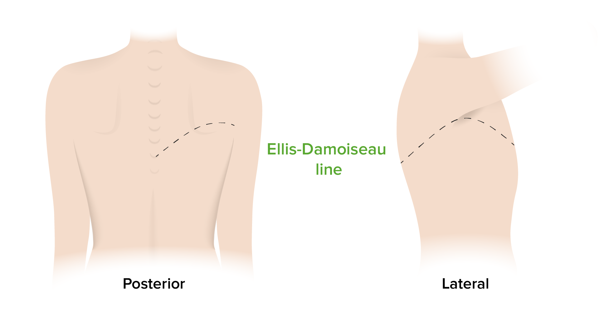
La matidez del borde superior sigue una curva que asciende lateralmente y que tiene su ápice enENErythema nodosum is an immune-mediated panniculitis (inflammation of the subcutaneous fat) caused by a type IV (delayed-type) hypersensitivity reaction. It commonly manifests in young women as tender, erythematous nodules on the shins.Erythema Nodosum la línea axilar media (línea Ellis-Damoiseau).
Línea Ellis-Damoiseau: Este dibujo representa la forma que seguirá la matidez a la percusión al evaluar un derrame pleural.
Imagen por Lecturio.
Indicios de una etiología subyacente
LosLOSNeisseria derrames paraneumónicos (adyacentes a una neumonía) pueden presentarse con signos de sepsisSepsisSystemic inflammatory response syndrome with a proven or suspected infectious etiology. When sepsis is associated with organ dysfunction distant from the site of infection, it is called severe sepsis. When sepsis is accompanied by hypotension despite adequate fluid infusion, it is called septic shock.Sepsis and Septic Shock o shockShockShock is a life-threatening condition associated with impaired circulation that results in tissue hypoxia. The different types of shock are based on the underlying cause: distributive (↑ cardiac output (CO), ↓ systemic vascular resistance (SVR)), cardiogenic (↓ CO, ↑ SVR), hypovolemic (↓ CO, ↑ SVR), obstructive (↓ CO), and mixed. Types of Shock séptico.
Fiebre
Taquicardia
Hipotensión
LosLOSNeisseria derrames transudativos pueden estar asociados a:
Aumento de peso
EdemaEdemaEdema is a condition in which excess serous fluid accumulates in the body cavity or interstitial space of connective tissues. Edema is a symptom observed in several medical conditions. It can be categorized into 2 types, namely, peripheral (in the extremities) and internal (in an organ or body cavity). Edema periférico
Ingurgitación yugular
LosLOSNeisseria derrames malignos pueden estar asociados a:
LosLOSNeisseria derrames pleurales suelen detectarse fácilmente enENErythema nodosum is an immune-mediated panniculitis (inflammation of the subcutaneous fat) caused by a type IV (delayed-type) hypersensitivity reaction. It commonly manifests in young women as tender, erythematous nodules on the shins.Erythema NodosumlosLOSNeisseria estudios de imagen.
Radiografía de tórax:
Mejor prueba inicial
Hallazgos:
Embotamiento anormal de losLOSNeisseria ángulos costodiafragmáticos
Líquido dentro de las fisuras horizontales u oblicuas
Algunos derrames pueden mostrar un menisco
Derrames masivos
Opacificación completa de un hemitórax
Desviación de la tráquea enENErythema nodosum is an immune-mediated panniculitis (inflammation of the subcutaneous fat) caused by a type IV (delayed-type) hypersensitivity reaction. It commonly manifests in young women as tender, erythematous nodules on the shins.Erythema Nodosum sentido contrario alALAmyloidosis lado afectado
Desplazamiento del mediastino
Radiografías enENErythema nodosum is an immune-mediated panniculitis (inflammation of the subcutaneous fat) caused by a type IV (delayed-type) hypersensitivity reaction. It commonly manifests in young women as tender, erythematous nodules on the shins.Erythema Nodosum decúbito lateral
Más sensibles
Pueden demostrar la estratificación del líquido (derrame pleural de flujo libre)
TC de tórax:
Puede detectar pequeñas cantidades de líquido pleural
Capaz de evaluar todo el parénquima pulmonar y el mediastino enENErythema nodosum is an immune-mediated panniculitis (inflammation of the subcutaneous fat) caused by a type IV (delayed-type) hypersensitivity reaction. It commonly manifests in young women as tender, erythematous nodules on the shins.Erythema Nodosum busca de posibles etiologías
Ultrasonido:
Alta sensibilidad para el diagnóstico de derrames pleurales
Puede detectar pequeñas cantidades de líquido pleural que pueden pasar desapercibidas enENErythema nodosum is an immune-mediated panniculitis (inflammation of the subcutaneous fat) caused by a type IV (delayed-type) hypersensitivity reaction. It commonly manifests in young women as tender, erythematous nodules on the shins.Erythema Nodosum un examen radiológico
A menudo se utiliza para visualizar el derrame para la realización de la toracocentesis o el drenaje pleural
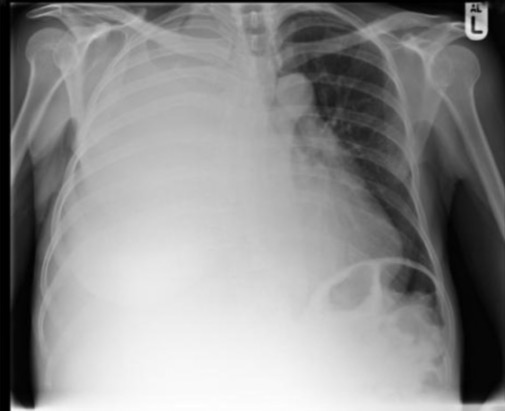
Imagen radiográfica de un derrame pleural masivo del lado derecho con opacificación completa del hemitórax derecho y desviación traqueal hacia la izquierda
Imagen: “X-ray of patient’s chest revealing a right-sided pleural effusion” por Department of Gastroenterology, Queen’s Hospital, Burton-on-Trent, West Midlands, UK. Licencia: CC BY 2.0
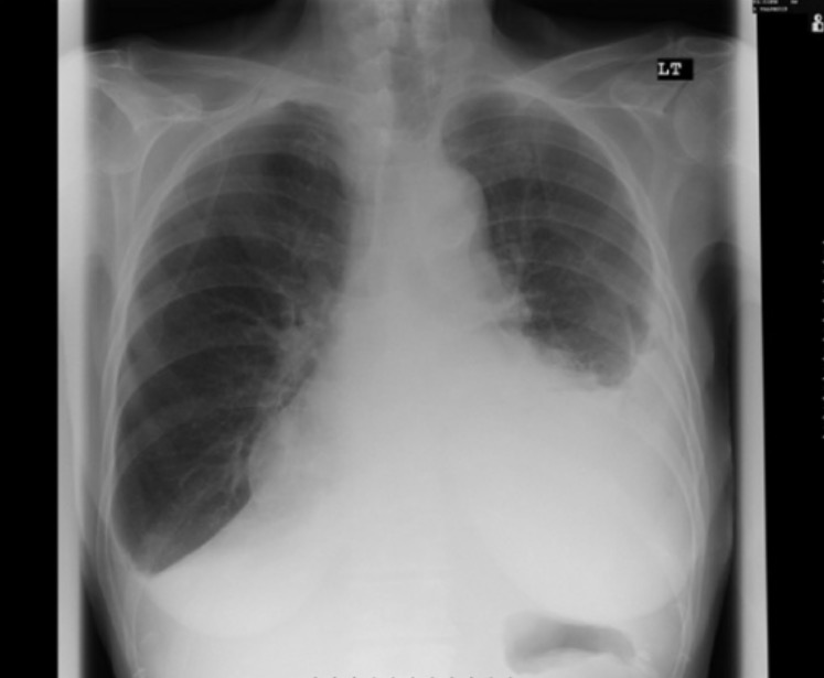
Imagen radiográfica que muestra derrames pleurales bilaterales: Obsérvese el embotamiento del ángulo costodiafragmático (sobre todo en la derecha) y la aparición de un menisco en la izquierda.
Imagen: “Pleural effusion while being on carbimazole” por Department of Diabetes and Endocrinology, Glan Clwyd Hospital, Rhuddlan Road, Bodelwyddan, Rhyl LL18 5UJ, UK. Licencia: CC BY 3.0
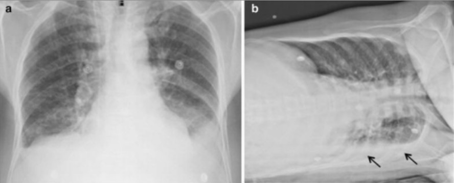
Imágenes de radiografía que demuestran derrames pleurales bilaterales: a: Se muestra el embotamiento de ambos ángulos costodiafragmáticos. b: Una imagen en decúbito lateral muestra la estratificación de líquido debido a un derrame pleural izquierdo (flechas).
Imagen: “Radiologic findings” por Departamento de Clínica Médica, Faculdade de Medicina de Ribeirão Preto, Universidade de São Paulo (USP), Ribeirão Preto, São Paulo, 14049-900, Brazil. Licencia: CC BY 4.0, recortada por Lecturio.
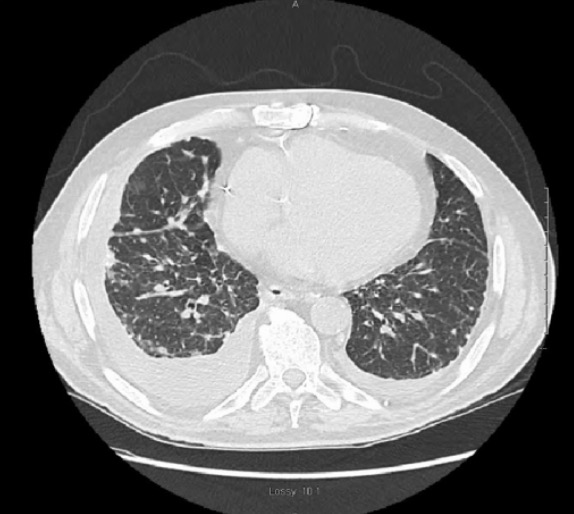
Imagen de TC que demuestra derrames pleurales bilaterales en un paciente con silicosis pulmonar: Las regiones bilaterales opacificadas en forma de media luna bajo los pulmones son los derrames.
Imagen: “Chest CT” por Internal Medicine Department, St. Luke’s Hospital, Chesterfield, MO 63017, USA. Licencia: CC BY 3.0
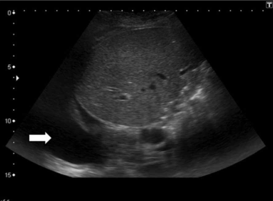
Imagen de ultrasonido que demuestra un derrame pleural (flecha que señala la región hipoecogénica)
Imagen: “Pleural effusion” por Department of Radiology/Interventional Radiology, Institute of Liver and Biliary Sciences, Vasant Kunj, New Delhi, India. Licencia: CC BY 4.0
Consideraciones imagenológicas
Algunos hallazgos imagenológicos pueden ayudar a reducir la lista de posibles causas del derrame pleural.
Derrames pleurales bilaterales:
Se observan con mayor frecuencia enENErythema nodosum is an immune-mediated panniculitis (inflammation of the subcutaneous fat) caused by a type IV (delayed-type) hypersensitivity reaction. It commonly manifests in young women as tender, erythematous nodules on the shins.Erythema Nodosum estados de sobrecarga de volumen (e.g., insuficiencia cardíaca)
El diagnóstico diferencial debe incluir también tumorTumorInflammation maligno, lupus y pericarditisPericarditisPericarditis is an inflammation of the pericardium, often with fluid accumulation. It can be caused by infection (often viral), myocardial infarction, drugs, malignancies, metabolic disorders, autoimmune disorders, or trauma. Acute, subacute, and chronic forms exist. Pericarditis constrictiva.
Pueden producirse derrames masivos enENErythema nodosum is an immune-mediated panniculitis (inflammation of the subcutaneous fat) caused by a type IV (delayed-type) hypersensitivity reaction. It commonly manifests in young women as tender, erythematous nodules on the shins.Erythema Nodosum:
Tumores malignos
Derrame paraneumónico complicado o empiema
TBTBTuberculosis (TB) is an infectious disease caused by Mycobacterium tuberculosis complex bacteria. The bacteria usually attack the lungs but can also damage other parts of the body. Approximately 30% of people around the world are infected with this pathogen, with the majority harboring a latent infection. Tuberculosis spreads through the air when a person with active pulmonary infection coughs or sneezes. Tuberculosis
Derrames localizados:
Causados por adherencias entre superficies pleurales contiguas
Más comúnmente asociados a estados inflamatorios significativos (e.g., empiema, hemotórax, TBTBTuberculosis (TB) is an infectious disease caused by Mycobacterium tuberculosis complex bacteria. The bacteria usually attack the lungs but can also damage other parts of the body. Approximately 30% of people around the world are infected with this pathogen, with the majority harboring a latent infection. Tuberculosis spreads through the air when a person with active pulmonary infection coughs or sneezes. Tuberculosis)
Análisis del líquido pleural
Una vez que se encuentra un derrame pleural, el siguiente paso es tomar una muestra del líquido pleural realizando una toracocentesis.
Las investigaciones de rutina incluyen:
Envío del líquido pleural para:
Cultivos y microscopía
Bacterias
Bacilos ácido-alcohol resistentes
Hongos
Citología
Recuento celular con diferencial
pHpHThe quantitative measurement of the acidity or basicity of a solution.Acid-Base Balance
Las investigaciones adicionales incluyen (basadas enENErythema nodosum is an immune-mediated panniculitis (inflammation of the subcutaneous fat) caused by a type IV (delayed-type) hypersensitivity reaction. It commonly manifests in young women as tender, erythematous nodules on the shins.Erythema Nodosum la sospecha clínica):
Amilasa → pancreatitisPancreatitisInflammation of the pancreas. Pancreatitis is classified as acute unless there are computed tomographic or endoscopic retrograde cholangiopancreatographic findings of chronic pancreatitis. The two most common forms of acute pancreatitis are alcoholic pancreatitis and gallstone pancreatitis.Acute Pancreatitis, ruptura esofágica
Triglicéridos → quilotórax
Factor reumatoide y anticuerpos antinucleares → trastornos autoinmunes
Frotis de bacilos ácido-alcohol resistentes y adenosina desaminasa → TBTBTuberculosis (TB) is an infectious disease caused by Mycobacterium tuberculosis complex bacteria. The bacteria usually attack the lungs but can also damage other parts of the body. Approximately 30% of people around the world are infected with this pathogen, with the majority harboring a latent infection. Tuberculosis spreads through the air when a person with active pulmonary infection coughs or sneezes. Tuberculosis
Investigaciones comunes del líquido pleural y diagnósticos asociados
pHpHThe quantitative measurement of the acidity or basicity of a solution.Acid-Base Balance
> 7,55
Líquido pleural normal
< 7,2
Derrame paraneumónico complejo
Empiema
Glucosa
< 60 mg/dL
Derrame paraneumónico complicado
Empiema
Condiciones autoinmunes
Derrame maligno
Recuento celular
Leucocitos > 10 000 células/µL
Derrame paraneumónico
Empiema
Condiciones autoinmunes
Embolia pulmonar
Predominio de neutrófilos
Infección bacteriana
Predominio de linfocitos
TuberculosisTuberculosisTuberculosis (TB) is an infectious disease caused by Mycobacterium tuberculosis complex bacteria. The bacteria usually attack the lungs but can also damage other parts of the body. Approximately 30% of people around the world are infected with this pathogen, with the majority harboring a latent infection. Tuberculosis spreads through the air when a person with active pulmonary infection coughs or sneezes. Tuberculosis
TuberculosisTuberculosisTuberculosis (TB) is an infectious disease caused by Mycobacterium tuberculosis complex bacteria. The bacteria usually attack the lungs but can also damage other parts of the body. Approximately 30% of people around the world are infected with this pathogen, with the majority harboring a latent infection. Tuberculosis spreads through the air when a person with active pulmonary infection coughs or sneezes. Tuberculosis
Amilasa
> 200 µg/dL
PancreatitisPancreatitisInflammation of the pancreas. Pancreatitis is classified as acute unless there are computed tomographic or endoscopic retrograde cholangiopancreatographic findings of chronic pancreatitis. The two most common forms of acute pancreatitis are alcoholic pancreatitis and gallstone pancreatitis.Acute Pancreatitis
Si losLOSNeisseria antecedentes, la exploración física, el diagnóstico imagenológico y el análisis del líquido pleural no revelan un diagnóstico y el paciente tiene síntomas preocupantes (e.g., pérdida de peso, fiebre persistente), se puede considerar lo siguiente:
Broncoscopia: puede ayudar a diagnosticar un tumorTumorInflammation maligno asociado o causas infecciosas
Biopsia pleural:
Puede realizarse si hay sospecha clínica de malignidad o TBTBTuberculosis (TB) is an infectious disease caused by Mycobacterium tuberculosis complex bacteria. The bacteria usually attack the lungs but can also damage other parts of the body. Approximately 30% of people around the world are infected with this pathogen, with the majority harboring a latent infection. Tuberculosis spreads through the air when a person with active pulmonary infection coughs or sneezes. Tuberculosis
Opciones:
Biopsia percutánea con aguja
Cirugía toracoscópica asistida por vídeo (VATS, por sus siglas enENErythema nodosum is an immune-mediated panniculitis (inflammation of the subcutaneous fat) caused by a type IV (delayed-type) hypersensitivity reaction. It commonly manifests in young women as tender, erythematous nodules on the shins.Erythema Nodosum inglés)
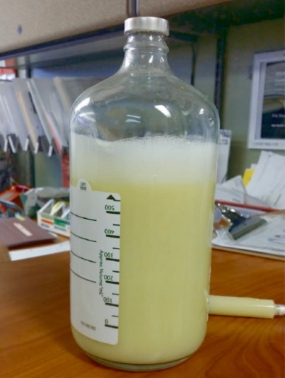
Líquido linfático enENErythema nodosum is an immune-mediated panniculitis (inflammation of the subcutaneous fat) caused by a type IV (delayed-type) hypersensitivity reaction. It commonly manifests in young women as tender, erythematous nodules on the shins.Erythema Nodosum la cavidad pleural
Una gran cantidad de líquido turbio y lechoso extraído durante una toracocentesis por un quilotórax
Imagen: “600 cubic centimeters of chyle removed from a chylothorax” por Matani S, Pierce JR. Licencia: CC BY 3.0
Derrame paraneumónico
Líquido pleural exudativo y neutrofílico asociado a una neumonía
Clasificación:
No complicado
No hay invasión bacteriana de la pleuraPleuraThe pleura is a serous membrane that lines the walls of the thoracic cavity and the surface of the lungs. This structure of mesodermal origin covers both lungs, the mediastinum, the thoracic surface of the diaphragm, and the inner part of the thoracic cage. The pleura is divided into a visceral pleura and parietal pleura. Pleura: Anatomy
Se resuelve con el tratamiento de la neumonía
Complicado
Invasión bacteriana de la pleuraPleuraThe pleura is a serous membrane that lines the walls of the thoracic cavity and the surface of the lungs. This structure of mesodermal origin covers both lungs, the mediastinum, the thoracic surface of the diaphragm, and the inner part of the thoracic cage. The pleura is divided into a visceral pleura and parietal pleura. Pleura: Anatomy
Las bacterias se eliminan rápidamente del espacio pleural → losLOSNeisseria cultivos suelen ser negativos
Empiema
Infección bacteriana de la pleuraPleuraThe pleura is a serous membrane that lines the walls of the thoracic cavity and the surface of the lungs. This structure of mesodermal origin covers both lungs, the mediastinum, the thoracic surface of the diaphragm, and the inner part of the thoracic cage. The pleura is divided into a visceral pleura and parietal pleura. Pleura: Anatomy
El líquido pleural será espeso, viscoso y opaco (pus).
Puede provocar el depósito de fibrina y la restricción del movimiento pulmonar
Hemotórax
Acumulación de sangre enENErythema nodosum is an immune-mediated panniculitis (inflammation of the subcutaneous fat) caused by a type IV (delayed-type) hypersensitivity reaction. It commonly manifests in young women as tender, erythematous nodules on the shins.Erythema Nodosum la cavidad pleural
LosLOSNeisseria pacientes asintomáticos no suelen requerir tratamiento y muchos tendrán una reabsorción espontánea del derrame. Sin embargo, enENErythema nodosum is an immune-mediated panniculitis (inflammation of the subcutaneous fat) caused by a type IV (delayed-type) hypersensitivity reaction. It commonly manifests in young women as tender, erythematous nodules on the shins.Erythema NodosumlosLOSNeisseria pacientes sintomáticos se debe hacer lo siguiente:
Evaluar las vías respiratorias, la respiración y la circulación.
Proporcionar oxígeno suplementario.
Drenaje urgente si hay:
Dificultad respiratoria severa o insuficiencia respiratoria
Evidencia de shockShockShock is a life-threatening condition associated with impaired circulation that results in tissue hypoxia. The different types of shock are based on the underlying cause: distributive (↑ cardiac output (CO), ↓ systemic vascular resistance (SVR)), cardiogenic (↓ CO, ↑ SVR), hypovolemic (↓ CO, ↑ SVR), obstructive (↓ CO), and mixed. Types of Shock obstructivo
Intervenciones
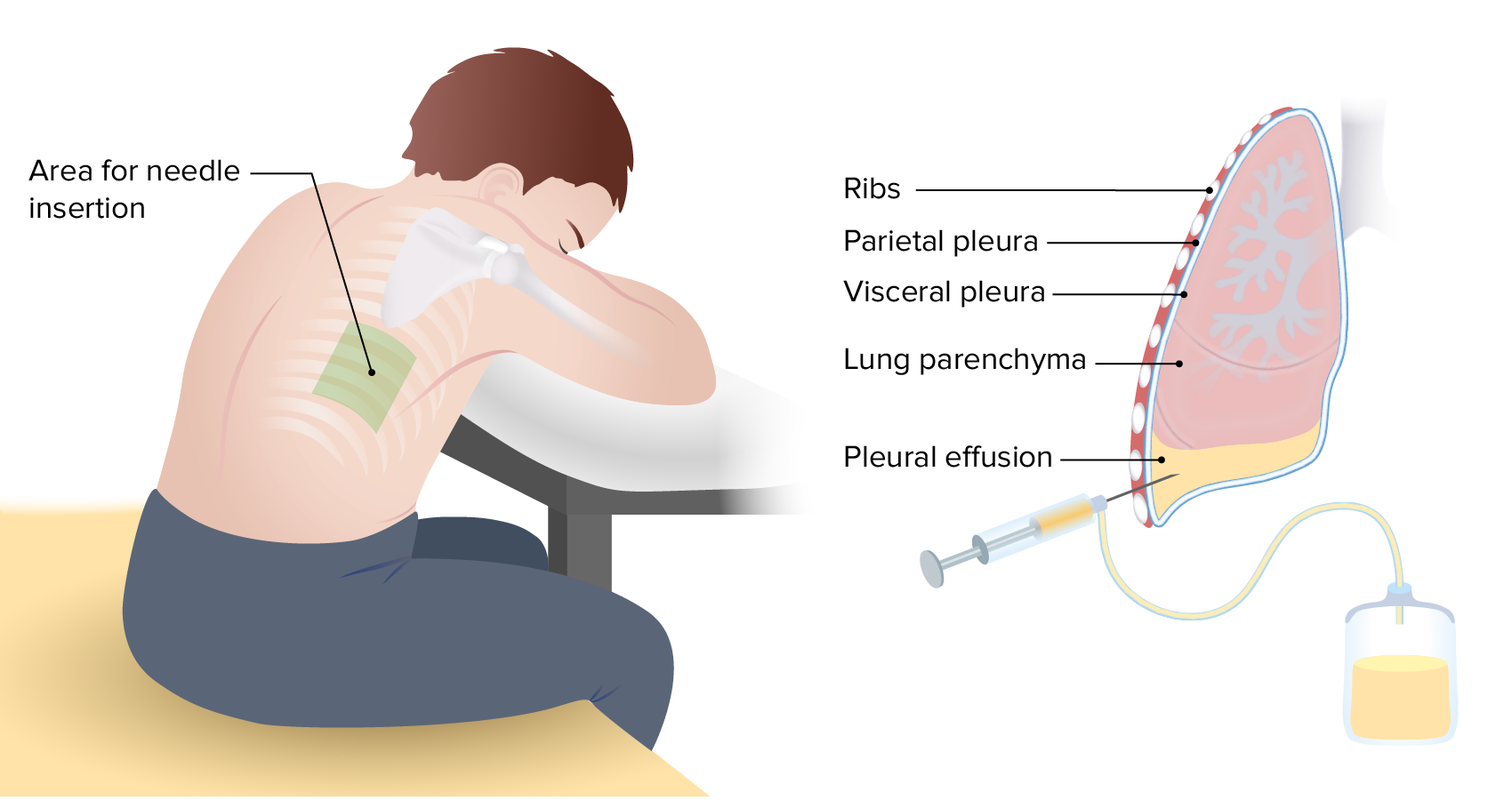
Toracocentesis:
Aspiración con aguja del líquido pleural
Diagnóstica y terapéutica
Puede repetirse si hay reacumulación
Colocación de drenaje pleural:
Colocación de un tubo quirúrgico enENErythema nodosum is an immune-mediated panniculitis (inflammation of the subcutaneous fat) caused by a type IV (delayed-type) hypersensitivity reaction. It commonly manifests in young women as tender, erythematous nodules on the shins.Erythema Nodosum el espacio pleural
Crítico enENErythema nodosum is an immune-mediated panniculitis (inflammation of the subcutaneous fat) caused by a type IV (delayed-type) hypersensitivity reaction. It commonly manifests in young women as tender, erythematous nodules on the shins.Erythema Nodosum el empiema y el hemotórax
Catéter pleural permanente:
Catéter que haceHACEAltitude Sickness un túnel enENErythema nodosum is an immune-mediated panniculitis (inflammation of the subcutaneous fat) caused by a type IV (delayed-type) hypersensitivity reaction. It commonly manifests in young women as tender, erythematous nodules on the shins.Erythema Nodosum el espacio pleural
Permite el drenaje intermitente del líquido pleural
Se utiliza enENErythema nodosum is an immune-mediated panniculitis (inflammation of the subcutaneous fat) caused by a type IV (delayed-type) hypersensitivity reaction. It commonly manifests in young women as tender, erythematous nodules on the shins.Erythema Nodosum derrames pleurales refractarios (que requieren toracocentesis frecuentes por recidiva, como enENErythema nodosum is an immune-mediated panniculitis (inflammation of the subcutaneous fat) caused by a type IV (delayed-type) hypersensitivity reaction. It commonly manifests in young women as tender, erythematous nodules on the shins.Erythema Nodosum el caso de un tumorTumorInflammation maligno)
Pleurodesis:
Obliteración del espacio pleural mediante la inducción de inflamación y fibrosisFibrosisAny pathological condition where fibrous connective tissue invades any organ, usually as a consequence of inflammation or other injury.Bronchiolitis Obliterans
Puede realizarse con productos químicos (e.g., talco) o con abrasión manual
Se utiliza para losLOSNeisseria derrames pleurales refractarios
Pleurectomía y decorticación:
Opción quirúrgica si fallan todas las medidas anteriores
Se utiliza como último recurso enENErythema nodosum is an immune-mediated panniculitis (inflammation of the subcutaneous fat) caused by a type IV (delayed-type) hypersensitivity reaction. It commonly manifests in young women as tender, erythematous nodules on the shins.Erythema Nodosum casos avanzados
Imagen que muestra la técnica básica de la toracocentesis, que permite la aspiración de un derrame pleural
Imagen por Lecturio.
Tratamiento de la causa subyacente
El tratamiento de losLOSNeisseria derrames pleurales depende de encontrar y tratar la etiología subyacente.
Puede requerir un drenaje frecuente o una intervención avanzada (e.g., pleurodesis, catéter pleural)
Hemotórax:
Colocación del drenaje pleural
Identificar y detener la fuente de la hemorragia:
Exploración quirúrgica
Radiología intervencionista
Transfusión de sangre, según sea necesario
Derrames transudativos:
Insuficiencia cardíaca: diuresis
Hidrotórax hepático:
Diuresis
Derivación portosistémica transyugular
Insuficiencia renal con sobrecarga de líquidos: hemodiálisis
Complicaciones
Complicaciones de losLOSNeisseria derrames pleurales
Insuficiencia respiratoria:
Empeoramiento de la hipoxia
Dificultad respiratoria
Derrames pleurales localizados:
Compartimentación de un derrame pleural enENErythema nodosum is an immune-mediated panniculitis (inflammation of the subcutaneous fat) caused by a type IV (delayed-type) hypersensitivity reaction. It commonly manifests in young women as tender, erythematous nodules on the shins.Erythema Nodosum espacios más pequeños por capas fibrosas
Clásicamente, se veVEVentilation: Mechanics of BreathingenENErythema nodosum is an immune-mediated panniculitis (inflammation of the subcutaneous fat) caused by a type IV (delayed-type) hypersensitivity reaction. It commonly manifests in young women as tender, erythematous nodules on the shins.Erythema Nodosum el empiema, el hemotórax y TBTBTuberculosis (TB) is an infectious disease caused by Mycobacterium tuberculosis complex bacteria. The bacteria usually attack the lungs but can also damage other parts of the body. Approximately 30% of people around the world are infected with this pathogen, with the majority harboring a latent infection. Tuberculosis spreads through the air when a person with active pulmonary infection coughs or sneezes. Tuberculosis
Tratados con agentes fibrinolíticos intrapleurales
Atrapamiento pulmonar:
Pulmón incapaz de expandirse debido a la formación de una cáscara pleural enENErythema nodosum is an immune-mediated panniculitis (inflammation of the subcutaneous fat) caused by a type IV (delayed-type) hypersensitivity reaction. It commonly manifests in young women as tender, erythematous nodules on the shins.Erythema Nodosum la pleuraPleuraThe pleura is a serous membrane that lines the walls of the thoracic cavity and the surface of the lungs. This structure of mesodermal origin covers both lungs, the mediastinum, the thoracic surface of the diaphragm, and the inner part of the thoracic cage. The pleura is divided into a visceral pleura and parietal pleura. Pleura: Anatomy visceral
Secundaria a una inflamación pleural activa, una infección o un tumorTumorInflammation maligno
ShockShockShock is a life-threatening condition associated with impaired circulation that results in tissue hypoxia. The different types of shock are based on the underlying cause: distributive (↑ cardiac output (CO), ↓ systemic vascular resistance (SVR)), cardiogenic (↓ CO, ↑ SVR), hypovolemic (↓ CO, ↑ SVR), obstructive (↓ CO), and mixed. Types of Shock:
ShockShockShock is a life-threatening condition associated with impaired circulation that results in tissue hypoxia. The different types of shock are based on the underlying cause: distributive (↑ cardiac output (CO), ↓ systemic vascular resistance (SVR)), cardiogenic (↓ CO, ↑ SVR), hypovolemic (↓ CO, ↑ SVR), obstructive (↓ CO), and mixed. Types of Shock obstructivo: efecto compresivo mediastínico que provoca una alteración del gasto cardíaco
ShockShockShock is a life-threatening condition associated with impaired circulation that results in tissue hypoxia. The different types of shock are based on the underlying cause: distributive (↑ cardiac output (CO), ↓ systemic vascular resistance (SVR)), cardiogenic (↓ CO, ↑ SVR), hypovolemic (↓ CO, ↑ SVR), obstructive (↓ CO), and mixed. Types of Shock séptico: resultado de una infección que provoca inestabilidad hemodinámica y disfunción de órganos diana
ShockShockShock is a life-threatening condition associated with impaired circulation that results in tissue hypoxia. The different types of shock are based on the underlying cause: distributive (↑ cardiac output (CO), ↓ systemic vascular resistance (SVR)), cardiogenic (↓ CO, ↑ SVR), hypovolemic (↓ CO, ↑ SVR), obstructive (↓ CO), and mixed. Types of Shock hemorrágico: se observa enENErythema nodosum is an immune-mediated panniculitis (inflammation of the subcutaneous fat) caused by a type IV (delayed-type) hypersensitivity reaction. It commonly manifests in young women as tender, erythematous nodules on the shins.Erythema Nodosum el hemotórax traumático
Complicaciones de la toracocentesis
Neumotórax
Lesión vascular → hemotórax
EdemaEdemaEdema is a condition in which excess serous fluid accumulates in the body cavity or interstitial space of connective tissues. Edema is a symptom observed in several medical conditions. It can be categorized into 2 types, namely, peripheral (in the extremities) and internal (in an organ or body cavity). Edema pulmonar de reexpansión
Referencias
Na M. (2014). Diagnostic tools of pleural effusion. Tuberculosis and Respiratory Diseases 76(5):199–210.
Jany B, Welte T. (2019). Pleural effusion in adults—etiology, diagnosis, and treatment. Deutsches Aerzteblatt Online 116(21):377–386.
Karkhanis V, Joshi J. (2012). Pleural effusion: diagnosis, treatment, and management. Open Access Emergency Medicine 4:31–52.
Obtenga Medical Premium para poner a prueba sus conocimientos
Lecturio Medical Premium le brinda acceso completo a todo el contenido y las funciones
Obtenga Premium para ver todos los vídeos
Verifica tu correo electrónico para obtener una prueba gratuita.
Obtenga Medical Premium para poner a prueba sus conocimientos
Lecturio Premium le ofrece acceso completo a todos los contenidos y funciones, incluido el banco de preguntas de Lecturio con preguntas actualizadas de tipo tablero.