Domina Conceptos Médicos

Estudia para la escuela de medicina y tus examenes con Lecturio.

Pérdida de la Audición

La pérdida de la audición, también conocida como deterioro auditivo, es cualquier grado de impedimento en EN Erythema nodosum is an immune-mediated panniculitis (inflammation of the subcutaneous fat) caused by a type IV (delayed-type) hypersensitivity reaction. It commonly manifests in young women as tender, erythematous nodules on the shins. Erythema Nodosum la capacidad de captar el sonido según lo determinado por la audiometría por debajo de los LOS Neisseria umbrales auditivos normales. La presentación clínica puede ocurrir al AL Amyloidosis nacimiento o como una pérdida gradual de la audición con la edad, incluida una pérdida repentina o a corto plazo en EN Erythema nodosum is an immune-mediated panniculitis (inflammation of the subcutaneous fat) caused by a type IV (delayed-type) hypersensitivity reaction. It commonly manifests in young women as tender, erythematous nodules on the shins. Erythema Nodosum cualquier momento. La evaluación diagnóstica se basa en EN Erythema nodosum is an immune-mediated panniculitis (inflammation of the subcutaneous fat) caused by a type IV (delayed-type) hypersensitivity reaction. It commonly manifests in young women as tender, erythematous nodules on the shins. Erythema Nodosum los LOS Neisseria antecedentes, el examen físico (incluyendo el examen otoscópico y diapasón) y las pruebas de audiología. El tratamiento se dirige hacia la etiología subyacente de la pérdida de la audición para elegir el apropiado curso del tratamiento.

Last updated: Dec 15, 2025

Editorial responsibility: Stanley Oiseth, Lindsay Jones, Evelin Maza

Descripción General

Definición

La pérdida de la audición, también conocida como deterioro auditivo, es cualquier grado de impedimento en EN Erythema nodosum is an immune-mediated panniculitis (inflammation of the subcutaneous fat) caused by a type IV (delayed-type) hypersensitivity reaction. It commonly manifests in young women as tender, erythematous nodules on the shins. Erythema Nodosum la capacidad de captar el sonido según lo determinado por la audiometría por debajo de los LOS Neisseria umbrales auditivos normales.

Anatomía

  • Oído externo: pabellón auricular y conducto auditivo externo
  • Oído medio:
    • Membrana timpánica
    • Osículos: martillo, yunque y estribo
  • Oído interno:
    • Cóclea
    • Canales semicirculares
    • Nervio vestibulococlear (nervio craneal VIII)
  • SNC:
    • Vías auditivas del tronco encefálico
    • Corteza auditiva
Anatomía del oído externo, medio e interno

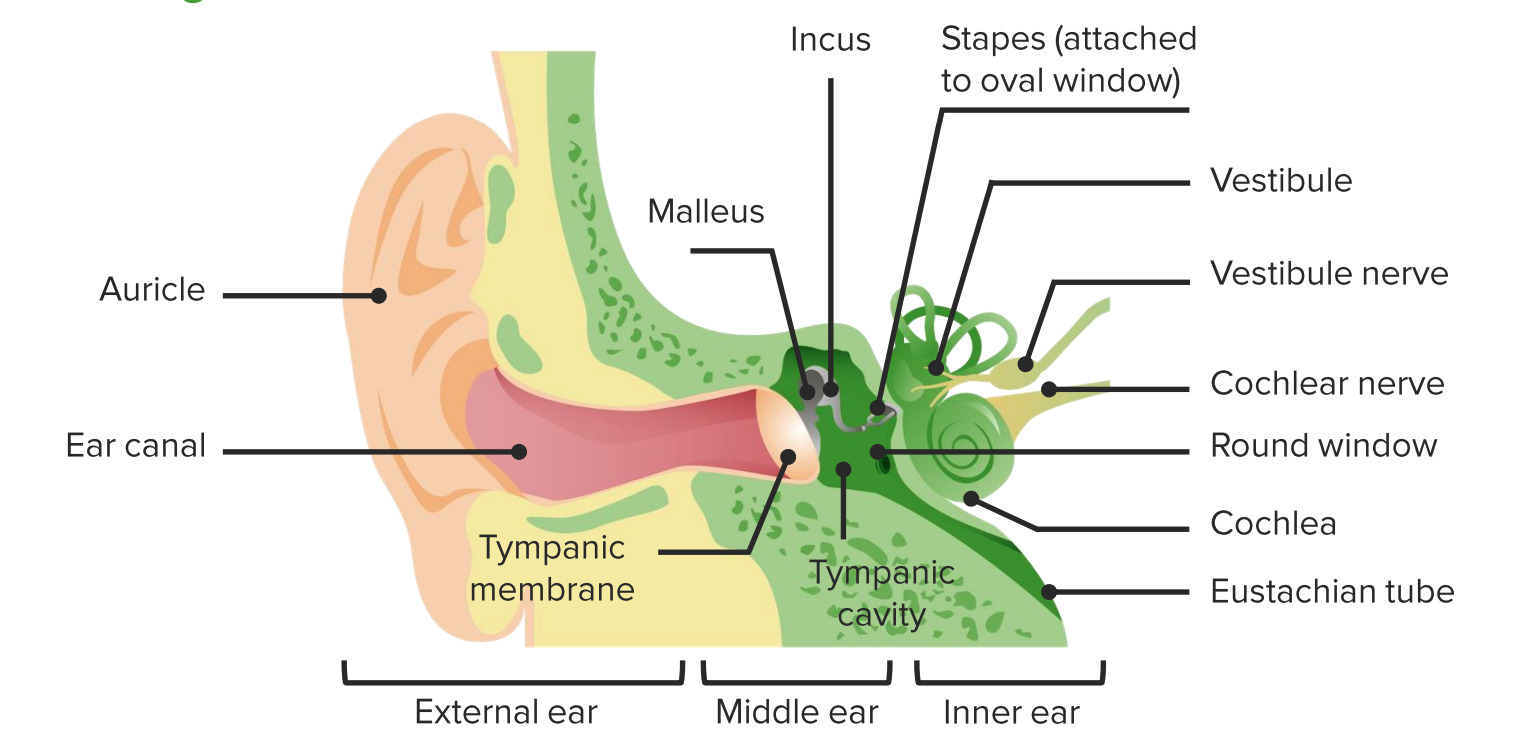
Diagrama de la anatomía del oído externo, medio e interno

Imagen por Lecturio.

Epidemiología

  • Incidencia de la pérdida de la audición: 2 por cada 1 000 en EN Erythema nodosum is an immune-mediated panniculitis (inflammation of the subcutaneous fat) caused by a type IV (delayed-type) hypersensitivity reaction. It commonly manifests in young women as tender, erythematous nodules on the shins. Erythema Nodosum niños pequeños
  • Prevalencia en EN Erythema nodosum is an immune-mediated panniculitis (inflammation of the subcutaneous fat) caused by a type IV (delayed-type) hypersensitivity reaction. It commonly manifests in young women as tender, erythematous nodules on the shins. Erythema Nodosum adultos:
    • 21–34 años de edad: 3%
    • 35–44 años de edad: 6%
    • 44–54 años de edad: 11%
    • 55–64 años de edad: 25%
    • 65–84 años de edad: 43%

Clasificación de la pérdida de la audición

  • Pérdida de audición conductiva: incapacidad del oído para conducir el sonido a través de las estructuras del oído (cadena osicular y membrana timpánica)
  • Pérdida de audición neurosensorial: repercusión en EN Erythema nodosum is an immune-mediated panniculitis (inflammation of the subcutaneous fat) caused by a type IV (delayed-type) hypersensitivity reaction. It commonly manifests in young women as tender, erythematous nodules on the shins. Erythema Nodosum la cóclea, el nervio craneal VIII o la vía auditiva central
  • Mixto: componentes conductivos y neurosensoriales
  • Trastorno del procesamiento auditivo central: daño a nivel del nervio craneal VIII, el tronco del encéfalo auditivo o la corteza cerebral

Etiología

Pérdida de audición conductiva:

  • Otitis media (aguda o crónica)
  • Otitis externa Otitis externa Otitis externa (also known as external otitis or swimmer’s ear) is an infection of the external auditory canal that is most often caused by acute bacterial infection and is frequently associated with hot, humid weather and water exposure. Patients commonly present with ear pain, pruritus, discharge, and hearing loss. Otitis Externa (aguda o crónica)
  • Disfunción de la trompa de Eustaquio/retención de líquidos en EN Erythema nodosum is an immune-mediated panniculitis (inflammation of the subcutaneous fat) caused by a type IV (delayed-type) hypersensitivity reaction. It commonly manifests in young women as tender, erythematous nodules on the shins. Erythema Nodosum el oído medio
  • Colesteatoma
  • Otoesclerosis
  • Impactación de cerumen Cerumen The yellow or brown waxy secretions produced by vestigial apocrine sweat glands in the external ear canal. Otitis Externa
  • Neoplasia:
    • Tumores del conducto auditivo externo
    • Paraganglioma Paraganglioma A neural crest tumor usually derived from the chromoreceptor tissue of a paraganglion, such as the carotid body, or medulla of the adrenal gland (usually called a chromaffinoma or pheochromocytoma). It is more common in women than in men. Hearing Loss
    • Adenoma del oído medio
  • Timpanoesclerosis
  • Traumatismo:
    • Fractura osicular
    • Perforación de la membrana timpánica
  • Malformaciones congénitas:

Pérdida de audición neurosensorial:

  • Presbiacusia
  • Exposición prolongada a ruidos fuertes
  • Infecciones por toxoplasmosis Toxoplasmosis Toxoplasmosis is an infectious disease caused by Toxoplasma gondii, an obligate intracellular protozoan parasite. Felines are the definitive host, but transmission to humans can occur through contact with cat feces or the consumption of contaminated foods. The clinical presentation and complications depend on the host’s immune status. Toxoplasma/Toxoplasmosis prenatal, otros agentes, rubéola, citomegalovirus y herpes simple:
    • Toxoplasmosis Toxoplasmosis Toxoplasmosis is an infectious disease caused by Toxoplasma gondii, an obligate intracellular protozoan parasite. Felines are the definitive host, but transmission to humans can occur through contact with cat feces or the consumption of contaminated foods. The clinical presentation and complications depend on the host’s immune status. Toxoplasma/Toxoplasmosis
    • Rubéola
    • Citomegalovirus (puede causar pérdida asintomática de la audición)
    • Herpes Simple
  • Infecciones posnatales:
    • Meningitis Meningitis Meningitis is inflammation of the meninges, the protective membranes of the brain, and spinal cord. The causes of meningitis are varied, with the most common being bacterial or viral infection. The classic presentation of meningitis is a triad of fever, altered mental status, and nuchal rigidity. Meningitis por Neisseria meningitidis Neisseria meningitidis A species of gram-negative, aerobic bacteria. It is a commensal and pathogen only of humans, and can be carried asymptomatically in the nasopharynx. When found in cerebrospinal fluid it is the causative agent of cerebrospinal meningitis. It is also found in venereal discharges and blood. There are at least 13 serogroups based on antigenic differences in the capsular polysaccharides; the ones causing most meningitis infections being a, b, c, y, and w-135. Each serogroup can be further classified by serotype, serosubtype, and immunotype. Neisseria, Haemophilus Haemophilus Haemophilus is a genus of Gram-negative coccobacilli, all of whose strains require at least 1 of 2 factors for growth (factor V [NAD] and factor X [heme]); therefore, it is most often isolated on chocolate agar, which can supply both factors. The pathogenic species are H. influenzae and H. ducreyi. Haemophilus influenza Influenza Influenza viruses are members of the Orthomyxoviridae family and the causative organisms of influenza, a highly contagious febrile respiratory disease. There are 3 primary influenza viruses (A, B, and C) and various subtypes, which are classified based on their virulent surface antigens, hemagglutinin (HA) and neuraminidase (NA). Influenza typically presents with a fever, myalgia, headache, and symptoms of an upper respiratory infection. Influenza Viruses/Influenza o Streptococcus Streptococcus Streptococcus is one of the two medically important genera of gram-positive cocci, the other being Staphylococcus. Streptococci are identified as different species on blood agar on the basis of their hemolytic pattern and sensitivity to optochin and bacitracin. There are many pathogenic species of streptococci, including S. pyogenes, S. agalactiae, S. pneumoniae, and the viridans streptococci. Streptococcus pneumoniae
    • La meningitis Meningitis Meningitis is inflammation of the meninges, the protective membranes of the brain, and spinal cord. The causes of meningitis are varied, with the most common being bacterial or viral infection. The classic presentation of meningitis is a triad of fever, altered mental status, and nuchal rigidity. Meningitis por otros muchos organismos puede causar pérdida de la audición.
  • Causas infecciosas adicionales de pérdida auditiva:
    • Paperas
    • Sífilis
  • Causas genéticas:
    • > 400 síndromes genéticos (e.g., síndrome de Alport)
    • Existen muchas causas genéticas no sindrómicas autosómicas dominantes, autosómicas recesivas y ligadas al AL Amyloidosis cromosoma X.
  • Neoplasias:
    • Neurinoma del acústico
    • Meningioma Meningioma Meningiomas are slow-growing tumors that arise from the meninges of the brain and spinal cord. The vast majority are benign. These tumors commonly occur in individuals with a history of high doses of skull radiation, head trauma, and neurofibromatosis 2. Meningioma
    • Tumor Tumor Inflammation del saco endolinfático
  • Traumatismo:
    • Traumatismo cerrado o penetrante en EN Erythema nodosum is an immune-mediated panniculitis (inflammation of the subcutaneous fat) caused by a type IV (delayed-type) hypersensitivity reaction. It commonly manifests in young women as tender, erythematous nodules on the shins. Erythema Nodosum el oído
    • Lesión por explosión o fractura del hueso temporal
    • Barotrauma Barotrauma Injury following pressure changes; includes injury to the eustachian tube, ear drum, lung and stomach. Invasive Mechanical Ventilation por bucear o volar
  • Medicamentos ototóxicos:
    • Antibióticos aminoglucósidos
    • Diuréticos de asa ASA Anterior Cord Syndrome
    • Aspirina
    • Agentes para la quimioterapia a base de platino
  • Radioterapia
  • Enfermedad de Ménière

Fisiología de la audición

  1. El sonido se recibe en EN Erythema nodosum is an immune-mediated panniculitis (inflammation of the subcutaneous fat) caused by a type IV (delayed-type) hypersensitivity reaction. It commonly manifests in young women as tender, erythematous nodules on the shins. Erythema Nodosum el canal auditivo externo y se canaliza al AL Amyloidosis oído medio.
  2. La membrana timpánica vibra → conduce las vibraciones sonoras a través de los LOS Neisseria osículos → ventana oval → oído interno
  3. El sonido viaja a través de la perilinfa del oído interno ( en EN Erythema nodosum is an immune-mediated panniculitis (inflammation of the subcutaneous fat) caused by a type IV (delayed-type) hypersensitivity reaction. It commonly manifests in young women as tender, erythematous nodules on the shins. Erythema Nodosum la cámara superior e inferior de la cóclea) → detectado por las células ciliadas en EN Erythema nodosum is an immune-mediated panniculitis (inflammation of the subcutaneous fat) caused by a type IV (delayed-type) hypersensitivity reaction. It commonly manifests in young women as tender, erythematous nodules on the shins. Erythema Nodosum la membrana basilar en EN Erythema nodosum is an immune-mediated panniculitis (inflammation of the subcutaneous fat) caused by a type IV (delayed-type) hypersensitivity reaction. It commonly manifests in young women as tender, erythematous nodules on the shins. Erythema Nodosum el órgano de Corti (cámara media de la cóclea), que está llena de endolinfa
  4. Las señales se transportan a través de la división coclear del nervio craneal VIII → se procesan en EN Erythema nodosum is an immune-mediated panniculitis (inflammation of the subcutaneous fat) caused by a type IV (delayed-type) hypersensitivity reaction. It commonly manifests in young women as tender, erythematous nodules on the shins. Erythema Nodosum el núcleo del nervio craneal VIII y la corteza auditiva → percepción del sonido (audición)

Relevancia cultural

  • Los LOS Neisseria miembros de la comunidad sorda de los LOS Neisseria Estados Unidos son sordos, lo que implica una pérdida importante de la audición
  • Los LOS Neisseria miembros utilizan el lenguaje de señas americano.
  • No se considera que los LOS Neisseria miembros tengan un deterioro auditivo o una pérdida de la audición
  • La sordera no es una enfermedad para ser tratada o curada.

Antecedentes de la Enfermedad Actual

Información que se debe recopilar del paciente o del cuidador con respecto a la pérdida de la audición:

  • Inicio y duración de la pérdida de la audición:
    • Prelingual o poslingual (antes o después de que un niño aprenda a hablar)
    • Repentino o crónico
    • Fluctuante o progresivo
  • Lateralidad: unilateral o bilateral
  • Gravedad:
    • Las formas más leves pueden pasar desapercibidas para el paciente
    • Las formas más graves pueden requerir auxiliares auditivos para la amplificación del sonido
  • Síntomas de alarma:
    • Inicio repentino o instauración rápida
    • Rápidamente progresivo (posiblemente por medicamentos ototóxicos)
    • Tinnitus Tinnitus A nonspecific symptom of hearing disorder characterized by the sensation of buzzing, ringing, clicking, pulsations, and other noises in the ear. Objective tinnitus refers to noises generated from within the ear or adjacent structures that can be heard by other individuals. The term subjective tinnitus is used when the sound is audible only to the affected individual. Tinnitus may occur as a manifestation of cochlear diseases; vestibulocochlear nerve diseases; intracranial hypertension; craniocerebral trauma; and other conditions. Cranial Nerve Palsies asociado con la pérdida auditiva aguda
    • El vértigo puede indicar una patología del nervio craneal VIII:
      • Enfermedad de Ménière
      • Laberintitis
      • Neoplasia
      • Isquemia/infarto del tronco encefálico
      • Fístula perilinfática
  • Antecedentes recientes:
    • Infección de las vías respiratorias altas
    • Uso de antibióticos aminoglucósidos
    • Cirugía otológica reciente:
      • Tubos para la compensación de la presión
      • Timpanoplastia
      • Mastoidectomía

Examen Físico

Se utiliza un otoscopio para examinar el oído externo y medio y un diapasón para determinar el tipo de la pérdida auditiva.

Examen otoscópico

  • Oído externo y medio:
    • Inspección del canal auditivo (e.g., eritema, drenaje, inflamación, impactación de cerumen Cerumen The yellow or brown waxy secretions produced by vestigial apocrine sweat glands in the external ear canal. Otitis Externa y cuerpo extraño)
    • Exploración de la membrana timpánica (e.g., perforación, abultamiento, retracción y eritema)
  • Otoscopia neumática (ráfaga de aire en EN Erythema nodosum is an immune-mediated panniculitis (inflammation of the subcutaneous fat) caused by a type IV (delayed-type) hypersensitivity reaction. It commonly manifests in young women as tender, erythematous nodules on the shins. Erythema Nodosum el canal auditivo):
    • Se utiliza un insuflador para determinar la movilidad de la membrana timpánica.
    • Si existe líquido en EN Erythema nodosum is an immune-mediated panniculitis (inflammation of the subcutaneous fat) caused by a type IV (delayed-type) hypersensitivity reaction. It commonly manifests in young women as tender, erythematous nodules on the shins. Erythema Nodosum el oído medio, este puede inhibir el movimiento de la membrana timpánica
Membrana timpánica normal

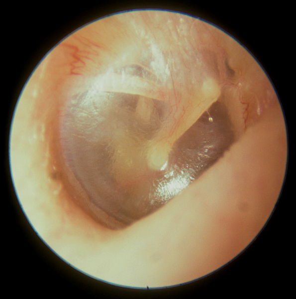
Vista de una membrana timpánica normal en un examen otoscópico:
Obsérvese el martillo óseo adherido a la membrana timpánica.

Imagen: “Normal tympanic membrane” por Michael Hawke, MD.  Licencia: CC BY 4.0

Pruebas de diapasón

  • Diapasón de 512 Hz
  • Prueba de Rinne para la pérdida de audición conductiva:
    • Procedimiento: Se golpea el diapasón y se coloca detrás de la oreja sobre el hueso mastoideo; una vez que ya no se escucha, el diapasón se aleja a 2,5 cm del oído externo.
    • Normal: conducción aérea > conducción ósea (todavía se escucha la vibración cerca del oído externo)
    • Pérdida de audición conductiva: conducción ósea > conducción aérea (la vibración ya no se escucha una vez que el diapasón se mueve frente al AL Amyloidosis canal auditivo)
  • Prueba de Weber:
    • El diapasón se golpea y se coloca en EN Erythema nodosum is an immune-mediated panniculitis (inflammation of the subcutaneous fat) caused by a type IV (delayed-type) hypersensitivity reaction. It commonly manifests in young women as tender, erythematous nodules on the shins. Erythema Nodosum la parte superior de la cabeza.
    • Normal: el sonido se escucha por igual en EN Erythema nodosum is an immune-mediated panniculitis (inflammation of the subcutaneous fat) caused by a type IV (delayed-type) hypersensitivity reaction. It commonly manifests in young women as tender, erythematous nodules on the shins. Erythema Nodosum ambos oídos.
    • Pérdida de audición conductiva: El sonido es más fuerte en EN Erythema nodosum is an immune-mediated panniculitis (inflammation of the subcutaneous fat) caused by a type IV (delayed-type) hypersensitivity reaction. It commonly manifests in young women as tender, erythematous nodules on the shins. Erythema Nodosum el oído afectado.
    • Pérdida de audición neurosensorial: El sonido es menor en EN Erythema nodosum is an immune-mediated panniculitis (inflammation of the subcutaneous fat) caused by a type IV (delayed-type) hypersensitivity reaction. It commonly manifests in young women as tender, erythematous nodules on the shins. Erythema Nodosum el oído afectado.
Pruebas de Rinne y Weber

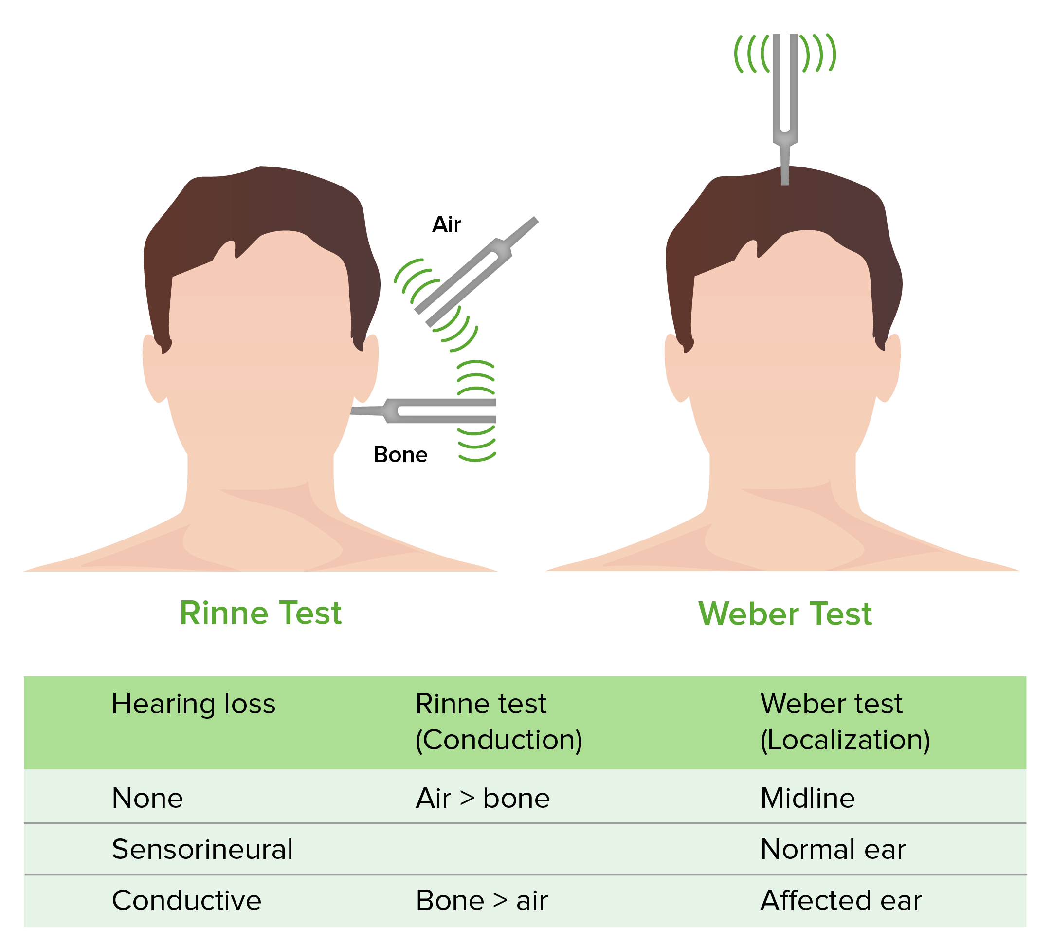
Prueba de Weber y Rinne

Imagen por Lecturio.

Diagnóstico y Tratamiento

El diagnóstico de la pérdida de la audición se basa en EN Erythema nodosum is an immune-mediated panniculitis (inflammation of the subcutaneous fat) caused by a type IV (delayed-type) hypersensitivity reaction. It commonly manifests in young women as tender, erythematous nodules on the shins. Erythema Nodosum pruebas fisiológicas y audiométricas. También se pueden necesitar pruebas de laboratorio e imagenología para descartar otras patologías.

Pruebas fisiológicas

  • Prueba de respuesta auditiva del tronco encefálico:
    • Se utiliza un sonido de “clic” para evocar respuestas fisiológicas en EN Erythema nodosum is an immune-mediated panniculitis (inflammation of the subcutaneous fat) caused by a type IV (delayed-type) hypersensitivity reaction. It commonly manifests in young women as tender, erythematous nodules on the shins. Erythema Nodosum el nervio craneal VIII y el tronco encefálico auditivo.
    • Es registrado mediante electrodos de superficie
    • No evalúa sonidos de baja frecuencia < 1 500 Hz
  • Prueba de respuesta auditiva de estado estable:
    • Potenciales evocados auditivos de estado estable (como la respuesta auditiva del tronco encefálico)
    • Mide varias frecuencias y proporciona información más específica (a diferencia de la respuesta auditiva del tronco encefálico)
  • Emisiones otoacústicas evocadas: evalúan la actividad de las células ciliadas cocleares
  • Timpanometría: evalúa la movilidad de la membrana timpánica y los LOS Neisseria osículos del oído medio

Audiometría

  • Pruebas de conducta para niños pequeños:
    • Indicado para niños con retraso en EN Erythema nodosum is an immune-mediated panniculitis (inflammation of the subcutaneous fat) caused by a type IV (delayed-type) hypersensitivity reaction. It commonly manifests in young women as tender, erythematous nodules on the shins. Erythema Nodosum el desarrollo del habla
    • Audiometría por observación de la conducta: desde el nacimiento hasta los LOS Neisseria 6 meses de edad
    • Audiometría por refuerzo visual: 6 meses a los LOS Neisseria 2,5 años de edad
  • Audiometría de tonos puros:
    • Los LOS Neisseria auriculares se utilizan para identificar el umbral escuchado para un conjunto definido de frecuencias.
    • Se utiliza en EN Erythema nodosum is an immune-mediated panniculitis (inflammation of the subcutaneous fat) caused by a type IV (delayed-type) hypersensitivity reaction. It commonly manifests in young women as tender, erythematous nodules on the shins. Erythema Nodosum adultos y niños lo suficientemente maduros para colaborar en EN Erythema nodosum is an immune-mediated panniculitis (inflammation of the subcutaneous fat) caused by a type IV (delayed-type) hypersensitivity reaction. It commonly manifests in young women as tender, erythematous nodules on the shins. Erythema Nodosum la audiometría
    • Indicada en EN Erythema nodosum is an immune-mediated panniculitis (inflammation of the subcutaneous fat) caused by a type IV (delayed-type) hypersensitivity reaction. It commonly manifests in young women as tender, erythematous nodules on the shins. Erythema Nodosum cualquier paciente con pérdida auditiva unilateral o bilateral.
    • Normal: dentro de 15 dB del umbral normal
    • Los LOS Neisseria sonidos de alta frecuencia son los LOS Neisseria primeros en EN Erythema nodosum is an immune-mediated panniculitis (inflammation of the subcutaneous fat) caused by a type IV (delayed-type) hypersensitivity reaction. It commonly manifests in young women as tender, erythematous nodules on the shins. Erythema Nodosum perderse con el envejecimiento.
  • Pérdida de la audición: el rango audible normal es de 0–180 dB (> 85 dB es perjudicial):
    • Gravedad:
      • Leve: 26–40 dB
      • Moderado: 41–55 dB
      • Moderadamente grave: 56–70 dB
      • Grave: 71–90 dB
      • Severo: >90 dB
    • Frecuencias: la audición normal es de 20–20 000 Hz:
      • Pérdida de audición de baja frecuencia: sonidos < 500 Hz
      • Pérdida de audición de frecuencias medias: sonidos de 501 a 2 000 Hz
      • Pérdida de audición de alta frecuencia: sonidos > 2 000 Hz
  • Deterioro funcional:
    • Puede ser confuso si se basa solo en EN Erythema nodosum is an immune-mediated panniculitis (inflammation of the subcutaneous fat) caused by a type IV (delayed-type) hypersensitivity reaction. It commonly manifests in young women as tender, erythematous nodules on the shins. Erythema Nodosum el promedio de los LOS Neisseria tonos puros:
      • 30% de deterioro = 70% de audición residual a 45 dB
      • El habla conversacional es de aproximadamente 50–60 dB → impacto significativo en EN Erythema nodosum is an immune-mediated panniculitis (inflammation of the subcutaneous fat) caused by a type IV (delayed-type) hypersensitivity reaction. It commonly manifests in young women as tender, erythematous nodules on the shins. Erythema Nodosum la capacidad funcional normal
    • Se utiliza una escala diferente de clasificación para los LOS Neisseria niños pequeños → las deficiencias leves pueden causar una dificultad significativa en EN Erythema nodosum is an immune-mediated panniculitis (inflammation of the subcutaneous fat) caused by a type IV (delayed-type) hypersensitivity reaction. It commonly manifests in young women as tender, erythematous nodules on the shins. Erythema Nodosum el desarrollo del lenguaje
Audiograma

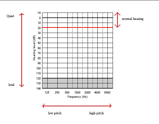
Gráfico del audiograma

Imagen: “Audiogram” por Audiology6. Licencia: Dominio Público

Pruebas adicionales

  • Pruebas de laboratorio:
    • Glucosa en EN Erythema nodosum is an immune-mediated panniculitis (inflammation of the subcutaneous fat) caused by a type IV (delayed-type) hypersensitivity reaction. It commonly manifests in young women as tender, erythematous nodules on the shins. Erythema Nodosum sangre: la vasculopatía diabética puede causar isquemia coclear.
    • Hemograma: verificar presencia de anemia Anemia Anemia is a condition in which individuals have low Hb levels, which can arise from various causes. Anemia is accompanied by a reduced number of RBCs and may manifest with fatigue, shortness of breath, pallor, and weakness. Subtypes are classified by the size of RBCs, chronicity, and etiology. Anemia: Overview and Types y el recuento leucocitario
    • Pruebas de función tiroidea
    • Prueba serológica para sífilis
    • Pruebas para enfermedades autoinmunes (si se sospechan):
      • Velocidad de sedimentación globular (VES)
      • ANA
      • Factor reumatoideo
  • Imagenología:
    • RM: recomendada en EN Erythema nodosum is an immune-mediated panniculitis (inflammation of the subcutaneous fat) caused by a type IV (delayed-type) hypersensitivity reaction. It commonly manifests in young women as tender, erythematous nodules on the shins. Erythema Nodosum casos de pérdida de audición neurosensorial asimétrica o unilateral
    • TC del hueso temporal:
      • Identifica hallazgos óseos anormales
      • Localiza derrames y anormalidades en EN Erythema nodosum is an immune-mediated panniculitis (inflammation of the subcutaneous fat) caused by a type IV (delayed-type) hypersensitivity reaction. It commonly manifests in young women as tender, erythematous nodules on the shins. Erythema Nodosum tejidos blandos

Tratamiento

  • Tratamiento médico:
    • Proporcionar antibióticos si hay una infección presente
    • Evacuación de la obstrucción: cerumen Cerumen The yellow or brown waxy secretions produced by vestigial apocrine sweat glands in the external ear canal. Otitis Externa u otro cuerpo extraño
    • Tratamiento de las afecciones subyacentes:
      • Enfermedad autoinmune
      • Enfermedad cerebrovascular
      • Meningitis Meningitis Meningitis is inflammation of the meninges, the protective membranes of the brain, and spinal cord. The causes of meningitis are varied, with the most common being bacterial or viral infection. The classic presentation of meningitis is a triad of fever, altered mental status, and nuchal rigidity. Meningitis
      • Esclerosis múltiple
      • Sífilis
  • Referencia a la especialidad de otorrinolaringología:
    • Implante coclear (procedimiento quirúrgico):
      • Indicado para la pérdida de audición neurosensorial
      • > 6 meses de edad
    • Emergencia otológica:
      • Pérdida súbita de audición neurosensorial
      • Traumatismo
    • El tratamiento quirúrgico depende de la etiología:
      • Reparación de la membrana timpánica perforada
      • Estapedectomía por otosclerosis Otosclerosis Formation of spongy bone in the labyrinth capsule which can progress toward the stapes (stapedial fixation) or anteriorly toward the cochlea leading to conductive, sensorineural, or mixed hearing loss. Several genes are associated with familial otosclerosis with varied clinical signs. Vertigo
      • Reconstrucción de la cadena osícular
      • Tumor Tumor Inflammation (e.g., neurinoma del acústico)

Relevancia Clínica

Afecciones comunes que causan pérdida de la audición:

  • Otitis media: infección en EN Erythema nodosum is an immune-mediated panniculitis (inflammation of the subcutaneous fat) caused by a type IV (delayed-type) hypersensitivity reaction. It commonly manifests in young women as tender, erythematous nodules on the shins. Erythema Nodosum el oído medio caracterizada por la inflamación de las mucosas y secreción. Los LOS Neisseria patógenos más comunes son S. pneumoniae, H. influenzae H. influenzae A species of Haemophilus found on the mucous membranes of humans and a variety of animals. The species is further divided into biotypes I through VIII. Haemophilus y Moraxella catarrhalis Moraxella catarrhalis Gram-negative aerobic cocci of low virulence that colonize the nasopharynx and occasionally cause meningitis; bacteremia; empyema; pericarditis; and pneumonia. Moraxella. La otitis media puede presentarse con fiebre, otalgia Otalgia Acute Otitis Media y pérdida de la audición. El diagnóstico se realiza mediante los LOS Neisseria antecedentes clínicos y el examen físico. El tratamiento es con antibióticos orales. Es posible que se necesiten procedimientos específicos para las infecciones recurrentes.
  • Otitis externa Otitis externa Otitis externa (also known as external otitis or swimmer’s ear) is an infection of the external auditory canal that is most often caused by acute bacterial infection and is frequently associated with hot, humid weather and water exposure. Patients commonly present with ear pain, pruritus, discharge, and hearing loss. Otitis Externa (oído de nadador): infección del conducto auditivo externo. La otitis externa Otitis externa Otitis externa (also known as external otitis or swimmer’s ear) is an infection of the external auditory canal that is most often caused by acute bacterial infection and is frequently associated with hot, humid weather and water exposure. Patients commonly present with ear pain, pruritus, discharge, and hearing loss. Otitis Externa es causada con mayor frecuencia por una infección bacteriana aguda y con frecuencia se asocia con el clima cálido, húmedo y la exposición al AL Amyloidosis agua. Los LOS Neisseria pacientes suelen presentar otalgia Otalgia Acute Otitis Media, prurito, secreción y pérdida de la audición. El diagnóstico se realiza mediante los LOS Neisseria antecedentes clínicos y el examen físico. El tratamiento es con antibióticos tópicos, alivio del dolor Dolor Inflammation y protección del oído.
  • Meningitis Meningitis Meningitis is inflammation of the meninges, the protective membranes of the brain, and spinal cord. The causes of meningitis are varied, with the most common being bacterial or viral infection. The classic presentation of meningitis is a triad of fever, altered mental status, and nuchal rigidity. Meningitis: inflamación de las meninges Meninges The brain and the spinal cord are enveloped by 3 overlapping layers of connective tissue called the meninges. The layers are, from the most external layer to the most internal layer, the dura mater, arachnoid mater, and pia mater. Between these layers are 3 potential spaces called the epidural, subdural, and subarachnoid spaces. Meninges: Anatomy (las membranas protectoras del cerebro y la médula espinal). La presentación clásica de la meningitis Meningitis Meningitis is inflammation of the meninges, the protective membranes of the brain, and spinal cord. The causes of meningitis are varied, with the most common being bacterial or viral infection. The classic presentation of meningitis is a triad of fever, altered mental status, and nuchal rigidity. Meningitis es una tríada de fiebre, alteración del estado mental y rigidez nucal. La meningitis Meningitis Meningitis is inflammation of the meninges, the protective membranes of the brain, and spinal cord. The causes of meningitis are varied, with the most common being bacterial or viral infection. The classic presentation of meningitis is a triad of fever, altered mental status, and nuchal rigidity. Meningitis puede causar pérdida de la audición neurosensorial debido a la propagación de la infección hacia la cóclea. El diagnóstico de meningitis Meningitis Meningitis is inflammation of the meninges, the protective membranes of the brain, and spinal cord. The causes of meningitis are varied, with the most common being bacterial or viral infection. The classic presentation of meningitis is a triad of fever, altered mental status, and nuchal rigidity. Meningitis se realiza clínicamente con un examen neurológico completo y un análisis del LCR. El tratamiento incluye antibióticos de amplio espectro inmediatos y atención de soporte.
  • Esclerosis múltiple: una enfermedad autoinmune crónica inflamatoria que conduce a la desmielinización del SNC. La presentación clínica varía ampliamente dependiendo del sitio de las lesiones. La audición puede verse comprometida con una implicación significativa del nervio craneal VIII. El diagnóstico se realiza mediante exámenes clínicos y de LCR e imagenología como la RM. El tratamiento involucra corticosteroides para las exacerbaciones agudas y agentes modificadores de la enfermedad para retrasar la progresión de la enfermedad.
  • Sífilis: una infección bacteriana causada por el Treponema pallidum Treponema pallidum The causative agent of venereal and non-venereal syphilis as well as yaws. Treponema, una espiroqueta, que generalmente se propaga a través del contacto sexual. Las presentaciones clínicas pueden variar desde una úlcera genital indolora (sífilis primaria) hasta una erupción (sífilis secundaria) y un deterioro neurológico grave (sífilis terciaria). Los LOS Neisseria pacientes pueden experimentar pérdida de la audición y vértigo debido a la afectación del oído en EN Erythema nodosum is an immune-mediated panniculitis (inflammation of the subcutaneous fat) caused by a type IV (delayed-type) hypersensitivity reaction. It commonly manifests in young women as tender, erythematous nodules on the shins. Erythema Nodosum la sífilis secundaria. El diagnóstico es mediante pruebas de laboratorio. El tratamiento es con penicilina y la duración depende del estadio de la enfermedad.
  • Enfermedad de Ménière: trastorno del oído interno caracterizado por pérdida de la audición, síntomas auditivos fluctuantes (e.g., tinnitus Tinnitus A nonspecific symptom of hearing disorder characterized by the sensation of buzzing, ringing, clicking, pulsations, and other noises in the ear. Objective tinnitus refers to noises generated from within the ear or adjacent structures that can be heard by other individuals. The term subjective tinnitus is used when the sound is audible only to the affected individual. Tinnitus may occur as a manifestation of cochlear diseases; vestibulocochlear nerve diseases; intracranial hypertension; craniocerebral trauma; and other conditions. Cranial Nerve Palsies) y episodios espontáneos de vértigo. El diagnóstico se realiza con una toma de antecedentes clínicos completos, un examen físico y un examen otológico completo. El audiograma es un componente clave de la evaluación. El tratamiento incluye la modificación de la dieta y el estilo de vida, vasodilatadores, diuréticos, antihistamínicos, benzodiacepinas, antieméticos, glucocorticoides, intervención quirúrgica y/o auxiliares auditivos.

Referencias

  1. Shapiro, S.B., Noij, K.S., Naples, J.G., & Samy, R.N. (2021). Hearing Loss and Tinnitus. Medical Clinics of North America, 105(5), 799–811. https://doi.org/http://dx.doi.org/10.1016/j.mcna.2021.05.003 
  2. Molina, F.J. (2012). Chapter 18. Hearing Loss. In M. C. Henderson, L. M. Tierney, & G. W. Smetana (Eds.), The Patient History: An Evidence-Based Approach to Differential Diagnosis, 2nd ed. The McGraw-Hill Companies. http://accessmedicine.mhmedical.com/content.aspx?aid=56852049 
  3. Berkowitz, A.L. (2016). The auditory and vestibular pathways and approach to hearing loss and dizziness/vertigo: Cranial nerve 8. In Clinical Neurology and Neuroanatomy: A Localization-Based Approach. McGraw-Hill Education. http://accessmedicine.mhmedical.com/content.aspx?aid=1160204039 
  4. Sutton, A. E., Krogmann, R. J., & Khalili, Y. A. (2025). Cochlear implants. In StatPearls. StatPearls Publishing. Retrieved July 19, 2025, from https://www.ncbi.nlm.nih.gov/books/NBK544280/
  5. Weber, P.C. (2025). Etiology of hearing loss. UpToDate. Retrieved July 19, 2025, from https://www.uptodate.com/contents/etiology-of-hearing-loss-in-adults
  6. Weber, P.C. (2025). Evaluation of hearing loss in adults. UpToDate. Retrieved July 19, 2025, from https://www.uptodate.com/contents/evaluation-of-hearing-loss-in-adults
  7. Smith, R.J.H. & Gooi, A. (2025). Hearing loss in children: Etiology. UpToDate. Retrieved July 19, 2025, from https://www.uptodate.com/contents/hearing-loss-in-children-etiology

¡Crea tu cuenta gratis o inicia una sesión para seguir leyendo!

Regístrate ahora y obtén acceso gratuito a Lecturio con páginas de concepto, videos médicos y cuestionarios para tu educación médica.

User Reviews

Que tengas una sesión de estudio alegre y navideña 🎁 Ahorra 50% en todos los planes >>

Details