La amebiasis Amebiasis Amebiasis, or amoebic dysentery, is an infection caused by the parasite Entamoeba histolytica. Transmission is through the fecal-oral route or by consumption of contaminated food and water. Most patients infected with E. histolytica are asymptomatic, but about 10% may develop dysentery. Entamoeba spp./Amebiasis o disentería amebiana, es una infección causada por el parásito Entamoeba histolytica Entamoeba Histolytica A species of parasitic protozoa causing entamoebiasis and amebic dysentery (dysentery, amebic). Characteristics include a single nucleus containing a small central karyosome and peripheral chromatin that is finely and regularly beaded. Amebicides. La transmisión se produce por la vía fecal-oral o por el consumo de agua y alimentos contaminados. La mayoría de los LOS Neisseria pacientes infectados por E. histolytica son asintomáticos, pero alrededor del 10% pueden desarrollar disentería. Las infecciones invasivas se caracterizan por dolor Dolor Inflammation abdominal, fiebre y diarrea sanguinolenta, las cuales pueden provocar complicaciones graves, como abscesos hepáticos, fístulas intestinales o colitis Colitis Inflammation of the colon section of the large intestine, usually with symptoms such as diarrhea (often with blood and mucus), abdominal pain, and fever. Pseudomembranous Colitis fulminante. El diagnóstico suele realizarse mediante el estudio de las heces o por la detección de marcadores inmunológicos. El tratamiento consiste en EN Erythema nodosum is an immune-mediated panniculitis (inflammation of the subcutaneous fat) caused by a type IV (delayed-type) hypersensitivity reaction. It commonly manifests in young women as tender, erythematous nodules on the shins. Erythema Nodosum administrar un agente amebicida absorbible como el metronidazol y un agente amebicida intraluminal como la paromomicina.
Last updated: Dec 15, 2025
Agente causal: parásitos protozoarios anaerobios del género Entamoeba Entamoeba A genus of ameboid protozoa characterized by the presence of beaded chromatin on the inner surface of the nuclear membrane. Its organisms are parasitic in invertebrates and vertebrates, including humans. Nitroimidazoles
La transmisión se produce mediante la ingestión de quistes:
Factores de riesgo y poblaciones de alto riesgo:
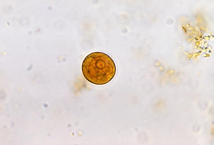
Microfotografía de Entamoeba histolytica, en la fase de quiste
Imagen: “Entamoeba histolytica cyst” por Yasser. Licencia: CC BY 2.0
Trofozoítos de Entamoeba histolytica con eritrocitos ingeridos teñidos con tricrómico:
Los eritrocitos ingeridos aparecen como inclusiones oscuras. Los parásitos contienen núcleos que tienen el típico cariosoma pequeño, situado en el centro, con una cromatina periférica fina y uniforme. La eritrofagocitosis o la ingestión de eritrocitos, es la característica que distingue a E. histolytica de E. dispar.
El ciclo de vida de la Entamoeba Entamoeba A genus of ameboid protozoa characterized by the presence of beaded chromatin on the inner surface of the nuclear membrane. Its organisms are parasitic in invertebrates and vertebrates, including humans. Nitroimidazoles spp. depende de la infección de un huésped, porque la transición a través de las distintas etapas de vida se producen dentro del tracto intestinal del huésped.
Fase de quiste:
Fase de trofozoíto:
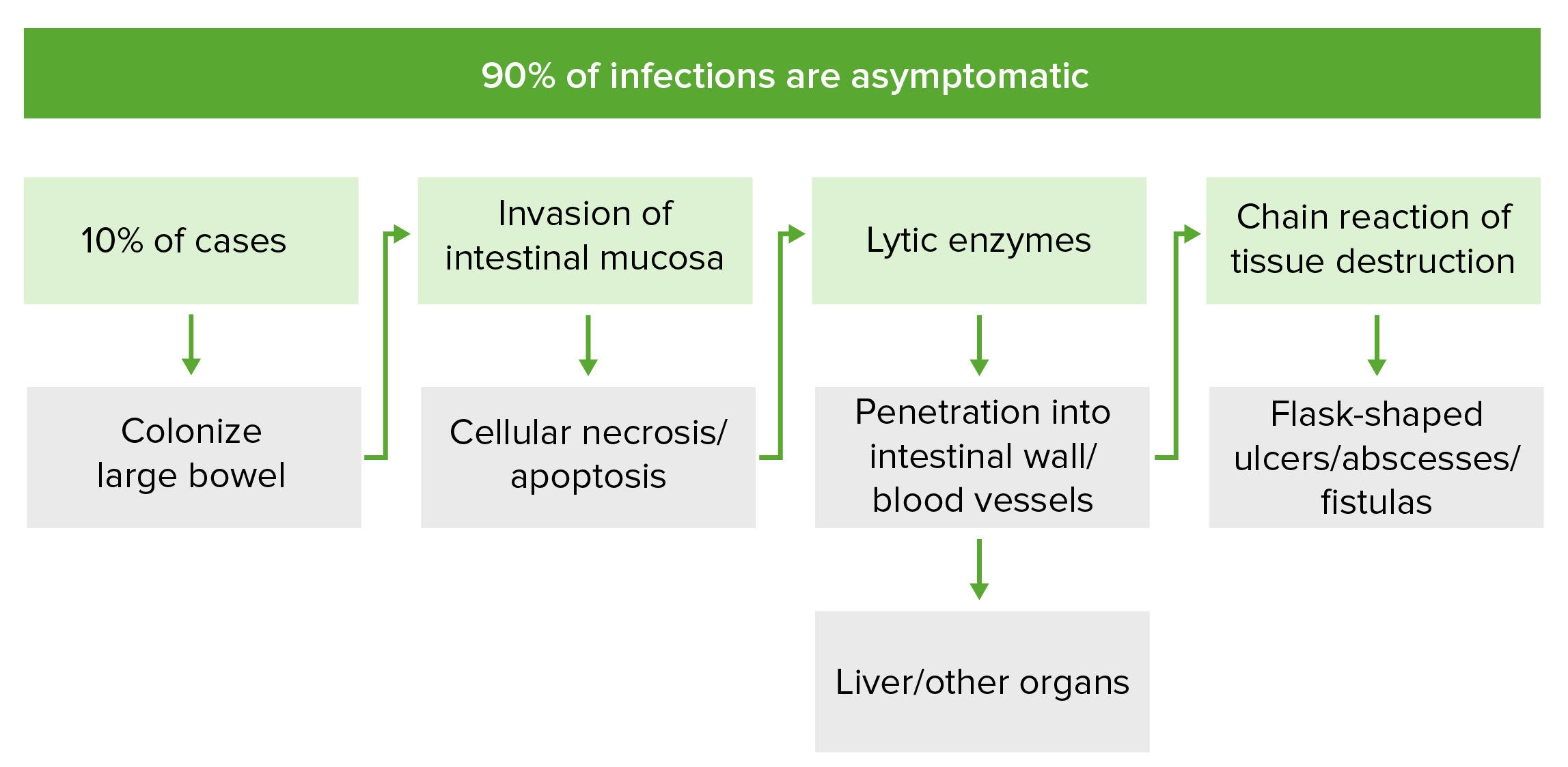
Patogénesis de la infección invasiva por Entamoeba histolytica:
En el 10% de los casos, E. histolytica coloniza la mucosa del intestino grueso e invade mediante la secreción de proteinasas y enzimas líticas. Esto provoca la necrosis celular y la lisis de las membranas, respectivamente. Esta cadena de acontecimientos inducen la apoptosis de las células de la mucosa y alteran las estrechas uniones entre las células, lo que permiten la formación de úlceras en forma de botella, abscesos y fístulas. La invasión puede alcanzar el sistema venoso portal, a través del cual E. histolytica puede extenderse a otros órganos.
El periodo de incubación suele ser de 2–4 semanas una vez ingerido, pero los LOS Neisseria síntomas pueden desarrollarse hasta 1 año después de la infección.
El 90% de las infecciones por Entamoeba Entamoeba A genus of ameboid protozoa characterized by the presence of beaded chromatin on the inner surface of the nuclear membrane. Its organisms are parasitic in invertebrates and vertebrates, including humans. Nitroimidazoles son asintomáticas:
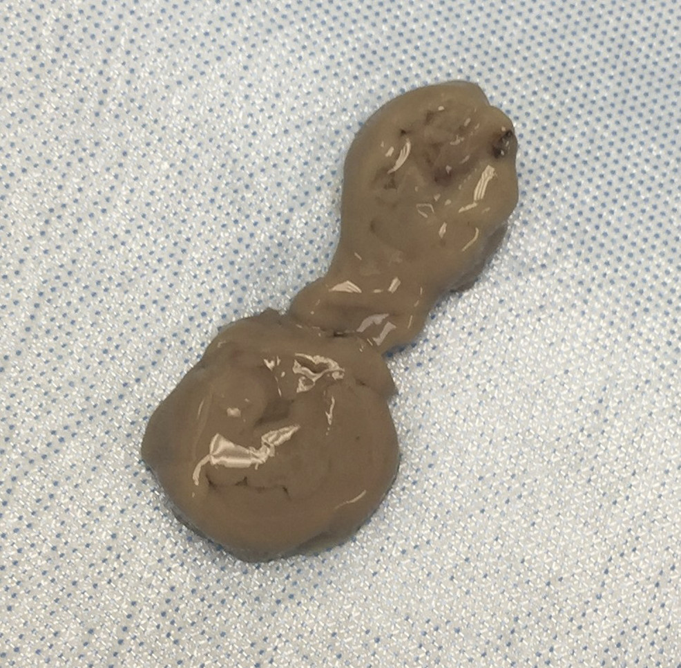
Aspirado de “pasta de anchoas” del absceso hepático de un paciente con una infección invasiva por E. histolytica
Imagen: “Aspirate of “anchovy paste” por Palak Patel et al. Licencia: CC BY 4.0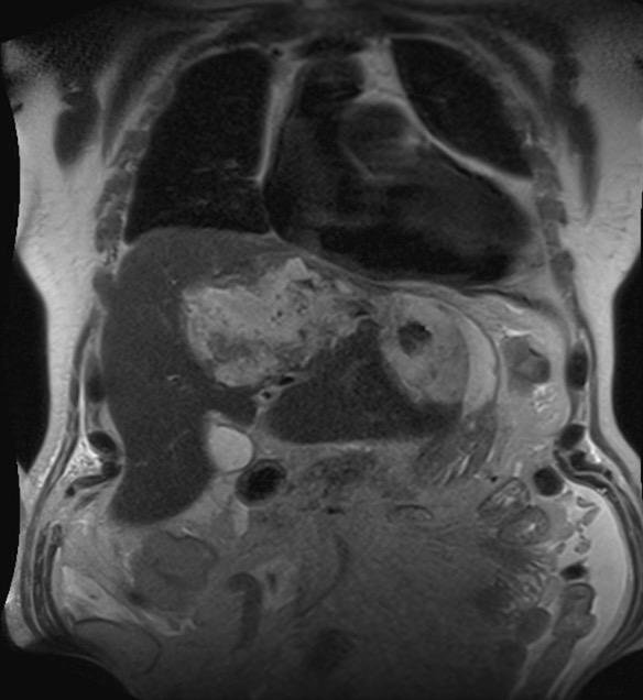
Absceso amebiano en el lóbulo izquierdo del hígado:
Colangiopancreatografía por resonancia magnética coronal ponderada en T2 que muestra una colección de material heterogéneo en el lóbulo izquierdo del hígado
El diagnóstico de la amebiasis Amebiasis Amebiasis, or amoebic dysentery, is an infection caused by the parasite Entamoeba histolytica. Transmission is through the fecal-oral route or by consumption of contaminated food and water. Most patients infected with E. histolytica are asymptomatic, but about 10% may develop dysentery. Entamoeba spp./Amebiasis se basa en EN Erythema nodosum is an immune-mediated panniculitis (inflammation of the subcutaneous fat) caused by a type IV (delayed-type) hypersensitivity reaction. It commonly manifests in young women as tender, erythematous nodules on the shins. Erythema Nodosum la sospecha clínica y en EN Erythema nodosum is an immune-mediated panniculitis (inflammation of the subcutaneous fat) caused by a type IV (delayed-type) hypersensitivity reaction. It commonly manifests in young women as tender, erythematous nodules on the shins. Erythema Nodosum las pruebas confirmatorias. Se pueden utilizar distintas modalidades de pruebas.
Todas las infecciones por E. histolytica deben ser tratadas independientemente de la ausencia o gravedad de los LOS Neisseria síntomas; sin embargo, las infecciones por E. dispar E. dispar Entamoeba spp./Amebiasis no necesitan ser tratadas.
Todas las acciones que causan diarrea inespecífica o sanguinolenta son diagnósticos diferenciales para la amebiasis Amebiasis Amebiasis, or amoebic dysentery, is an infection caused by the parasite Entamoeba histolytica. Transmission is through the fecal-oral route or by consumption of contaminated food and water. Most patients infected with E. histolytica are asymptomatic, but about 10% may develop dysentery. Entamoeba spp./Amebiasis.