El carcinoma de células renales (CCR) es un tumor Tumor Inflammation que surge del revestimiento del sistema tubular renal dentro de la corteza renal. El carcinoma de células renales es responsable del 80%–85% de todas las neoplasias renales primarias. La mayoría de los LOS Neisseria CCR surgen de forma esporádica, pero el tabaquismo, la hipertensión y la obesidad están relacionados con su desarrollo. La enfermedad suele presentarse de manera asintomática. Cuando finalmente aparecen los LOS Neisseria síntomas, el tumor Tumor Inflammation ya ha HA Hemolytic anemia (HA) is the term given to a large group of anemias that are caused by the premature destruction/hemolysis of circulating red blood cells (RBCs). Hemolysis can occur within (intravascular hemolysis) or outside the blood vessels (extravascular hemolysis). Hemolytic Anemia crecido considerablemente y/o se ha HA Hemolytic anemia (HA) is the term given to a large group of anemias that are caused by the premature destruction/hemolysis of circulating red blood cells (RBCs). Hemolysis can occur within (intravascular hemolysis) or outside the blood vessels (extravascular hemolysis). Hemolytic Anemia extendido a otros tejidos. La tríada clínica clásica del CCR es el dolor Dolor Inflammation de costado, la hematuria Hematuria Presence of blood in the urine. Renal Cell Carcinoma y una masa renal abdominal palpable, pero esta tríada solo aparece en EN Erythema nodosum is an immune-mediated panniculitis (inflammation of the subcutaneous fat) caused by a type IV (delayed-type) hypersensitivity reaction. It commonly manifests in young women as tender, erythematous nodules on the shins. Erythema Nodosum un 9% de los LOS Neisseria casos. Los LOS Neisseria individuos afectados también suelen presentar fiebre y/o anemia Anemia Anemia is a condition in which individuals have low Hb levels, which can arise from various causes. Anemia is accompanied by a reduced number of RBCs and may manifest with fatigue, shortness of breath, pallor, and weakness. Subtypes are classified by the size of RBCs, chronicity, and etiology. Anemia: Overview and Types. El carcinoma de células renales suele diagnosticarse mediante una TC de abdomen y pelvis Pelvis The pelvis consists of the bony pelvic girdle, the muscular and ligamentous pelvic floor, and the pelvic cavity, which contains viscera, vessels, and multiple nerves and muscles. The pelvic girdle, composed of 2 "hip" bones and the sacrum, is a ring-like bony structure of the axial skeleton that links the vertebral column with the lower extremities. Pelvis: Anatomy. Los LOS Neisseria casos localizados de CCR suelen tratarse y curarse con cirugía, y los LOS Neisseria casos avanzados se tratan con una combinación de inmunoterapia y/o terapia molecular dirigida. El pronóstico a largo plazo del CCR localmente avanzado o metastásico suele ser malo.
Last updated: Dec 15, 2025
El carcinoma de células renales (CCR), también llamado hipernefroma y adenocarcinoma de células renales, es un tipo de cáncer que se origina en EN Erythema nodosum is an immune-mediated panniculitis (inflammation of the subcutaneous fat) caused by a type IV (delayed-type) hypersensitivity reaction. It commonly manifests in young women as tender, erythematous nodules on the shins. Erythema Nodosum el revestimiento de los LOS Neisseria túbulos pequeños de la corteza renal.
La diferencia entre masas benignas y malignas en EN Erythema nodosum is an immune-mediated panniculitis (inflammation of the subcutaneous fat) caused by a type IV (delayed-type) hypersensitivity reaction. It commonly manifests in young women as tender, erythematous nodules on the shins. Erythema Nodosum el riñón se determina por criterios histológicos. Según la morfología, el patrón de crecimiento, la célula de origen y la base histoquímica y molecular, los LOS Neisseria CCR se clasifican en EN Erythema nodosum is an immune-mediated panniculitis (inflammation of the subcutaneous fat) caused by a type IV (delayed-type) hypersensitivity reaction. It commonly manifests in young women as tender, erythematous nodules on the shins. Erythema Nodosum varios tipos:
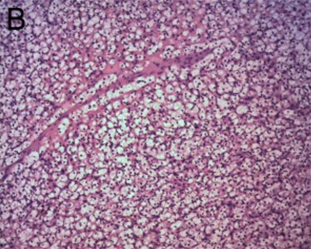
Carcinoma renal de células claras:
El tumor está formado por células claras, sólidas y en forma de lámina. H&E × 100
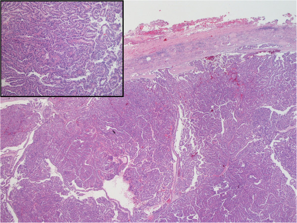
Muestra teñida con H&E que muestra la histología característica del carcinoma de células renales papilar
Imagen: “Hematoxylin and Eosin staining of the tumor tissue showing characteristic histology of papillary renal cell carcinoma” por Alanee S, Dynda DI, Hemmer P, Schwartz B. Licencia: CC BY 4.0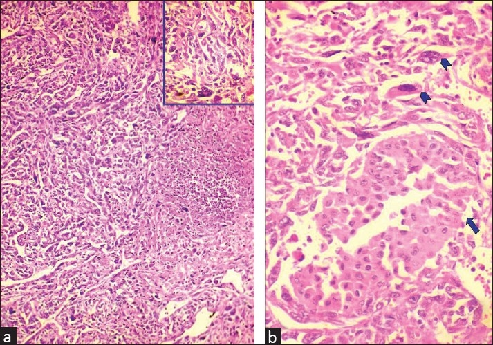
Carcinoma de células renales cromófobo sarcomatoide
a. Microfotografía que muestra un carcinoma renal sarcomatoide con tumores de células gigantes y necrosis (H&E x 100) (el recuadro muestra un tumor de células gigantes entre células pleomórficas (H&E x 400))
b. Microfotografía que muestra un carcinoma renal cromófobo (flecha) mezclado con áreas sarcomatoides con células bizarras y células gigantes (cabezas de flecha)
Tumor renal oncocítico de alto grado
Imagen: “High-grade oncocytic renal tumour — high mag” por Nephron. Licencia: CC BY-SA 4.0 es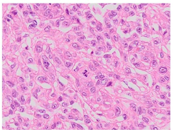
Secciones de tejido teñidas con H&E que muestran un carcinoma del conducto colector
Imagen: “Hematoxylin and eosin staining of tissue sections from the nephrectomy specimens demonstrating collecting duct carcinoma” por Tazi EM, Essadi I, Tazi MF, Ahellal Y, M’rabti H, Errihani H. Licencia: CC BY 2.0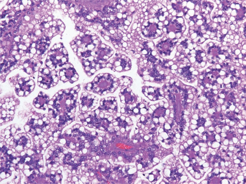
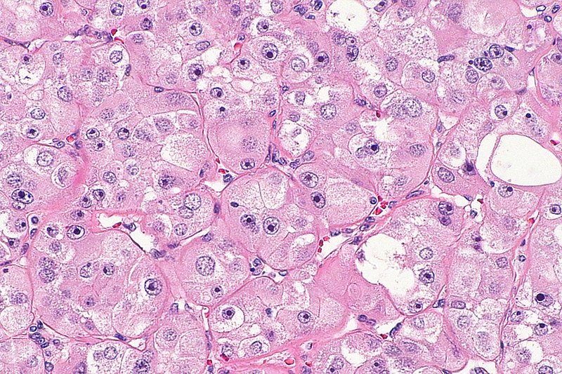
Carcinoma de células renales con translocación, compuesto por células claras con citoplasma voluminoso y bordes celulares definidos que muestran una arquitectura papilar típica y cordones vasculares fibrosos hialinizados
Imagen: “Translocation renal cell carcinoma” por Kmetec A, Jeruc J. Licencia: CC BY 3.0Muchos casos se presentan de forma asintomática, lo que conduce a una mayor progresión de las enfermedades. Los LOS Neisseria síntomas aparecen cuando el tumor Tumor Inflammation ha HA Hemolytic anemia (HA) is the term given to a large group of anemias that are caused by the premature destruction/hemolysis of circulating red blood cells (RBCs). Hemolysis can occur within (intravascular hemolysis) or outside the blood vessels (extravascular hemolysis). Hemolytic Anemia alcanzado un tamaño considerable, ha HA Hemolytic anemia (HA) is the term given to a large group of anemias that are caused by the premature destruction/hemolysis of circulating red blood cells (RBCs). Hemolysis can occur within (intravascular hemolysis) or outside the blood vessels (extravascular hemolysis). Hemolytic Anemia invadido estructuras adyacentes o ha HA Hemolytic anemia (HA) is the term given to a large group of anemias that are caused by the premature destruction/hemolysis of circulating red blood cells (RBCs). Hemolysis can occur within (intravascular hemolysis) or outside the blood vessels (extravascular hemolysis). Hemolytic Anemia desarrollado metástasis a distancia.
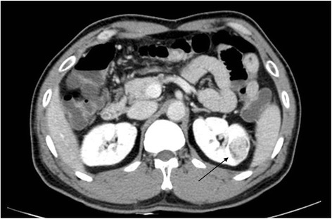
Una TC que muestra un carcinoma de células renales (flecha)
Imagen: “Fig1: CT scan of renal cell carcinoma (Arrow)” por June-Hee Lee et al. Licencia: CC BY 4.0 CC BY 1.0Los LOS Neisseria CCR varían mucho en EN Erythema nodosum is an immune-mediated panniculitis (inflammation of the subcutaneous fat) caused by a type IV (delayed-type) hypersensitivity reaction. It commonly manifests in young women as tender, erythematous nodules on the shins. Erythema Nodosum cuanto a la extensión de la enfermedad en EN Erythema nodosum is an immune-mediated panniculitis (inflammation of the subcutaneous fat) caused by a type IV (delayed-type) hypersensitivity reaction. It commonly manifests in young women as tender, erythematous nodules on the shins. Erythema Nodosum el momento de su presentación.
| Estadio | Definición | Subdivisión |
|---|---|---|
| Estadio del tumor Tumor Inflammation | ||
| T0 | No hay evidencia de tumor Tumor Inflammation primario | |
| T1 | < 7 cm en EN Erythema nodosum is an immune-mediated panniculitis (inflammation of the subcutaneous fat) caused by a type IV (delayed-type) hypersensitivity reaction. It commonly manifests in young women as tender, erythematous nodules on the shins. Erythema Nodosum su mayor dimensión, confinado en EN Erythema nodosum is an immune-mediated panniculitis (inflammation of the subcutaneous fat) caused by a type IV (delayed-type) hypersensitivity reaction. It commonly manifests in young women as tender, erythematous nodules on the shins. Erythema Nodosum el riñón |
|
| T2 | > 7 cm en EN Erythema nodosum is an immune-mediated panniculitis (inflammation of the subcutaneous fat) caused by a type IV (delayed-type) hypersensitivity reaction. It commonly manifests in young women as tender, erythematous nodules on the shins. Erythema Nodosum su mayor dimensión, confinado en EN Erythema nodosum is an immune-mediated panniculitis (inflammation of the subcutaneous fat) caused by a type IV (delayed-type) hypersensitivity reaction. It commonly manifests in young women as tender, erythematous nodules on the shins. Erythema Nodosum el riñón |
|
| T3 T3 A T3 thyroid hormone normally synthesized and secreted by the thyroid gland in much smaller quantities than thyroxine (T4). Most T3 is derived from peripheral monodeiodination of T4 at the 5′ position of the outer ring of the iodothyronine nucleus. The hormone finally delivered and used by the tissues is mainly t3. Thyroid Hormones | Se extiende hacia las venas principales o los LOS Neisseria tejidos perinéfricos, pero no hacia la glándula suprarrenal o más allá de la fascia Fascia Layers of connective tissue of variable thickness. The superficial fascia is found immediately below the skin; the deep fascia invests muscles, nerves, and other organs. Cellulitis de Gerota ( fascia Fascia Layers of connective tissue of variable thickness. The superficial fascia is found immediately below the skin; the deep fascia invests muscles, nerves, and other organs. Cellulitis perirrenal anterior) |
|
| T4 T4 The major hormone derived from the thyroid gland. Thyroxine is synthesized via the iodination of tyrosines (monoiodotyrosine) and the coupling of iodotyrosines (diiodotyrosine) in the thyroglobulin. Thyroxine is released from thyroglobulin by proteolysis and secreted into the blood. Thyroxine is peripherally deiodinated to form triiodothyronine which exerts a broad spectrum of stimulatory effects on cell metabolism. Thyroid Hormones | Invade más allá de la fascia Fascia Layers of connective tissue of variable thickness. The superficial fascia is found immediately below the skin; the deep fascia invests muscles, nerves, and other organs. Cellulitis de Gerota y/o la glándula suprarrenal | |
| Ganglios linfáticos regionales | ||
| N0 | No hay metástasis en EN Erythema nodosum is an immune-mediated panniculitis (inflammation of the subcutaneous fat) caused by a type IV (delayed-type) hypersensitivity reaction. It commonly manifests in young women as tender, erythematous nodules on the shins. Erythema Nodosum los LOS Neisseria ganglios linfáticos regionales | |
| N1 | Metástasis en EN Erythema nodosum is an immune-mediated panniculitis (inflammation of the subcutaneous fat) caused by a type IV (delayed-type) hypersensitivity reaction. It commonly manifests in young women as tender, erythematous nodules on the shins. Erythema Nodosum los LOS Neisseria ganglios linfáticos regionales | |
| Metástasis a distancia | ||
| M0 | No hay metástasis a distancia | |
| M1 | Metástasis en EN Erythema nodosum is an immune-mediated panniculitis (inflammation of the subcutaneous fat) caused by a type IV (delayed-type) hypersensitivity reaction. It commonly manifests in young women as tender, erythematous nodules on the shins. Erythema Nodosum ganglios linfáticos y/u órganos distantes | |