>

>

Cardiac Output: Equation & Normal Values

Nursing Knowledge

Cardiac Output: Equation & Normal Values

Cardiac output is a measure of heart function that is used to assess the adequacy of blood flow to the body’s tissues. Understanding the determinants of cardiac output is essential for nurses and other healthcare professionals who care for clients with cardiovascular disease. This knowledge can help them identify and manage clients who are at risk of developing complications related to poor cardiac output.
Last updated: December 4, 2023

Table of contents

What is cardiac output and how is it calculated? 

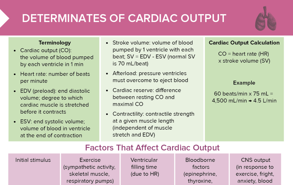
Cardiac output is the volume of blood pumped by each ventricle in one minute. It is calculated by multiplying heart rate by stroke volume.

Cardiac output (CO) = heart rate (HR) x stroke volume (SV)

What is cardiac reserve? 

Cardiac reserve is the difference between resting cardiac output and maximal cardiac output. 

What is preload? 

Preload (also known as end diastolic volume, EDV) is the degree to which the cardiac muscle is stretched before it contracts. 

What is the end systolic volume? 

The end systolic volume (ESV) is the volume of blood in the ventricle at the end of contraction. 

What is afterload? 

Afterload is the pressure ventricles must overcome to eject blood. 

What is contractility? 

Contractility refers to the contractile strength at a given muscle length (independent of muscle stretch and EDV).

What is stroke volume? 

Stroke volume is the volume of blood pumped by one ventricle with each beat.

Stroke volume = EDV – ESV

What are normal cardiac output values? 

  • Normal cardiac output: 4–8 L/min
    • Maximum CO: 20–25 L/min in non-athletes; can reach 35 L/min in elite athletes
  • Normal stroke volume: 50–100 mL
  • Normal heart rate: 60–100 beats per minute
  • Normal EDV (preload): 70–244 mL (dependent on age, sex, ventricle)
  • Normal ESV: 15–117 mL (dependent on age, sex, ventricle) 
  • Normal cardiac reserve: up to 4–5x greater than resting value 

Factors that affect cardiac output 

  • Exercise → ↑ Venous return → ↑ EDV (preload) or ↓ afterload → ↑ stroke volume
  • Ventricular filling time (due to heart rate) → ↑ venous return → ↑ EDV (preload) or ↓ afterload → ↑ stroke volume
  • Blood-borne factors (epinephrine, thyroxine, excess Ca2+) → ↑ contractility → ↓ ESV → ↑ stroke volume
  • CNS output (in response to exercise, fright, blood pressure) → ↑ sympathetic activity → ↑ heart rate 

Result: Increased cardiac output (CO = SV x HR). The opposite relationship also occurs, resulting in decreased cardiac output. 

Decreased cardiac output: nursing diagnosis & interventions 

Decreased cardiac output occurs when the heart’s function as a pump is compromised. 

Potential nursing diagnoses include: 

  • Fluid volume excess: monitor fluid balance, administer diuretics as prescribed, monitor for signs of pulmonary congestion, provide low-sodium diet
  • Activity intolerance: schedule activities with rest periods, encourage gradual activity increase 
  • Anxiety: provide calm environment and reassurance

FREE CHEAT SHEET

Cardiac Output: Equation & Normal Values

Free Download

Nursing Cheat Sheet

Review of factors that affect cardiac output, terminology, normal values, and calculations

Master the topic with a unique study combination of a concise summary paired with video lectures. 

User Reviews