La enfermedad de Chagas es una infección causada por el tripanosoma americano Trypanosoma cruzi Trypanosoma cruzi Chagas disease is an infection caused by the American trypanosome Trypanosoma cruzi. This parasitic protozoan is transmitted in the feces of reduviid bugs in South and Central America. Acute infection may present with inflammation at the inoculation site (chagoma), fever, and lymphadenopathy. Untreated, chronic infection can progress to severe complications. Trypanosoma cruzi/Chagas disease. Este protozoo parásito se transmite en EN Erythema nodosum is an immune-mediated panniculitis (inflammation of the subcutaneous fat) caused by a type IV (delayed-type) hypersensitivity reaction. It commonly manifests in young women as tender, erythematous nodules on the shins. Erythema Nodosum las heces de las chinches triatominas (familia Reduviidae, subfamilia Triatominae) en EN Erythema nodosum is an immune-mediated panniculitis (inflammation of the subcutaneous fat) caused by a type IV (delayed-type) hypersensitivity reaction. It commonly manifests in young women as tender, erythematous nodules on the shins. Erythema Nodosum América del Sur y Central. La infección aguda puede presentarse con inflamación en EN Erythema nodosum is an immune-mediated panniculitis (inflammation of the subcutaneous fat) caused by a type IV (delayed-type) hypersensitivity reaction. It commonly manifests in young women as tender, erythematous nodules on the shins. Erythema Nodosum el sitio de inoculación ( chagoma Chagoma Trypanosoma cruzi/Chagas disease), fiebre y linfadenopatía. Si no se trata, la infección crónica puede evolucionar a complicaciones graves, como megacolon Megacolon Megacolon is a severe, abnormal dilatation of the colon, and is classified as acute or chronic. There are many etiologies of megacolon, including neuropathic and dysmotility conditions, severe infections, ischemia, and inflammatory bowel disease. Megacolon, megaesófago y cardiomiopatía. El diagnóstico puede confirmarse con la identificación de los LOS Neisseria organismos en EN Erythema nodosum is an immune-mediated panniculitis (inflammation of the subcutaneous fat) caused by a type IV (delayed-type) hypersensitivity reaction. It commonly manifests in young women as tender, erythematous nodules on the shins. Erythema Nodosum el frotis de sangre, la serología o PCR PCR Polymerase chain reaction (PCR) is a technique that amplifies DNA fragments exponentially for analysis. The process is highly specific, allowing for the targeting of specific genomic sequences, even with minuscule sample amounts. The PCR cycles multiple times through 3 phases: denaturation of the template DNA, annealing of a specific primer to the individual DNA strands, and synthesis/elongation of new DNA molecules. Polymerase Chain Reaction (PCR). El tratamiento con benzinidazol o nifurtimox Nifurtimox A nitrofuran thiazine that has been used against trypanosomiasis. Trypanosoma cruzi/Chagas disease solo es eficaz en EN Erythema nodosum is an immune-mediated panniculitis (inflammation of the subcutaneous fat) caused by a type IV (delayed-type) hypersensitivity reaction. It commonly manifests in young women as tender, erythematous nodules on the shins. Erythema Nodosum la fase aguda.
Last updated: Dec 15, 2025
Microfotografía de un frotis de sangre que muestra 5 tripomastigotes parasitarios flagelados de Trypanosoma cruzi
Imagen: “Photomicrograph of a blood smear showing five parasitic, flagellated, Trypanosoma cruzi trypomastigotes” por CDC. Licencia: Dominio Público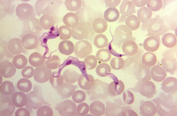
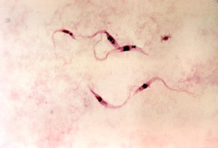
4 parásitos flagelados de Trypanosoma cruzi, denominados tripomastigotes
Imagen: “Under a magnification of 1200X, this photomicrograph of a blood sample specimen, revealed the presence of four flagellated, Trypanosoma cruzi parasites” por CDC. Licencia: Dominio PúblicoLa tripanosomiasis americana es llamada enfermedad de Chagas.
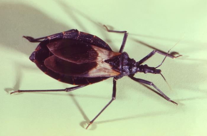
Una especie de Triatoma o chinche besucona:
El chinche del beso sirve como vector para transmitir el patógeno protozoario Trypanosoma cruzi, causante de la enfermedad de Chagas.
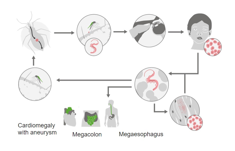
Ciclo de vida del tripanosoma americano, Trypanosoma cruzi:
Mientras se alimenta de sangre, el insecto redúvido defecará. El rascado de la zona permite la entrada de parásitos a través de la picadura o a través de la conjuntiva.
Una vez dentro del cuerpo, se produce la replicación y diseminación. Hay una preferencia particular por el miocardio y el plexo mientérico.
Con la infección crónica, el daño tisular puede conducir a cardiomiopatía, megacolon y megaesófago.
El periodo de incubación es de aproximadamente 1-2 semanas y la infección dura entre 8 y 12 semanas.
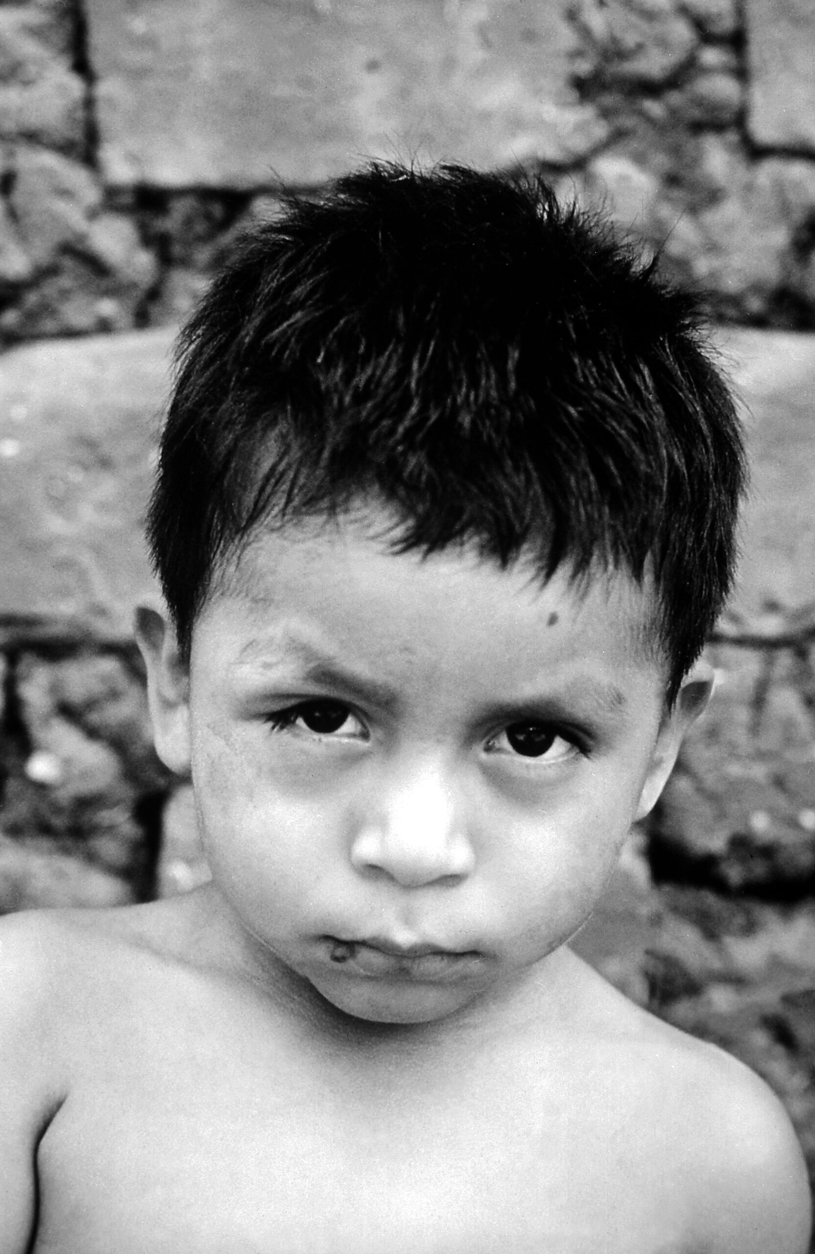
Fotografía de un paciente con infección aguda de la enfermedad de Chagas con edema del ojo derecho (signo de Romaña).
Imagen:“An acute Chagas disease infection with swelling of the right eye (Romaña’s sign)” por CDC. Licencia: Dominio PúblicoUna minoría de los LOS Neisseria pacientes desarrolla una infección crónica, que se presenta entre 10 y 20 años después del periodo de inoculación inicial.
La enfermedad congénita se produce en EN Erythema nodosum is an immune-mediated panniculitis (inflammation of the subcutaneous fat) caused by a type IV (delayed-type) hypersensitivity reaction. It commonly manifests in young women as tender, erythematous nodules on the shins. Erythema Nodosum la minoría de los LOS Neisseria bebés nacidos de madres infectadas.
Pruebas confirmatorias:
Evaluación de apoyo:
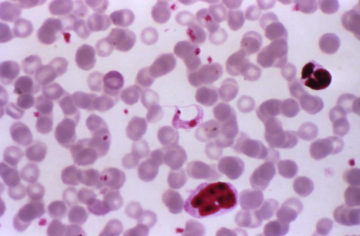
Fotomicrografía de una muestra de frotis de sangre teñida con Giemsa que revela la presencia de un tripomastigote de Trypanosoma cruzi
Imagen: “Under a magnification of 1000X, this photomicrograph of a Giemsa stained blood smear specimen revealed the presence of a parasitic Trypanosoma cruzi protozoan” por CDC. Licencia: Dminio Público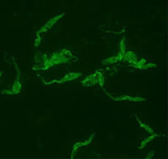
Prueba indirecta de anticuerpos fluorescentes en un frotis de sangre de un paciente infectado por Trypanosoma cruzi.
Imagen por Lecturio. Licencia: CC BY-NC-SA 4.0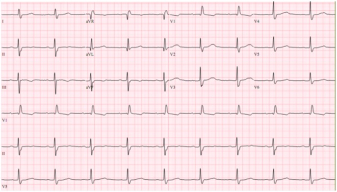
ECG que muestra un bloqueo de rama derecha con bloqueo fascicular anterior izquierdo en un paciente con enfermedad de Chagas
Imagen: “ECG showing a right bundle branch block with left anterior fascicular block” por de Alencar MC et al. Licencia: CC BY 4.0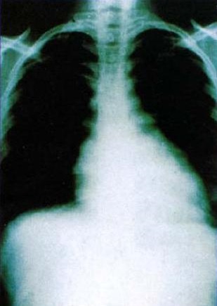
Radiografía de tórax que muestra un agrandamiento cardíaco debido a una cardiomiopatía chagásica crónica
Imagen: “Heart radiology Chagas disease” por CDC. Licencia: Dominio Público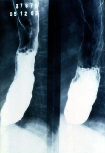
Esofagrama de bario que demuestra el megaesófago en un paciente con enfermedad de Chagas
Imagen: “Chagas megaseophagus” por CDC. Licencia: Dominio PúblicoInfección aguda:
Enfermedad crónica:
| Giardia Giardia A genus of flagellate intestinal eukaryotes parasitic in various vertebrates, including humans. Characteristics include the presence of four pairs of flagella arising from a complicated system of axonemes and cysts that are ellipsoidal to ovoidal in shape. Nitroimidazoles | Leishmania Leishmania Leishmania species are obligate intracellular parasites that are transmitted by an infected sandfly. The disease is endemic to Asia, the Middle East, Africa, the Mediterranean, and South and Central America. Clinical presentation varies, dependent on the pathogenicity of the species and the host’s immune response. Leishmania/Leishmaniasis | Trypanosoma | Trichomonas Trichomonas A genus of parasitic flagellate eukaryotes distinguished by the presence of four anterior flagella, an undulating membrane, and a trailing flagellum. Nitroimidazoles | |
|---|---|---|---|---|
| Características |
|
|
|
|
| Variantes |
|
|
|
|
| Transmisión |
|
|
|
Transmisión sexual |
| Cuadro Clínico | Giardiasis Giardiasis An infection of the small intestine caused by the flagellated protozoan giardia. It is spread via contaminated food and water and by direct person-to-person contact. Giardia/Giardiasis | Leishmaniasis Leishmaniasis Leishmania species are obligate intracellular parasites that are transmitted by an infected sandfly. The mildest form is cutaneous leishmaniasis (CL), characterized by painless skin ulcers. The mucocutaneous type involves more tissue destruction, causing deformities. Visceral leishmaniasis (VL), the most severe form, presents with hepatosplenomegaly, anemia, thrombocytopenia, and fever. Leishmania/Leishmaniasis |
|
Tricomoniasis |
| Diagnóstico |
|
|
|
|
| Tratamiento |
|
Depende del síndrome clínico:
|
Depende de la enfermedad clínica:
|
|
| Prevención |
|
|
|
|
Tinción de Giemsa de trofozoitos de Giardia lamblia
Imagen: “Trophozoites of Giardia lamblia” por Eva Nohýnková, Department of Tropical Medicine, 1st Faculty of Medicine, Charles University in Prague and Hospital Bulovka, Czech Republic. Licencia: CC BY 4.0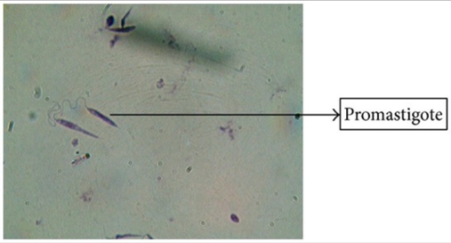
Tinción de Giemsa de promastigotes de Leishmania
Imagen: “Giemsa stain” por Arriyadh Community College, King Saud University, P.O. Box 28095, Riyadh 11437, Saudi Arabia. Licencia: CC BY 3.0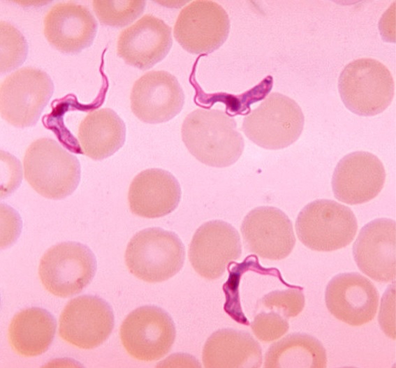
Frotis de sangre que demuestra la presencia de tripomastigotes de Trypanosoma
Imagen por Lecturio. License: CC BY-NC-SA 4.0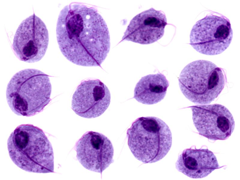
Imágenes microscópicas de trofozoitos de Trichomonas vaginalis
Imagen: “Trichomonas protozoa” por isis325. Licencia: CC BY 2.0.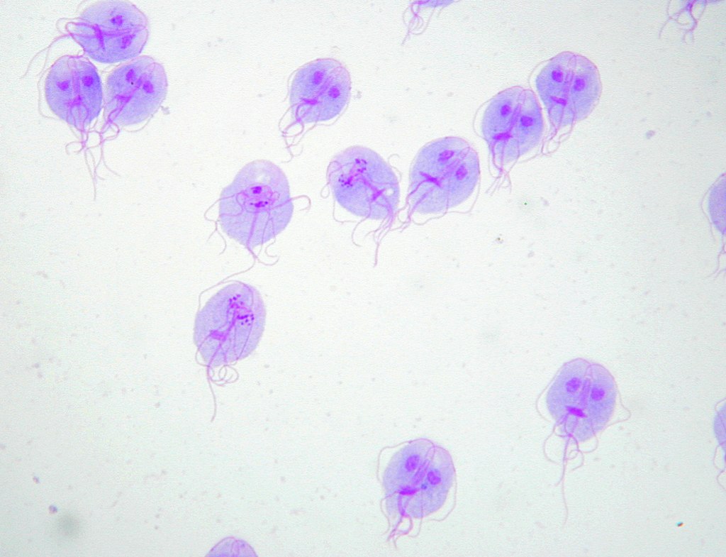