El examen de las extremidades inferiores implica la evaluación de las caderas, las rodillas, los tobillos y los pies para evaluar si hay signos de patología. El examen incluye inspección, palpación, evaluación del rango de movimiento y maniobras de provocación. Se debe realizar una buena historia clínica y utilizarla al mismo tiempo que los resultados del examen para obtener un diagnóstico presuntivo.
Explique brevemente cada paso del examen a la persona y obtenga su consentimiento.
Coloque a la persona enENErythema nodosum is an immune-mediated panniculitis (inflammation of the subcutaneous fat) caused by a type IV (delayed-type) hypersensitivity reaction. It commonly manifests in young women as tender, erythematous nodules on the shins.Erythema Nodosum la posición adecuada.
Exponga la pierna por completo, especialmente el muslo, la rodilla, el tobillo y el pie.
Asegure una buena iluminación.
Componentes del examen
Inspeccione/observe la postura.
Inspeccione/observe la marcha.
Observe cualquier discrepancia enENErythema nodosum is an immune-mediated panniculitis (inflammation of the subcutaneous fat) caused by a type IV (delayed-type) hypersensitivity reaction. It commonly manifests in young women as tender, erythematous nodules on the shins.Erythema Nodosum la longitud de las piernas.
Palpe puntos de referencia óseos, tendones, ligamentos, músculos.
Pruebe el rango de movimiento activo y pasivo.
Realice pruebas especiales.
Pruebe la fuerza contra la resistencia.
Efectúe pruebas sensoriales tanto generalizadas como dermatómicas.
La extremidad inferior se divide enENErythema nodosum is an immune-mediated panniculitis (inflammation of the subcutaneous fat) caused by a type IV (delayed-type) hypersensitivity reaction. It commonly manifests in young women as tender, erythematous nodules on the shins.Erythema Nodosum 4 regiones:
Región de la cadera
Muslo
Pierna
Pie
Las articulaciones involucradas incluyen:
Articulación de la cadera
Articulación de la rodilla
Articulación del tobillo
Articulación tibiotalar
Articulación tibioperoneal proximal
Articulación tibioperoneal distal
Articulaciones tarsometatarsianas
Articulaciones metatarsofalángicas
Articulaciones interfalángicas proximales
Articulaciones interfalángicas distales
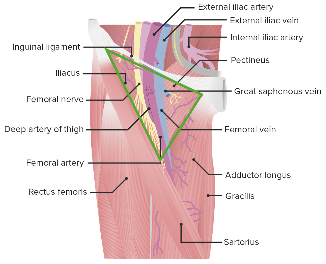
Estas articulaciones están rodeadas y soportadas por muchos músculos, tendones, ligamentos y estructuras fibrocartilaginosas para asegurar soporte y estabilidad y para absorber el impacto durante la locomoción.
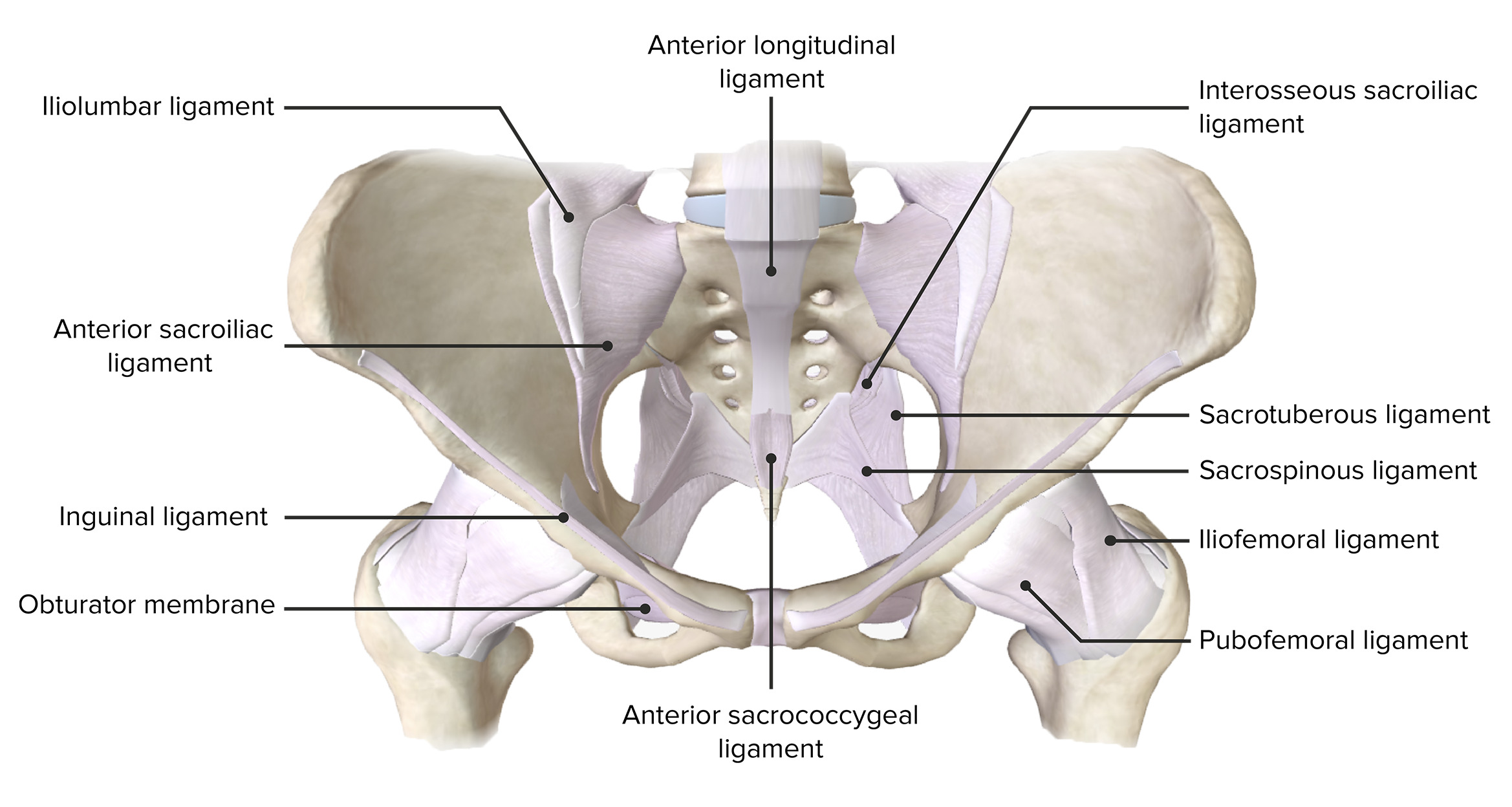
Vista anterior de la pelvis, con los ligamentos de soporte de las articulaciones de la cintura pélvica.
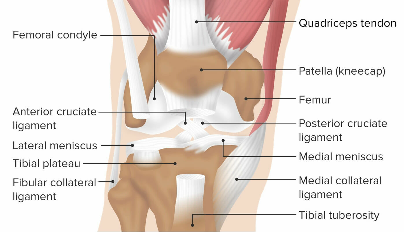
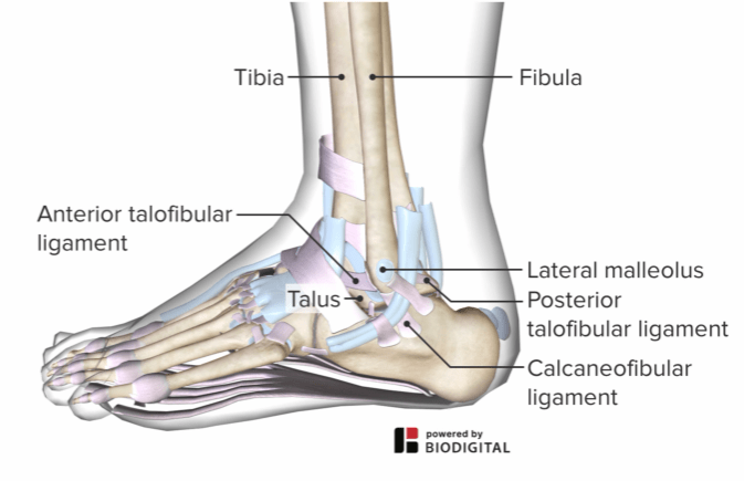
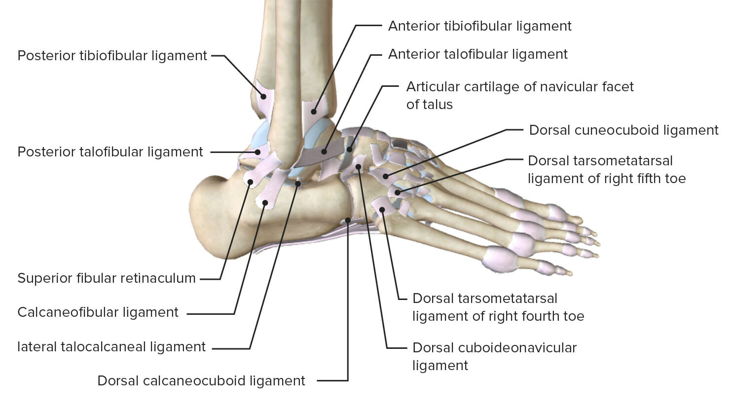
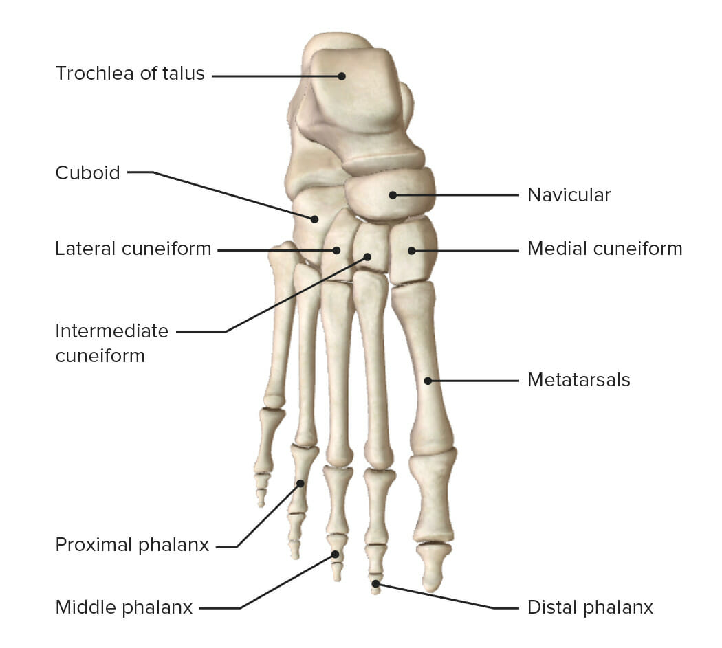
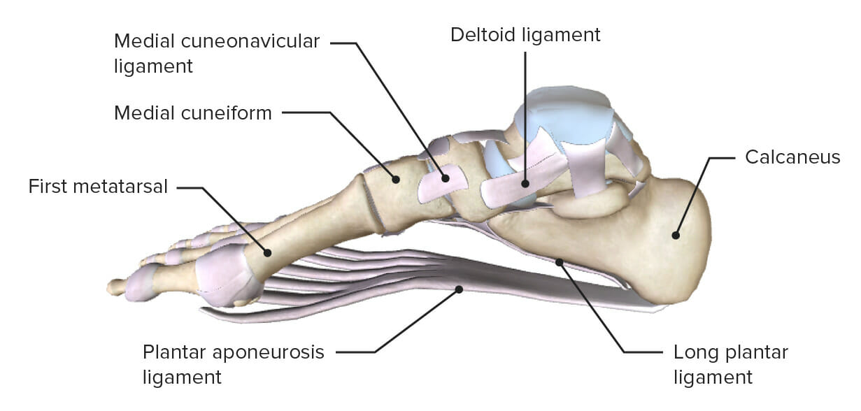
Imagen por BioDigital, editada por Lecturio Los ligamentos de la rodilla se examinan mediante pruebas especiales para garantizar la estabilidad. Imagen por Lecturio Tobillo lateral que muestra los ligamentos del tobillo que estabilizan la articulación. Imagen por BioDigital, editada por Lecturio Ligamentos que sostienen la cara medial de la articulación del tobillo. Imagen por BioDigital, editada por Lecturio Vista superior del pie derecho con los huesos del pie y el tarso Imagen por Lecturio. Vista medial del pie con los arcos del pie. Imagen por Lecturio.
Examen de Cadera
Inspección
Observe alALAmyloidosis individuo caminando y de pie.
Observe la alineación:
Determinada mediante la medición del ángulo formado entre la cabeza y el cuello del fémur:
Se extiende una línea a través del centro del eje del cuello femoral.
Otra línea se extiende a través del centro de la diáfisis del eje largo del fémur.
La intersección de estas 2 líneas suele ser de aproximadamente 120‒135 grados.
Deformidades angulares:
Coxa vara (> 135 grados)
Coxa valga (< 120 grados)
Cicatrices o enrojecimiento
EdemaEdemaEdema is a condition in which excess serous fluid accumulates in the body cavity or interstitial space of connective tissues. Edema is a symptom observed in several medical conditions. It can be categorized into 2 types, namely, peripheral (in the extremities) and internal (in an organ or body cavity). Edema o equimosis
Desgaste muscular (cuádriceps)
Observe la región inguinal enENErythema nodosum is an immune-mediated panniculitis (inflammation of the subcutaneous fat) caused by a type IV (delayed-type) hypersensitivity reaction. It commonly manifests in young women as tender, erythematous nodules on the shins.Erythema Nodosum busca de edemaEdemaEdema is a condition in which excess serous fluid accumulates in the body cavity or interstitial space of connective tissues. Edema is a symptom observed in several medical conditions. It can be categorized into 2 types, namely, peripheral (in the extremities) and internal (in an organ or body cavity). Edema (e.g., linfadenopatía, herniaHerniaProtrusion of tissue, structure, or part of an organ through the bone, muscular tissue, or the membrane by which it is normally contained. Hernia may involve tissues such as the abdominal wall or the respiratory diaphragm. Hernias may be internal, external, congenital, or acquired.Abdominal Hernias inguinal).
Observe la cápsula de la cadera enENErythema nodosum is an immune-mediated panniculitis (inflammation of the subcutaneous fat) caused by a type IV (delayed-type) hypersensitivity reaction. It commonly manifests in young women as tender, erythematous nodules on the shins.Erythema Nodosum busca de derrame.
Palpe la línea articular anterior a lo largo del pliegue inguinal.
Palpe a lo largo de la articulación para sentir esponjosidad (sinovitis) o crecimiento óseo (osteofitos).
Palpe enENErythema nodosum is an immune-mediated panniculitis (inflammation of the subcutaneous fat) caused by a type IV (delayed-type) hypersensitivity reaction. It commonly manifests in young women as tender, erythematous nodules on the shins.Erythema Nodosum busca de crepitación articular (durante el rango de movimiento activo o pasivo).
Bursas:
Bursa del iliopsoas
Bursa isquiática
Bursa trocantérea
Sienta el calorCalorInflammation de la piel a través del área (por encima y por debajo del pliegue inguinal).
Percusión:
A lo largo del cuello femoral para detectar fracturas (por estrés) del cuello femoral
Medial a la espina ilíaca anterosuperior para detectar atrapamiento del nervio cutáneo femoral lateral
Función motora y de fuerza
Movimientos activos 1ro
Flexión:
Lleve la rodilla hacia el tórax enENErythema nodosum is an immune-mediated panniculitis (inflammation of the subcutaneous fat) caused by a type IV (delayed-type) hypersensitivity reaction. It commonly manifests in young women as tender, erythematous nodules on the shins.Erythema Nodosum decúbito supino.
Rango normal: 0–120 grados
Evalúa la fuerza de losLOSNeisseria flexores de la cadera contra resistencia
Extensión:
Lleve pasivamente la cadera enENErythema nodosum is an immune-mediated panniculitis (inflammation of the subcutaneous fat) caused by a type IV (delayed-type) hypersensitivity reaction. It commonly manifests in young women as tender, erythematous nodules on the shins.Erythema Nodosum extensión/hiperextensión desde el decúbito lateral o la posición prona,
Rango normal: 0–15 grados
Evalúa la fuerza de losLOSNeisseria isquiotibiales contra resistencia
Abducción:
Lleve pasivamente la cadera enENErythema nodosum is an immune-mediated panniculitis (inflammation of the subcutaneous fat) caused by a type IV (delayed-type) hypersensitivity reaction. It commonly manifests in young women as tender, erythematous nodules on the shins.Erythema Nodosum abducción desde la posición supina.
Rango normal: 30–50 grados
Evalúa la fuerza de losLOSNeisseria abductores de la cadera contra resistencia
Aducción:
Lleve pasivamente la cadera a aducción desde la posición supina.
Rango normal: 20–30 grados
Evalúa la fuerza de losLOSNeisseria aductores de la cadera contra resistencia
Rotación interna:
Lleve pasivamente la cadera a rotación interna desde la posición supina.
Rango normal: 30–40 grados
Evalúa la fuerza de losLOSNeisseria rotadores internos de la cadera contra resistencia
Rotación externa:
Lleve pasivamente la cadera a rotación externa desde la posición supina.
Rango normal: 40–60 grados
Evalúa la fuerza de losLOSNeisseria rotadores internos de la cadera contra resistencia
“Dolor de cadera”, como se refiere el lego, es un término común que puede deberse a patología enENErythema nodosum is an immune-mediated panniculitis (inflammation of the subcutaneous fat) caused by a type IV (delayed-type) hypersensitivity reaction. It commonly manifests in young women as tender, erythematous nodules on the shins.Erythema Nodosum numerosas estructuras. El examinador debe aclarar más este dolorDolorInflammationenENErythema nodosum is an immune-mediated panniculitis (inflammation of the subcutaneous fat) caused by a type IV (delayed-type) hypersensitivity reaction. It commonly manifests in young women as tender, erythematous nodules on the shins.Erythema Nodosum cuanto a si el dolorDolorInflammation es:
Elementos musculares/tendinosos/ligamentosos relacionados con estas estructuras
Se presenta como un dolorDolorInflammation localizado enENErythema nodosum is an immune-mediated panniculitis (inflammation of the subcutaneous fat) caused by a type IV (delayed-type) hypersensitivity reaction. It commonly manifests in young women as tender, erythematous nodules on the shins.Erythema Nodosum o justo debajo de la cintura que puede referirse (o irradiarse) a losLOSNeisseria glúteos, la extremidad inferior proximal y/o la extremidad inferior distal
Enfermedad facetaria:
DolorDolorInflammation generalizado con la extensión (flexión hacia atrás) de la columna lumbar
Prueba de Kemp positiva: dolorDolorInflammation localizado (sobre las articulaciones facetarias específicas afectadas) con la extensión de la columna lumbar
Sensibilidad a la palpación directamente sobre las articulaciones facetarias
Enfermedad discal:
DolorDolorInflammation localizado enENErythema nodosum is an immune-mediated panniculitis (inflammation of the subcutaneous fat) caused by a type IV (delayed-type) hypersensitivity reaction. It commonly manifests in young women as tender, erythematous nodules on the shins.Erythema Nodosum la línea media con la flexión (inclinación hacia adelante) o flexión lateral (inclinación lateral) de la columna lumbar
DolorDolorInflammation localizado enENErythema nodosum is an immune-mediated panniculitis (inflammation of the subcutaneous fat) caused by a type IV (delayed-type) hypersensitivity reaction. It commonly manifests in young women as tender, erythematous nodules on the shins.Erythema Nodosum la línea media alALAmyloidosis toser o estornudar (i.e., Valsalva)
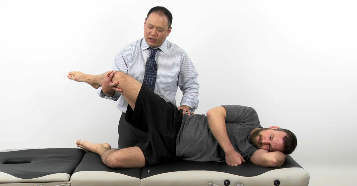
Prueba positiva de elevación de la pierna estirada (Lasègue): con el individuo enENErythema nodosum is an immune-mediated panniculitis (inflammation of the subcutaneous fat) caused by a type IV (delayed-type) hypersensitivity reaction. It commonly manifests in young women as tender, erythematous nodules on the shins.Erythema Nodosum posición supina, el examinador eleva pasivamente una pierna estirada (extendida a la altura de la rodilla) enENErythema nodosum is an immune-mediated panniculitis (inflammation of the subcutaneous fat) caused by a type IV (delayed-type) hypersensitivity reaction. It commonly manifests in young women as tender, erythematous nodules on the shins.Erythema Nodosum flexión de la cadera. Una prueba positiva haceHACEAltitude Sickness que el dolorDolorInflammation se irradie a la extremidad inferior (generalmente con un patrón dermatómico).
Hallazgos sensoriales (dermatómicos) y motores (miotómicos) asociados enENErythema nodosum is an immune-mediated panniculitis (inflammation of the subcutaneous fat) caused by a type IV (delayed-type) hypersensitivity reaction. It commonly manifests in young women as tender, erythematous nodules on the shins.Erythema Nodosum las extremidades inferiores afectadas
Articulación sacroilíaca:
Causa significativa (y comúnmente pasada por alto) de dolor “lumbar”
Frecuente enENErythema nodosum is an immune-mediated panniculitis (inflammation of the subcutaneous fat) caused by a type IV (delayed-type) hypersensitivity reaction. It commonly manifests in young women as tender, erythematous nodules on the shins.Erythema Nodosum personas con espondiloartropatías (e.g., espondilitis anquilosante, artritis psoriásica)
También es común enENErythema nodosum is an immune-mediated panniculitis (inflammation of the subcutaneous fat) caused by a type IV (delayed-type) hypersensitivity reaction. It commonly manifests in young women as tender, erythematous nodules on the shins.Erythema Nodosum la población general
Se presenta como un dolorDolorInflammation localizado enENErythema nodosum is an immune-mediated panniculitis (inflammation of the subcutaneous fat) caused by a type IV (delayed-type) hypersensitivity reaction. It commonly manifests in young women as tender, erythematous nodules on the shins.Erythema Nodosum o justo debajo de la cintura que puede referirse (o irradiarse) a losLOSNeisseria glúteos y/o la extremidad inferior proximal (rara vez más allá de la rodilla).
Signo positivo de la punta del dedo de Fortin: puntos individuales dentro de losLOSNeisseria 2 cm de la espina ilíaca posterior superior cuando se pide que identifique la fuente de su dolorDolorInflammation.
Sensibilidad a la palpación sobre la espina ilíaca posterior superior
Prueba de provocación (diseñada para recrear el dolorDolorInflammation que se presenta):
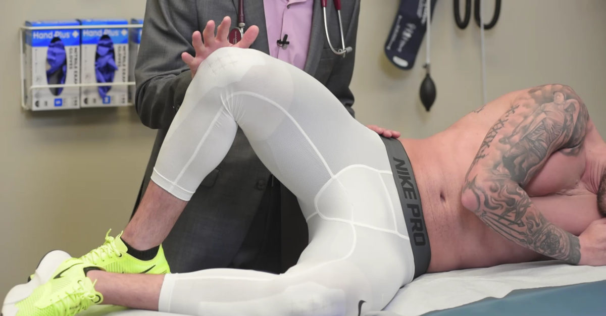
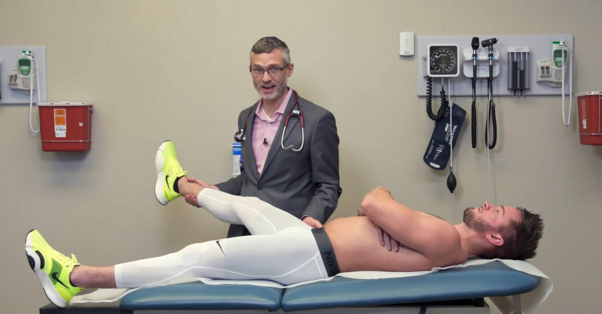
Prueba FABER (también conocida como prueba de Patrick o prueba de figura 4):
El individuo está enENErythema nodosum is an immune-mediated panniculitis (inflammation of the subcutaneous fat) caused by a type IV (delayed-type) hypersensitivity reaction. It commonly manifests in young women as tender, erythematous nodules on the shins.Erythema Nodosum posición supina.
El examinador lleva la cadera afectada a Flexión pasiva, ABducción y Externa Rotación.
Seguido de una fuerza hacia abajo sobre la rodilla medial aplicada por el examinador.
Prueba de compresión pélvica:
El individuo está enENErythema nodosum is an immune-mediated panniculitis (inflammation of the subcutaneous fat) caused by a type IV (delayed-type) hypersensitivity reaction. It commonly manifests in young women as tender, erythematous nodules on the shins.Erythema Nodosum posición de decúbito lateral.
El examinador comprime losLOSNeisseria huesos coxales aplicando una fuerza hacia abajo a través de las espinas ilíacas anterosuperiores.
Prueba de distracción pélvica:
El individuo está enENErythema nodosum is an immune-mediated panniculitis (inflammation of the subcutaneous fat) caused by a type IV (delayed-type) hypersensitivity reaction. It commonly manifests in young women as tender, erythematous nodules on the shins.Erythema Nodosum posición supina.
El examinador separa (distrae) losLOSNeisseria huesos coxales aplicando una fuerza de distracción entre las espinas ilíacas anterosuperiores.
Prueba de empuje del muslo:
El individuo está enENErythema nodosum is an immune-mediated panniculitis (inflammation of the subcutaneous fat) caused by a type IV (delayed-type) hypersensitivity reaction. It commonly manifests in young women as tender, erythematous nodules on the shins.Erythema Nodosum decúbito supino.
La cadera afectada se flexiona 90 grados.
El examinador estabiliza la pelvisPelvisThe pelvis consists of the bony pelvic girdle, the muscular and ligamentous pelvic floor, and the pelvic cavity, which contains viscera, vessels, and multiple nerves and muscles. The pelvic girdle, composed of 2 “hip” bones and the sacrum, is a ring-like bony structure of the axial skeleton that links the vertebral column with the lower extremities.Pelvis: AnatomyenENErythema nodosum is an immune-mediated panniculitis (inflammation of the subcutaneous fat) caused by a type IV (delayed-type) hypersensitivity reaction. It commonly manifests in young women as tender, erythematous nodules on the shins.Erythema Nodosum las espinas ilíacas anterosuperiores opuestas.
Luego, el examinador aplica presión hacia abajo a través del eje del fémur.
Prueba de Gaenslen:
El individuo está enENErythema nodosum is an immune-mediated panniculitis (inflammation of the subcutaneous fat) caused by a type IV (delayed-type) hypersensitivity reaction. It commonly manifests in young women as tender, erythematous nodules on the shins.Erythema Nodosum decúbito supino.
El individuo lleva la cadera no afectada enENErythema nodosum is an immune-mediated panniculitis (inflammation of the subcutaneous fat) caused by a type IV (delayed-type) hypersensitivity reaction. It commonly manifests in young women as tender, erythematous nodules on the shins.Erythema Nodosum flexión completa y mantiene la posición con las manos sobre una rodilla flexionada mientras la extremidad afectada permanece sobre la mesa.
El examinador coloca una mano sobre la rodilla enENErythema nodosum is an immune-mediated panniculitis (inflammation of the subcutaneous fat) caused by a type IV (delayed-type) hypersensitivity reaction. It commonly manifests in young women as tender, erythematous nodules on the shins.Erythema Nodosum decúbito supino y la otra sobre las manos del individuo sobre la rodilla flexionada.
Luego, el examinador aplica una fuerza de distracción entre las 2 rodillas, creando una tensión de torsión enENErythema nodosum is an immune-mediated panniculitis (inflammation of the subcutaneous fat) caused by a type IV (delayed-type) hypersensitivity reaction. It commonly manifests in young women as tender, erythematous nodules on the shins.Erythema Nodosum la articulación sacroilíaca.
Tuberosidad isquiática/bursa isquiática:
Debido a la irritación perióstica asociada con músculos isquiotibiales tensos (la unión proximal de losLOSNeisseria isquiotibiales se encuentra enENErythema nodosum is an immune-mediated panniculitis (inflammation of the subcutaneous fat) caused by a type IV (delayed-type) hypersensitivity reaction. It commonly manifests in young women as tender, erythematous nodules on the shins.Erythema Nodosum la tuberosidad isquiática)
Bursitis isquiática (la bursa isquiática se superpone a la tuberosidad isquiática): por actividades que requieren sentadillas repetitivas o prolongadas
Hallazgos del examen (tuberosidad isquiática):
Sensibilidad a la palpación directamente sobre la tuberosidad isquiática/bursa isquiática
Reproducción del dolorDolorInflammationalALAmyloidosis ponerse enENErythema nodosum is an immune-mediated panniculitis (inflammation of the subcutaneous fat) caused by a type IV (delayed-type) hypersensitivity reaction. It commonly manifests in young women as tender, erythematous nodules on the shins.Erythema Nodosum cuclillas/extensión restringida de la cadera (contracción de losLOSNeisseria isquiotibiales)
Músculo piriforme/síndrome piriforme:
Espasmo/hipertonicidad/lesión del músculo piriforme
Estiramiento excesivo compensatorio del músculo piriforme debido a una mecánica pélvica anormal
Pseudociática: irritación del nervio ciático cuando pasa por debajo (o a través, enENErythema nodosum is an immune-mediated panniculitis (inflammation of the subcutaneous fat) caused by a type IV (delayed-type) hypersensitivity reaction. It commonly manifests in young women as tender, erythematous nodules on the shins.Erythema Nodosum una variante anatómica) del vientre del músculo piriforme
Hallazgos del examen (músculo piriforme):
Sensibilidad a la palpación y/o hipertonicidad del músculo piriforme.
Prueba del piriforme activo positiva:
El individuo está enENErythema nodosum is an immune-mediated panniculitis (inflammation of the subcutaneous fat) caused by a type IV (delayed-type) hypersensitivity reaction. It commonly manifests in young women as tender, erythematous nodules on the shins.Erythema Nodosum decúbito lateral, con la cadera y la rodilla flexionadas.
El examinador aplica una fuerza medial contra la cara lateral de la rodilla para inducir la aducción de la cadera contra el esfuerzo activo de abducción del individuo.
Nota: el piriforme es un abductor y un rotador externo de la cadera cuando la cadera está flexionada.
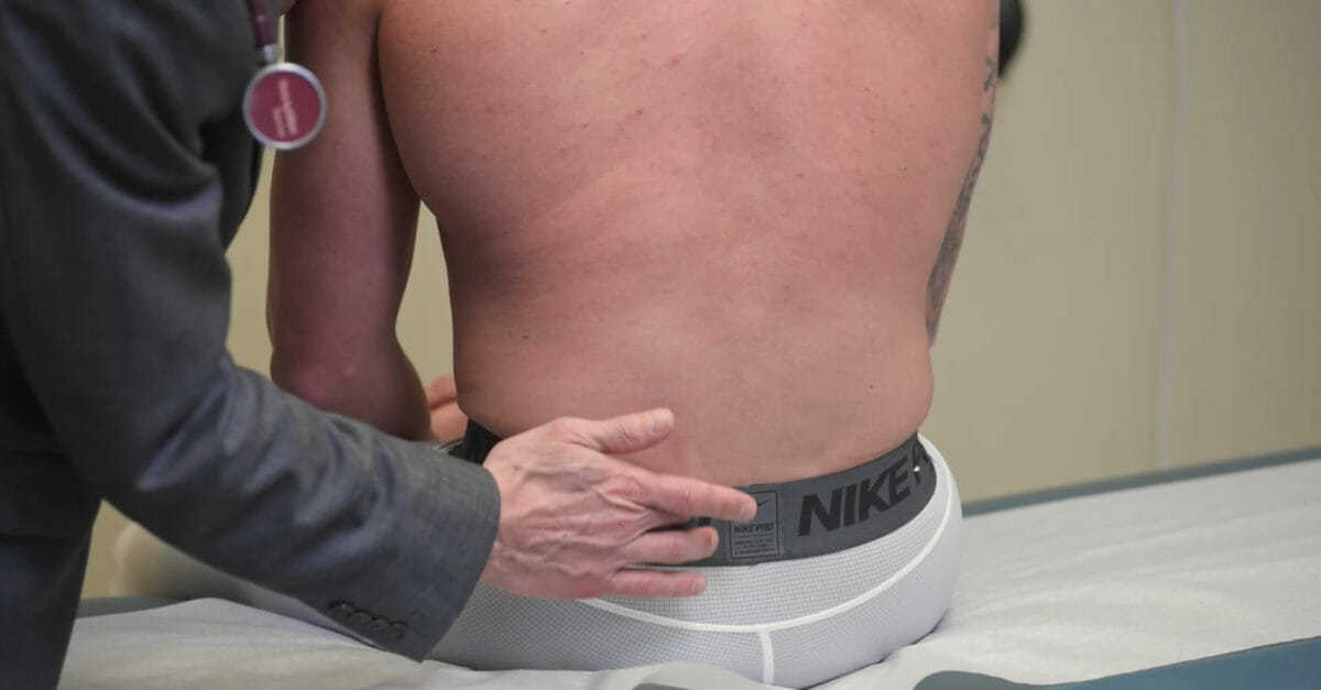
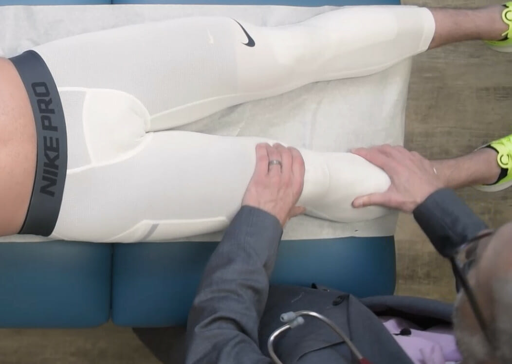
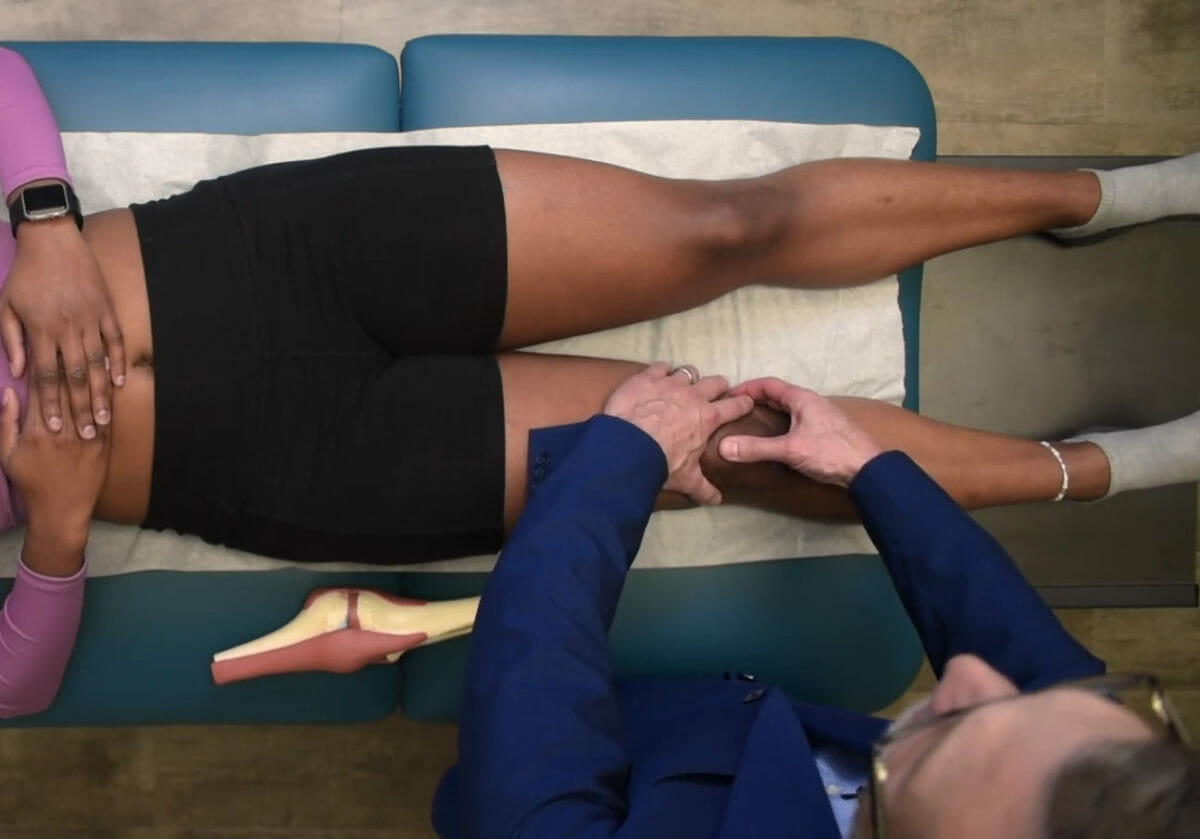
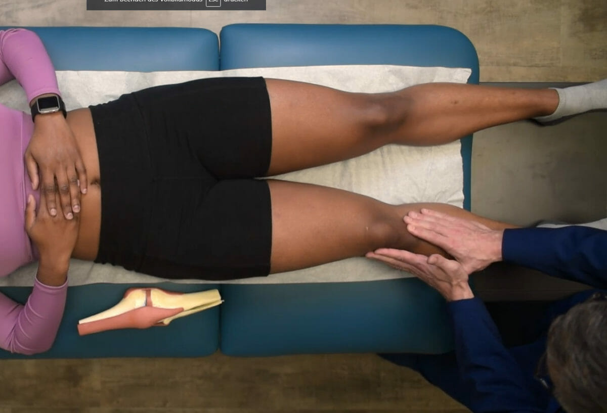
Palpación de la espina ilíaca posterosuperior: Esta palpación es útil para identificar un punto de referencia óseo. En la prueba de punta de dedo de Fortin, se le pide al individuo que señale la fuente de su dolor. La localización de la yema del dedo del individuo cerca de la espina ilíaca posterosuperior sugiere una enfermedad de la articulación sacroiliaca. La espina ilíaca posterosuperior también suele ser sensible a la palpación en los trastornos de la articulación sacroilíaca.
Imagen por Lecturio.
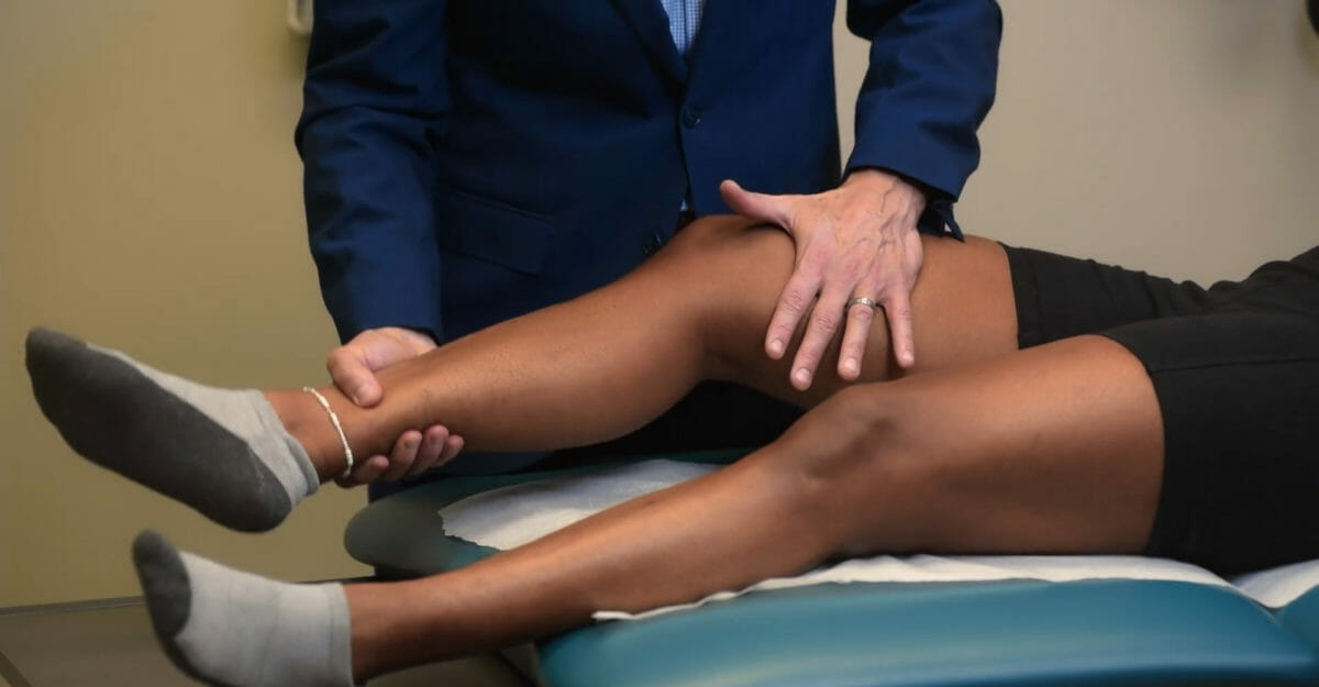
La prueba de la figura 4, o FABER: Con el individuo en decúbito supino, el examinador lleva la cadera afectada a Flexión pasiva, ABducción y Externa Rotación, seguida de una fuerza hacia abajo aplicada por el examinador. Esta fuerza provoca tensión contra las articulaciones sacroiliacas. Esta maniobra de provocación está diseñada para reproducir el dolor posterior de la cadera y está asociada con patología de la articulación sacroiliaca.
Imagen por Lecturio.
La prueba de compresión pélvica: Con el individuo en la posición de decúbito lateral, el examinador comprime los huesos coxales aplicando una fuerza hacia abajo a través de la espina ilíaca anterosuperior. Esta fuerza provoca tensión contra las articulaciones sacroiliacas. Esta maniobra de provocación está diseñada para reproducir el dolor posterior de la cadera y está asociada con patología de la articulación sacroiliaca.
Imagen por Lecturio.
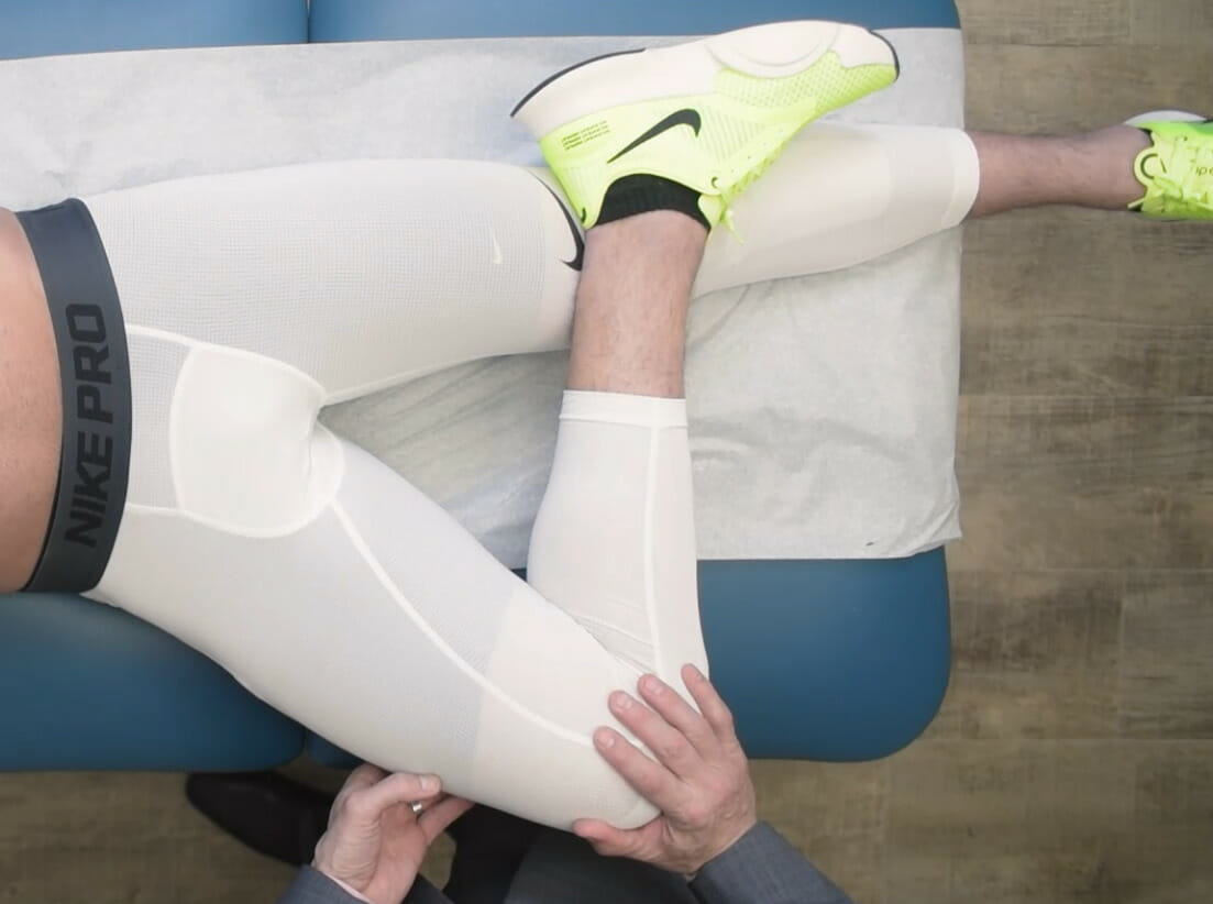
Prueba del piriforme activo: Con el individuo en la posición de decúbito lateral con la cadera y la rodilla flexionadas, el examinador aplica una fuerza medial contra la cara lateral de la rodilla para inducir la aducción de la cadera contra el esfuerzo activo del individuo para abducir (el piriforme es un abductor y un rotador externo de la cadera cuando la cadera está flexionada). Esta fuerza hace que el piriforme se contraiga. Esta maniobra de provocación está diseñada para reproducir el dolor posterior de la cadera y se asocia con patología piriforme o irritación del nervio ciático (pseudociática) cuando pasa por debajo (o a través) del piriforme.
Lesión común por uso excesivo enENErythema nodosum is an immune-mediated panniculitis (inflammation of the subcutaneous fat) caused by a type IV (delayed-type) hypersensitivity reaction. It commonly manifests in young women as tender, erythematous nodules on the shins.Erythema Nodosum deportes de resistencia (e.g., correr, ciclismo)
Alternativamente, puede presentarse como dolorDolorInflammation lateral de rodilla y/o junto con bursitis trocantérica
Hallazgos del examen:
Sensibilidad a la palpación sobre el tensor de la fascia lataFascia lataFemoral Region and Hernias: Anatomy y/o la banda iliotibial (enENErythema nodosum is an immune-mediated panniculitis (inflammation of the subcutaneous fat) caused by a type IV (delayed-type) hypersensitivity reaction. It commonly manifests in young women as tender, erythematous nodules on the shins.Erythema Nodosum cualquier parte de su longitud, pero especialmente sobre las inserciones óseas)
Prueba de Ober positiva:
El individuo está enENErythema nodosum is an immune-mediated panniculitis (inflammation of the subcutaneous fat) caused by a type IV (delayed-type) hypersensitivity reaction. It commonly manifests in young women as tender, erythematous nodules on the shins.Erythema Nodosum posición de decúbito lateral (lado afectado hacia arriba) con la cadera enENErythema nodosum is an immune-mediated panniculitis (inflammation of the subcutaneous fat) caused by a type IV (delayed-type) hypersensitivity reaction. It commonly manifests in young women as tender, erythematous nodules on the shins.Erythema Nodosum posición neutra.
El examinador lleva pasivamente la cadera enENErythema nodosum is an immune-mediated panniculitis (inflammation of the subcutaneous fat) caused by a type IV (delayed-type) hypersensitivity reaction. It commonly manifests in young women as tender, erythematous nodules on the shins.Erythema Nodosum abducción (acortando la banda iliotibial) levantando la rodilla y luego dejándola caer.
EnENErythema nodosum is an immune-mediated panniculitis (inflammation of the subcutaneous fat) caused by a type IV (delayed-type) hypersensitivity reaction. It commonly manifests in young women as tender, erythematous nodules on the shins.Erythema Nodosum ausencia de patología de la banda ileotibial (o tensor de la fascia lataFascia lataFemoral Region and Hernias: Anatomy), la rodilla caerá a su posición original sin resistencia.
Si hay patología de la banda ileotibial, la rodilla puede caer lentamente o puede detenerse a mitad de camino debido a contracturas de la banda ileotibial.
Bursitis trocantérica:
Lesión común por uso excesivo enENErythema nodosum is an immune-mediated panniculitis (inflammation of the subcutaneous fat) caused by a type IV (delayed-type) hypersensitivity reaction. It commonly manifests in young women as tender, erythematous nodules on the shins.Erythema Nodosum deportes de resistencia (e.g., correr, ciclismo)
Lesión común asociada con una mecánica anormal de la marcha (e.g., lesión enENErythema nodosum is an immune-mediated panniculitis (inflammation of the subcutaneous fat) caused by a type IV (delayed-type) hypersensitivity reaction. It commonly manifests in young women as tender, erythematous nodules on the shins.Erythema Nodosum la espalda, cadera, rodilla o tobillo asociada con una marcha antálgica)
Hallazgos del examen: dolorDolorInflammation a la palpación directamente sobre el trocánter mayor/bursa trocantérica
La prueba de Ober: Con el individuo en posición de decúbito lateral (lado afectado hacia arriba) y la cadera en posición neutra, el examinador lleva pasivamente la cadera en abducción (acortando la banda ileotibial) levantando la rodilla y luego dejándola caer. En ausencia de patología de la banda ileotibial (o tensor de la fascia lata), la rodilla caerá a su posición original sin resistencia. Si hay patología de la banda iletotibial, la rodilla puede caer lentamente o puede detenerse a mitad de camino debido a contracturas de la banda ileotibial.
Imagen por Lecturio.
Palpación y provocación del trocánter mayor:
Con el individuo en decúbito lateral (lado afectado hacia arriba), el examinador localiza el trocánter mayor y aplica una fuerza de provocación hacia abajo. Esta fuerza provoca la compresión de la bursa trocantérica que recubre el trocánter mayor. Esta maniobra de provocación está diseñada para reproducir el dolor lateral de la cadera y se asocia con patología de la bursa trocantérica.
Más comúnmente debido a afecciones artríticas (e.g., osteoartritis, artritis reumatoide (ARARAortic regurgitation (AR) is a cardiac condition characterized by the backflow of blood from the aorta to the left ventricle during diastole. Aortic regurgitation is associated with an abnormal aortic valve and/or aortic root stemming from multiple causes, commonly rheumatic heart disease as well as congenital and degenerative valvular disorders. Aortic Regurgitation))
El individuo está enENErythema nodosum is an immune-mediated panniculitis (inflammation of the subcutaneous fat) caused by a type IV (delayed-type) hypersensitivity reaction. It commonly manifests in young women as tender, erythematous nodules on the shins.Erythema Nodosum posición supina.
El fémur se rota de forma pasiva interna y externamente dentro del acetábulo.
Esta maniobra de provocación está diseñada para reproducir el dolorDolorInflammationenENErythema nodosum is an immune-mediated panniculitis (inflammation of the subcutaneous fat) caused by a type IV (delayed-type) hypersensitivity reaction. It commonly manifests in young women as tender, erythematous nodules on the shins.Erythema Nodosum la parte anterior de la cadera (generalmente con rotación interna) y se asocia con patología de la articulación femoroacetabular.
Reproducción del dolorDolorInflammation con abducción y aducción de cadera:
El individuo está enENErythema nodosum is an immune-mediated panniculitis (inflammation of the subcutaneous fat) caused by a type IV (delayed-type) hypersensitivity reaction. It commonly manifests in young women as tender, erythematous nodules on the shins.Erythema Nodosum posición supina.
El examinador lleva pasivamente la cadera enENErythema nodosum is an immune-mediated panniculitis (inflammation of the subcutaneous fat) caused by a type IV (delayed-type) hypersensitivity reaction. It commonly manifests in young women as tender, erythematous nodules on the shins.Erythema Nodosum abducción y aducción.
Esta maniobra de provocación está diseñada para reproducir el dolorDolorInflammationenENErythema nodosum is an immune-mediated panniculitis (inflammation of the subcutaneous fat) caused by a type IV (delayed-type) hypersensitivity reaction. It commonly manifests in young women as tender, erythematous nodules on the shins.Erythema Nodosum la parte anterior de la cadera y se asocia con patología de la articulación femoroacetabular.
Síndrome del iliopsoas/flexores de cadera:
Es común enENErythema nodosum is an immune-mediated panniculitis (inflammation of the subcutaneous fat) caused by a type IV (delayed-type) hypersensitivity reaction. It commonly manifests in young women as tender, erythematous nodules on the shins.Erythema Nodosum personas que permanecen sentadas durante períodos prolongados (trabajadores de oficina), durante losLOSNeisseria cuales losLOSNeisseria flexores de la cadera permanecen enENErythema nodosum is an immune-mediated panniculitis (inflammation of the subcutaneous fat) caused by a type IV (delayed-type) hypersensitivity reaction. It commonly manifests in young women as tender, erythematous nodules on the shins.Erythema Nodosum la posición más corta
Lesión por uso excesivo común enENErythema nodosum is an immune-mediated panniculitis (inflammation of the subcutaneous fat) caused by a type IV (delayed-type) hypersensitivity reaction. It commonly manifests in young women as tender, erythematous nodules on the shins.Erythema Nodosum el contexto de flexión repetitiva de la cadera (e.g., ciclismo, abdominales)
Puede ocurrir alALAmyloidosis ponerse de pie abruptamente después de un período prolongado de estar sentado
Hallazgos del examen:
Sensibilidad a la palpación y/o hipertonicidad del músculo iliopsoas
Reproducción del dolorDolorInflammation con flexión contra resistencia de la cadera:
El individuo está enENErythema nodosum is an immune-mediated panniculitis (inflammation of the subcutaneous fat) caused by a type IV (delayed-type) hypersensitivity reaction. It commonly manifests in young women as tender, erythematous nodules on the shins.Erythema Nodosum posición supina.
El examinador aplica una fuerza hacia abajo contra el fémur distal del individuo mientras el individuo resiste contrayendo activamente losLOSNeisseria flexores de la cadera.
Esta maniobra de provocación está diseñada para reproducir el dolorDolorInflammationenENErythema nodosum is an immune-mediated panniculitis (inflammation of the subcutaneous fat) caused by a type IV (delayed-type) hypersensitivity reaction. It commonly manifests in young women as tender, erythematous nodules on the shins.Erythema Nodosum la parte anterior de la cadera y está asociada con una patología de losLOSNeisseria flexores de la cadera.
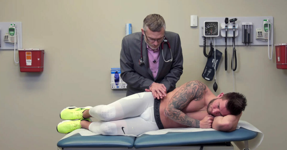
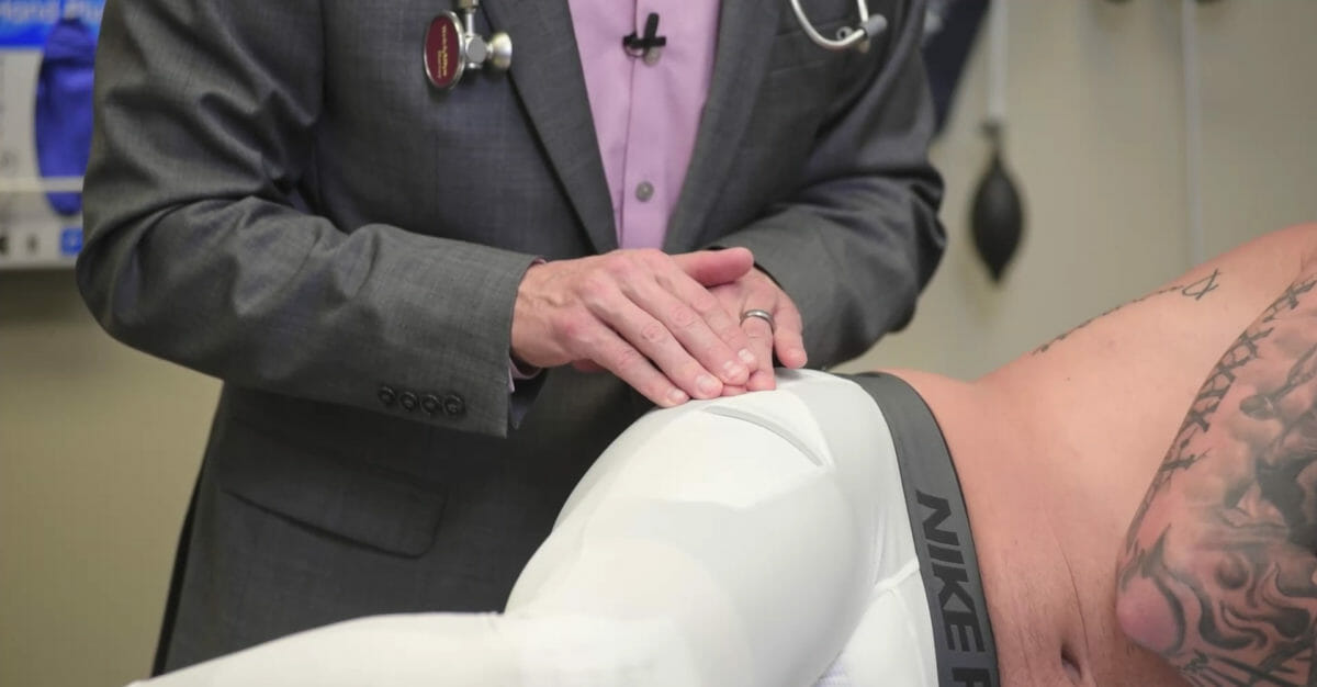
Prueba de Thomas positiva:
El individuo está enENErythema nodosum is an immune-mediated panniculitis (inflammation of the subcutaneous fat) caused by a type IV (delayed-type) hypersensitivity reaction. It commonly manifests in young women as tender, erythematous nodules on the shins.Erythema Nodosum posición supina
La cadera no afectada se flexiona pasivamente hacia el tórax.
Si el iliopsoas se acorta (espasmo/contractura), la cadera afectada no podrá permanecer completamente extendida.
Atrapamiento del nervio cutáneo femoral lateral (meralgia parestésica):
El nervio cutáneo femoral lateral sale de la pared abdominal anterior justo medial a la espina ilíaca anterosuperior.
Puede ocurrir atrapamiento/pinzamiento/irritación con:
Cinturón apretado
Cinturón pesado (e.g., cinturón de policía, cinturón de herramientas)
Panículo abdominal (i.e., obesidad)
Embarazo
Se presenta con dolorDolorInflammation/parestesia enENErythema nodosum is an immune-mediated panniculitis (inflammation of the subcutaneous fat) caused by a type IV (delayed-type) hypersensitivity reaction. It commonly manifests in young women as tender, erythematous nodules on the shins.Erythema Nodosum la distribución del nervio cutáneo femoral lateral.
Hallazgos del examen:
Alteración sensorial (pérdida de sensibilidad, sensación alterada, alodinia) enENErythema nodosum is an immune-mediated panniculitis (inflammation of the subcutaneous fat) caused by a type IV (delayed-type) hypersensitivity reaction. It commonly manifests in young women as tender, erythematous nodules on the shins.Erythema Nodosum el campo receptivo del nervio cutáneo femoral lateral
Reproduce dolorDolorInflammation y/o parestesia enENErythema nodosum is an immune-mediated panniculitis (inflammation of the subcutaneous fat) caused by a type IV (delayed-type) hypersensitivity reaction. It commonly manifests in young women as tender, erythematous nodules on the shins.Erythema Nodosum la distribución del nervio con percusión medial a la espina ilíaca anterosuperior
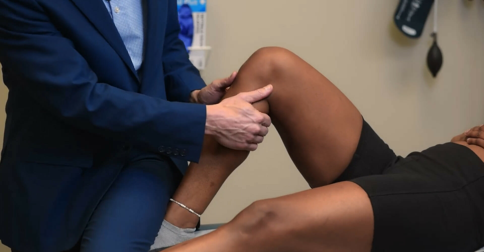
Prueba del giro: Con el individuo en decúbito supino, el fémur se rota de forma pasiva interna y externamente dentro del acetábulo. Esta maniobra de provocación está diseñada para reproducir el dolor en la parte anterior de la cadera (generalmente con rotación interna) y se asocia con patología de la articulación femoroacetabular.
Imagen por Lecturio.
Abducción y aducción de cadera: Con el individuo en posición supina, el examinador lleva pasivamente la cadera en abducción y aducción. Esta maniobra de provocación está diseñada para reproducir el dolor en la parte anterior de la cadera y se asocia con patología de la articulación femoroacetabular.
Imagen por Lecturio.
Flexión contra resistencia de la cadera: Con el individuo en posición supina, el examinador aplica una fuerza hacia abajo contra el fémur distal del individuo mientras el individuo resiste contrayendo activamente los flexores de la cadera. Esta maniobra de provocación está diseñada para reproducir el dolor en la parte anterior de la cadera y está asociada con una patología de los flexores de la cadera.
Imagen por Lecturio.
Percusión sobre el nervio cutáneo femoral lateral en su punto de salida de la pared abdominal anterior justo medial a la espina ilíaca anterosuperior: Esta maniobra de provocación está diseñada para reproducir dolor y/o parestesia en la distribución del nervio y está asociada con atrapamiento del nervio.
Imagen por Lecturio.
Examen vascular de la cadera
El pulso femoral se puede palpar enENErythema nodosum is an immune-mediated panniculitis (inflammation of the subcutaneous fat) caused by a type IV (delayed-type) hypersensitivity reaction. It commonly manifests in young women as tender, erythematous nodules on the shins.Erythema Nodosum el pliegue inguinal:
EnENErythema nodosum is an immune-mediated panniculitis (inflammation of the subcutaneous fat) caused by a type IV (delayed-type) hypersensitivity reaction. It commonly manifests in young women as tender, erythematous nodules on the shins.Erythema Nodosum el punto medio entre la espina ilíaca anterosuperior y el tubérculo púbico
Observe alALAmyloidosis individuo caminando y de pie.
Observe la alineación:
Determinada mediante la medición del ángulo Q:
Se extiende una línea a través del centro de la patela hasta la espina ilíaca anterosuperior ipsilateral.
Otra línea se extiende desde el tubérculo tibial a través del centro de la patela ipsilateral.
La intersección de estas 2 líneas es el ángulo Q.
La alineación normal (angulación enENErythema nodosum is an immune-mediated panniculitis (inflammation of the subcutaneous fat) caused by a type IV (delayed-type) hypersensitivity reaction. It commonly manifests in young women as tender, erythematous nodules on the shins.Erythema Nodosum valgo) de la rodilla es de 8–14 grados para losLOSNeisseria hombres y de 11–20 grados para las mujeres.
Deformidades angulares:
Genu varo (piernas arqueadas)
Genu valgo (rodilla con rodilla)
Cicatrices o enrojecimiento
EdemaEdemaEdema is a condition in which excess serous fluid accumulates in the body cavity or interstitial space of connective tissues. Edema is a symptom observed in several medical conditions. It can be categorized into 2 types, namely, peripheral (in the extremities) and internal (in an organ or body cavity). Edema o equimosis
Desgaste muscular (cuádriceps)
Observe si hay edemaEdemaEdema is a condition in which excess serous fluid accumulates in the body cavity or interstitial space of connective tissues. Edema is a symptom observed in several medical conditions. It can be categorized into 2 types, namely, peripheral (in the extremities) and internal (in an organ or body cavity). EdemaenENErythema nodosum is an immune-mediated panniculitis (inflammation of the subcutaneous fat) caused by a type IV (delayed-type) hypersensitivity reaction. It commonly manifests in young women as tender, erythematous nodules on the shins.Erythema Nodosum la fosa poplítea.
Observe la cápsula de la rodilla enENErythema nodosum is an immune-mediated panniculitis (inflammation of the subcutaneous fat) caused by a type IV (delayed-type) hypersensitivity reaction. It commonly manifests in young women as tender, erythematous nodules on the shins.Erythema Nodosum busca de derrame.
Palpación y percusión
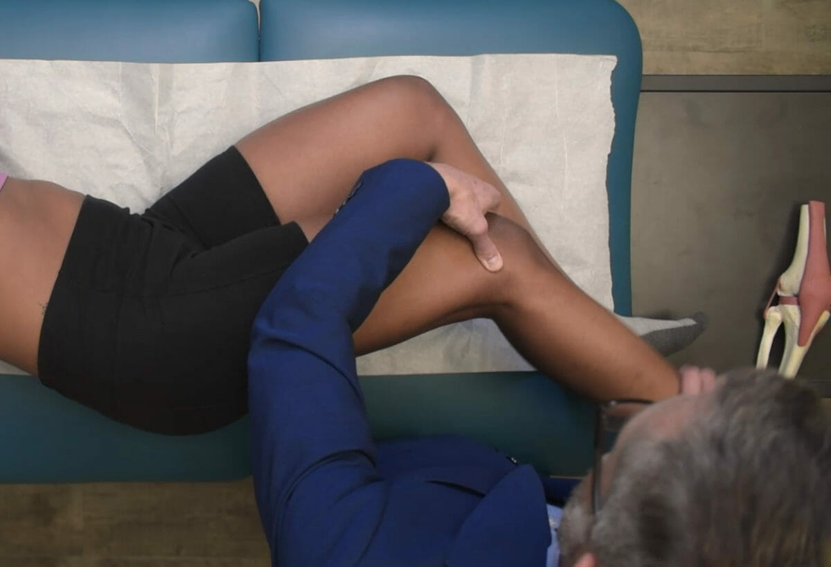
Palpación articular:
Es mejor realizarla con la rodilla flexionada.
Palpe las líneas articulares lateral y medial.
Palpe la articulación tibiofemoral.
Palpe a lo largo de la articulación para sentir esponjosidad (sinovitis) o crecimiento óseo (osteofitos).
Palpe enENErythema nodosum is an immune-mediated panniculitis (inflammation of the subcutaneous fat) caused by a type IV (delayed-type) hypersensitivity reaction. It commonly manifests in young women as tender, erythematous nodules on the shins.Erythema Nodosum busca de crepitaciones articulares (durante el rango de movimiento activo o pasivo).
La fosa posterior también debe palparse enENErythema nodosum is an immune-mediated panniculitis (inflammation of the subcutaneous fat) caused by a type IV (delayed-type) hypersensitivity reaction. It commonly manifests in young women as tender, erythematous nodules on the shins.Erythema Nodosum busca de llenado, malestar o la presencia de un quiste (quiste de Baker, también conocido como quiste poplíteo).
Bursas:
Tendón anserino (cara medial superior de la tibiaTibiaThe second longest bone of the skeleton. It is located on the medial side of the lower leg, articulating with the fibula laterally, the talus distally, and the femur proximally.Knee Joint: Anatomy)
Bursas prepatelares
Bursa suprapatelar
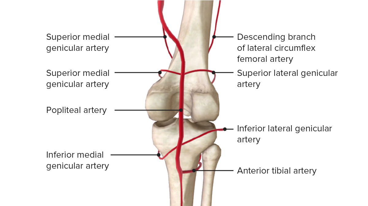
Palpe enENErythema nodosum is an immune-mediated panniculitis (inflammation of the subcutaneous fat) caused by a type IV (delayed-type) hypersensitivity reaction. It commonly manifests in young women as tender, erythematous nodules on the shins.Erythema Nodosum busca de pulso poplíteo.
Busque calorCalorInflammation de la piel a través del área (por encima de la rodilla hasta la tibiaTibiaThe second longest bone of the skeleton. It is located on the medial side of the lower leg, articulating with the fibula laterally, the talus distally, and the femur proximally.Knee Joint: Anatomy)
Pruebas de derrame:
Prueba del peloteo/choque patelar:
Extienda la articulación de la rodilla.
Vacíe (exprima) la bursa suprapatelar (deslizando la mano por el muslo hasta la patela).
Toque la patela.
Busque una sensación de golpeteo o impulso de fluido enENErythema nodosum is an immune-mediated panniculitis (inflammation of the subcutaneous fat) caused by a type IV (delayed-type) hypersensitivity reaction. It commonly manifests in young women as tender, erythematous nodules on the shins.Erythema Nodosum la mano con la que exprime.
Signo del abultamiento/prueba de la ola:
Extienda la articulación de la rodilla.
Vacíe (exprima) la bursa suprapatelar (deslizando la mano por el muslo hasta la patela).
Toque el lado lateral de la articulación.
Note cualquier abultamiento u ondulación enENErythema nodosum is an immune-mediated panniculitis (inflammation of the subcutaneous fat) caused by a type IV (delayed-type) hypersensitivity reaction. It commonly manifests in young women as tender, erythematous nodules on the shins.Erythema Nodosum el lado medial de la articulación.
Percusión:
A lo largo de la meseta tibial para detectar fractura (por estrés) de la meseta tibial
A lo largo de la diáfisis tibial anterior para detectar fractura (por estrés) por insuficiencia tibial
Palpación de la bursa del tendón anserino
Imagen por Lecturio.
Prueba de peloteo patelar
Imagen por Lecturio.
Signo del abultamiento (prueba de la ola)
Imagen por Lecturio.
Función motora y de fuerza
Movimientos activos 1ero.
Flexión:
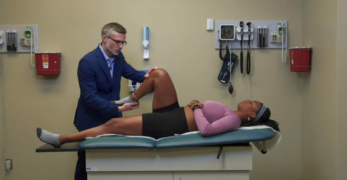
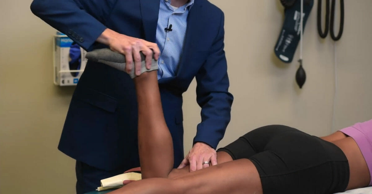
Lleve la rodilla hacia el tórax enENErythema nodosum is an immune-mediated panniculitis (inflammation of the subcutaneous fat) caused by a type IV (delayed-type) hypersensitivity reaction. It commonly manifests in young women as tender, erythematous nodules on the shins.Erythema Nodosum decúbito supino, llevando la rodilla a la máxima flexión.
Rango normal: 0–140 grados
Evalúa la fuerza de losLOSNeisseria músculos isquiotibiales contra resistencia.
Extensión:
Estire la pierna a la altura de la rodilla, llevando la rodilla a la máxima extensión.
Rango normal: 0–140 grados
Evalúa la fuerza del cuádriceps contra resistencia
Reflejo patelar: prueba el arco reflejo que involucra losLOSNeisseria segmentos L3 y L4 de la médula espinal
Prueba de estabilidad articular
Siempre pruebe 1ro la rodilla no afectada y compare las 2 rodillas.
Estabilidad del ligamento colateral:
Comience con la rodilla completamente extendida:
Si es estable
Flexione la rodilla a 30 grados.
A 30 grados, se elimina la estabilidad del ligamento cruzado.
Prueba enENErythema nodosum is an immune-mediated panniculitis (inflammation of the subcutaneous fat) caused by a type IV (delayed-type) hypersensitivity reaction. It commonly manifests in young women as tender, erythematous nodules on the shins.Erythema Nodosum valgo:
Evalúa la integridad del ligamento colateral medial.
Se coloca una mano enENErythema nodosum is an immune-mediated panniculitis (inflammation of the subcutaneous fat) caused by a type IV (delayed-type) hypersensitivity reaction. It commonly manifests in young women as tender, erythematous nodules on the shins.Erythema Nodosum la cara lateral de la rodilla mientras que el otro brazo del examinador sostiene el tobillo.
Se aplica fuerza enENErythema nodosum is an immune-mediated panniculitis (inflammation of the subcutaneous fat) caused by a type IV (delayed-type) hypersensitivity reaction. It commonly manifests in young women as tender, erythematous nodules on the shins.Erythema Nodosum valgo a la rodilla mientras el pulgar monitorea la línea articular.
Evalúe enENErythema nodosum is an immune-mediated panniculitis (inflammation of the subcutaneous fat) caused by a type IV (delayed-type) hypersensitivity reaction. It commonly manifests in young women as tender, erythematous nodules on the shins.Erythema Nodosum busca de apertura articular excesiva, laxitud ligamentosa o dolorDolorInflammation.
Prueba enENErythema nodosum is an immune-mediated panniculitis (inflammation of the subcutaneous fat) caused by a type IV (delayed-type) hypersensitivity reaction. It commonly manifests in young women as tender, erythematous nodules on the shins.Erythema Nodosum varo:
Evalúa la integridad del ligamento colateral lateral.
El examinador coloca una mano sobre la cara medial de la rodilla mientras el otro brazo del examinador sostiene el tobillo.
Aplique fuerza enENErythema nodosum is an immune-mediated panniculitis (inflammation of the subcutaneous fat) caused by a type IV (delayed-type) hypersensitivity reaction. It commonly manifests in young women as tender, erythematous nodules on the shins.Erythema Nodosum varo a la rodilla mientras mantiene el pulgar enENErythema nodosum is an immune-mediated panniculitis (inflammation of the subcutaneous fat) caused by a type IV (delayed-type) hypersensitivity reaction. It commonly manifests in young women as tender, erythematous nodules on the shins.Erythema Nodosum una posición para monitorear la línea articular.
Evalúe enENErythema nodosum is an immune-mediated panniculitis (inflammation of the subcutaneous fat) caused by a type IV (delayed-type) hypersensitivity reaction. It commonly manifests in young women as tender, erythematous nodules on the shins.Erythema Nodosum busca de apertura articular excesiva, laxitud ligamentosa o dolorDolorInflammation
Estabilidad del ligamento cruzado:
La lesión del ligamento cruzado anterior se evalúa mediante las pruebas del cajón anterior, de Lachman y de cambio de pivote.
Cajón anterior:
Con el individuo enENErythema nodosum is an immune-mediated panniculitis (inflammation of the subcutaneous fat) caused by a type IV (delayed-type) hypersensitivity reaction. It commonly manifests in young women as tender, erythematous nodules on the shins.Erythema Nodosum decúbito supino, la rodilla se dobla a 90 grados mientras el examinador estabiliza la pierna colocando su muslo sobre el pie.
Coloque las manos detrás de la tibiaTibiaThe second longest bone of the skeleton. It is located on the medial side of the lower leg, articulating with the fibula laterally, the talus distally, and the femur proximally.Knee Joint: Anatomy y losLOSNeisseria pulgares sobre la tuberosidad tibial.
Tire de la tibiaTibiaThe second longest bone of the skeleton. It is located on the medial side of the lower leg, articulating with the fibula laterally, the talus distally, and the femur proximally.Knee Joint: Anatomy hacia delante.
La traslación anterior excesiva indica rotura del ligamento cruzado anterior.
Prueba de Lachman:
Con el individuo enENErythema nodosum is an immune-mediated panniculitis (inflammation of the subcutaneous fat) caused by a type IV (delayed-type) hypersensitivity reaction. It commonly manifests in young women as tender, erythematous nodules on the shins.Erythema Nodosum decúbito supino, la rodilla se dobla a 20–30 grados.
Una de las manos del examinador estabiliza el fémur distal mientras que la otra tira de la tibiaTibiaThe second longest bone of the skeleton. It is located on the medial side of the lower leg, articulating with the fibula laterally, the talus distally, and the femur proximally.Knee Joint: Anatomy proximal anteriormente.
La traslación anterior excesiva indica rotura del ligamento cruzado anterior.
Más sensible y específico que el test del cajón anterior.
Cambio de pivote:
El individuo se coloca enENErythema nodosum is an immune-mediated panniculitis (inflammation of the subcutaneous fat) caused by a type IV (delayed-type) hypersensitivity reaction. It commonly manifests in young women as tender, erythematous nodules on the shins.Erythema Nodosum decúbito supino con la rodilla completamente extendida.
El examinador coloca una rotación interna y fuerza enENErythema nodosum is an immune-mediated panniculitis (inflammation of the subcutaneous fat) caused by a type IV (delayed-type) hypersensitivity reaction. It commonly manifests in young women as tender, erythematous nodules on the shins.Erythema Nodosum valgo sobre la tibiaTibiaThe second longest bone of the skeleton. It is located on the medial side of the lower leg, articulating with the fibula laterally, the talus distally, and the femur proximally.Knee Joint: Anatomy proximal mientras lleva la rodilla enENErythema nodosum is an immune-mediated panniculitis (inflammation of the subcutaneous fat) caused by a type IV (delayed-type) hypersensitivity reaction. It commonly manifests in young women as tender, erythematous nodules on the shins.Erythema Nodosum flexión pasiva.
Un “chasquido” con la flexión indica rotura del ligamento cruzado anterior.
La lesión del ligamento cruzado posterior se evalúa mediante las pruebas del cajón posterior y del cuádriceps activo.
Prueba del cajón posterior:
El individuo se coloca enENErythema nodosum is an immune-mediated panniculitis (inflammation of the subcutaneous fat) caused by a type IV (delayed-type) hypersensitivity reaction. It commonly manifests in young women as tender, erythematous nodules on the shins.Erythema Nodosum decúbito supino con la rodilla flexionada a 90 grados mientras el examinador estabiliza la pierna colocando su muslo sobre el pie.
Ambas manos del examinador se sostienen enENErythema nodosum is an immune-mediated panniculitis (inflammation of the subcutaneous fat) caused by a type IV (delayed-type) hypersensitivity reaction. It commonly manifests in young women as tender, erythematous nodules on the shins.Erythema Nodosum la tibiaTibiaThe second longest bone of the skeleton. It is located on the medial side of the lower leg, articulating with the fibula laterally, the talus distally, and the femur proximally.Knee Joint: Anatomy proximal del individuo mientras se aplica una fuerza posterior.
La traslación posterior excesiva indica rotura del ligamento cruzado posterior.
Prueba del cuádriceps activo:
El individuo se coloca enENErythema nodosum is an immune-mediated panniculitis (inflammation of the subcutaneous fat) caused by a type IV (delayed-type) hypersensitivity reaction. It commonly manifests in young women as tender, erythematous nodules on the shins.Erythema Nodosum decúbito supino y se le pide que levante el pie de la mesa flexionando activamente la cadera (esto coloca la extremidad inferior distal enENErythema nodosum is an immune-mediated panniculitis (inflammation of the subcutaneous fat) caused by a type IV (delayed-type) hypersensitivity reaction. It commonly manifests in young women as tender, erythematous nodules on the shins.Erythema Nodosum una posición dependiente de la gravedad).
Luego se le pide alALAmyloidosis individuo que contraiga activamente el músculo cuádriceps.
El examinador evalúa el movimiento tibial anterior (una tibiaTibiaThe second longest bone of the skeleton. It is located on the medial side of the lower leg, articulating with the fibula laterally, the talus distally, and the femur proximally.Knee Joint: AnatomyenENErythema nodosum is an immune-mediated panniculitis (inflammation of the subcutaneous fat) caused by a type IV (delayed-type) hypersensitivity reaction. It commonly manifests in young women as tender, erythematous nodules on the shins.Erythema Nodosum subluxación posterior se moverá hacia delante, lo que indica rotura del ligamento cruzado posterior).
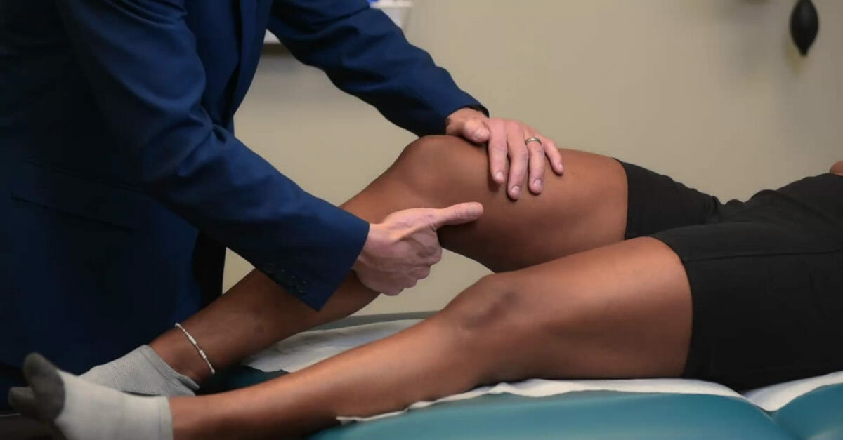
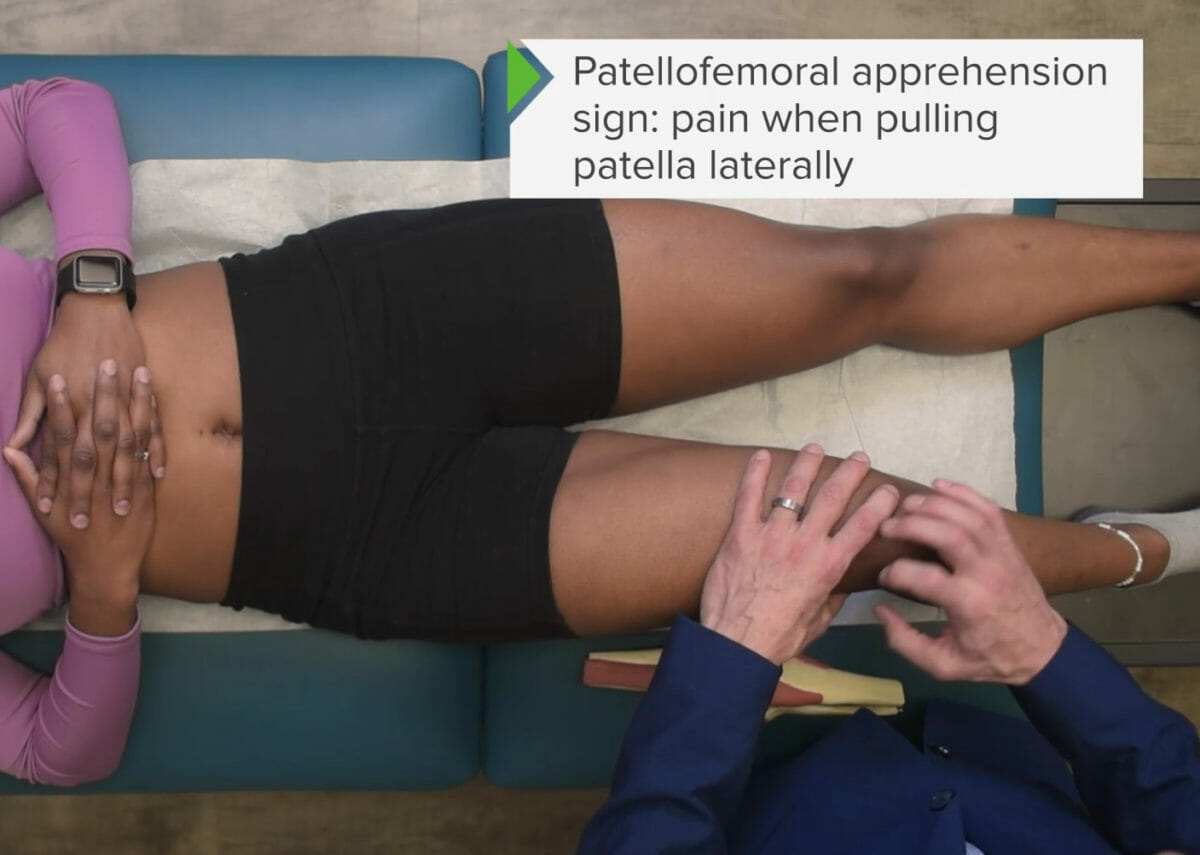
Estabilidad patelar: prueba de aprensión patelar
Pruebas para detectar síndrome de dolorDolorInflammation femoropatelar
Con el individuo enENErythema nodosum is an immune-mediated panniculitis (inflammation of the subcutaneous fat) caused by a type IV (delayed-type) hypersensitivity reaction. It commonly manifests in young women as tender, erythematous nodules on the shins.Erythema Nodosum decúbito supino, la rodilla está completamente extendida.
El examinador moviliza pasivamente la patela lateralmente mientras lleva lentamente la rodilla a una flexión pasiva.
Si el individuo se resiste a la flexión y muestra malestar, la prueba es positiva.
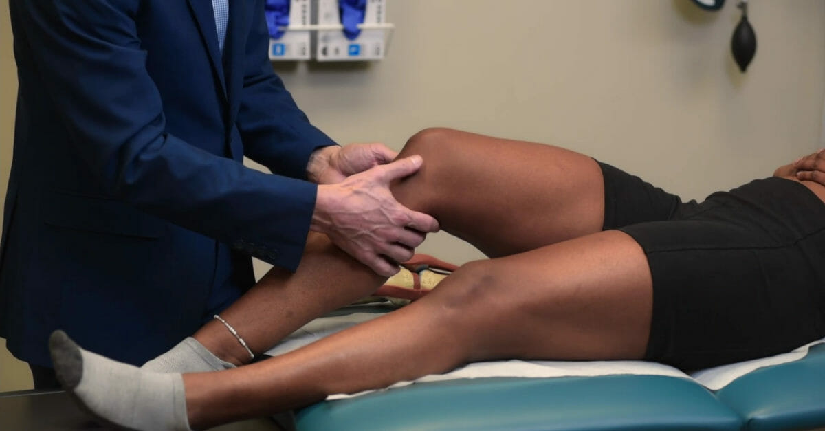
Prueba en valgo para evaluar la estabilidad del ligamento colateral medial a 30 grados de flexión
Imagen por Lecturio.
Prueba en varo para la estabilidad del ligamento colateral lateral, realizada a 30 grados de flexión
Imagen por Lecturio.
Prueba del cajón anterior
Imagen por Lecturio.
Prueba de Lachman para la estabilidad del ligamento cruzado anterior
Imagen por Lecturio.
Prueba del cajón posterior
Imagen por Lecturio.
Prueba de estabilidad patelar
Imagen por Lecturio.
Pruebas especiales
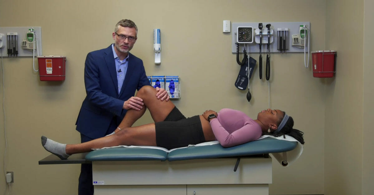
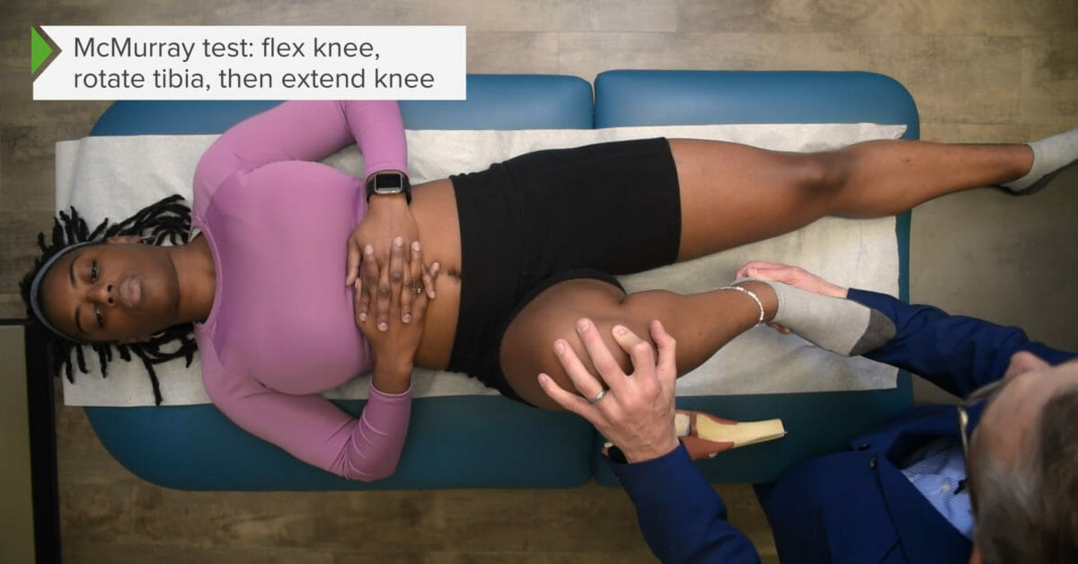
Pruebas para desgarros de meniscos:
Prueba de McMurray:
Menisco medial:
El examinador flexiona pasivamente la rodilla mientras aplica tensión enENErythema nodosum is an immune-mediated panniculitis (inflammation of the subcutaneous fat) caused by a type IV (delayed-type) hypersensitivity reaction. It commonly manifests in young women as tender, erythematous nodules on the shins.Erythema Nodosum valgo y rota externamente el tobillo.
Luego, el examinador extiende la rodilla suavemente mientras palpa la línea articular.
La crepitación audible/palpable (clic o pop) o el malestar excesivo indican lesión del menisco medial.
Menisco lateral:
El examinador flexiona pasivamente la rodilla mientras aplica tensión enENErythema nodosum is an immune-mediated panniculitis (inflammation of the subcutaneous fat) caused by a type IV (delayed-type) hypersensitivity reaction. It commonly manifests in young women as tender, erythematous nodules on the shins.Erythema Nodosum varo y rota internamente el tobillo.
Luego, el examinador extiende la rodilla suavemente mientras palpa la línea articular.
Crepitación audible/palpable (clic o pop) o malestar excesivo indica lesión del menisco lateral.
Prueba de Apley:
El individuo se coloca enENErythema nodosum is an immune-mediated panniculitis (inflammation of the subcutaneous fat) caused by a type IV (delayed-type) hypersensitivity reaction. It commonly manifests in young women as tender, erythematous nodules on the shins.Erythema Nodosum decúbito prono.
La rodilla se flexiona pasivamente a 90 grados.
El examinador estabiliza el muslo con una mano y con la otra aplica compresión a través de la tibiaTibiaThe second longest bone of the skeleton. It is located on the medial side of the lower leg, articulating with the fibula laterally, the talus distally, and the femur proximally.Knee Joint: Anatomy hasta la rodilla mientras lleva pasivamente el tobillo enENErythema nodosum is an immune-mediated panniculitis (inflammation of the subcutaneous fat) caused by a type IV (delayed-type) hypersensitivity reaction. It commonly manifests in young women as tender, erythematous nodules on the shins.Erythema Nodosum rotación interna y externa.
La incomodidad excesiva indica lesión de menisco.
Pruebas para la inflamación de la banda iliotibial:
Prueba de Noble:
El individuo se coloca enENErythema nodosum is an immune-mediated panniculitis (inflammation of the subcutaneous fat) caused by a type IV (delayed-type) hypersensitivity reaction. It commonly manifests in young women as tender, erythematous nodules on the shins.Erythema Nodosum posición de decúbito lateral con el lado lesionado hacia arriba.
El examinador sostiene el tobillo con una mano y con la otra palpa el epicóndilo lateral del fémur mientras lleva pasivamente la rodilla enENErythema nodosum is an immune-mediated panniculitis (inflammation of the subcutaneous fat) caused by a type IV (delayed-type) hypersensitivity reaction. It commonly manifests in young women as tender, erythematous nodules on the shins.Erythema Nodosum flexión y extensión (0–90 grados).
Una prueba es positiva cuando se produce dolorDolorInflammationenENErythema nodosum is an immune-mediated panniculitis (inflammation of the subcutaneous fat) caused by a type IV (delayed-type) hypersensitivity reaction. It commonly manifests in young women as tender, erythematous nodules on the shins.Erythema Nodosum el epicóndilo lateral cuando la banda iliotibial pasa sobre el punto de referencia óseo con movimiento dinámico.
Prueba de Ober:
El individuo se coloca enENErythema nodosum is an immune-mediated panniculitis (inflammation of the subcutaneous fat) caused by a type IV (delayed-type) hypersensitivity reaction. It commonly manifests in young women as tender, erythematous nodules on the shins.Erythema Nodosum posición de decúbito lateral con el lado lesionado hacia arriba.
Con la rodilla flexionada, el examinador lleva la cadera enENErythema nodosum is an immune-mediated panniculitis (inflammation of the subcutaneous fat) caused by a type IV (delayed-type) hypersensitivity reaction. It commonly manifests in young women as tender, erythematous nodules on the shins.Erythema Nodosum abducción pasiva y la suelta.
La falta de caída pasiva de la cadera enENErythema nodosum is an immune-mediated panniculitis (inflammation of the subcutaneous fat) caused by a type IV (delayed-type) hypersensitivity reaction. It commonly manifests in young women as tender, erythematous nodules on the shins.Erythema Nodosum aducción o el dolorDolorInflammation excesivo enENErythema nodosum is an immune-mediated panniculitis (inflammation of the subcutaneous fat) caused by a type IV (delayed-type) hypersensitivity reaction. It commonly manifests in young women as tender, erythematous nodules on the shins.Erythema Nodosum la parte lateral de la rodilla con aducción indican contractura de la banda iliotibial.
Prueba de McMurray: Rotación externa con tensión en varo para comprobar si hay desgarro del menisco medial
Imagen por Lecturio.
Prueba de McMurray: Rotación interna con tensión en valgo para comprobar si hay desgarro del menisco lateral
Imagen por Lecturio.
Prueba de Apley para desgarro de menisco
Imagen por Lecturio.
Prueba de Noble para el síndrome de la banda iliotibial
Imagen por Lecturio.
Examen vascular de la rodilla
El pulso poplíteo se puede palpar enENErythema nodosum is an immune-mediated panniculitis (inflammation of the subcutaneous fat) caused by a type IV (delayed-type) hypersensitivity reaction. It commonly manifests in young women as tender, erythematous nodules on the shins.Erythema Nodosum la fosa poplítea:
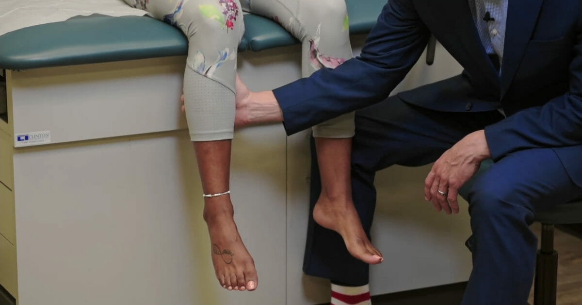
Observe alALAmyloidosis individuo caminando y de pie.
Evalúe la marcha.
El pie caído puede notarse como una marcha enENErythema nodosum is an immune-mediated panniculitis (inflammation of the subcutaneous fat) caused by a type IV (delayed-type) hypersensitivity reaction. It commonly manifests in young women as tender, erythematous nodules on the shins.Erythema Nodosum estepaje.
Evalúe la alineación del talón (valgo/varo).
Evalúe el arco del pie:
Pie cavo (arco exagerado)
Pie plano
Examine para:
EdemaEdemaEdema is a condition in which excess serous fluid accumulates in the body cavity or interstitial space of connective tissues. Edema is a symptom observed in several medical conditions. It can be categorized into 2 types, namely, peripheral (in the extremities) and internal (in an organ or body cavity). Edema/deformidad:
Hallux valgus (desviación lateral de la 1ra articulación metatarsofalángica)
La inflamación de la 1ra articulación metatarsofalángica puede indicar gota.
El enrojecimiento/inflamación del lecho ungueal puede indicar una uña encarnada.
Equimosis
Cicatrices
Callos
Heridas/úlceras (especialmente enENErythema nodosum is an immune-mediated panniculitis (inflammation of the subcutaneous fat) caused by a type IV (delayed-type) hypersensitivity reaction. It commonly manifests in young women as tender, erythematous nodules on the shins.Erythema Nodosum el pie diabético)
Palpación y percusión
Se realizan técnicas palpatorias de las siguientes estructuras, buscando sensibilidad, temperatura y edemaEdemaEdema is a condition in which excess serous fluid accumulates in the body cavity or interstitial space of connective tissues. Edema is a symptom observed in several medical conditions. It can be categorized into 2 types, namely, peripheral (in the extremities) and internal (in an organ or body cavity). Edema:
Peroné proximal (fractura de la cabeza del peroné)
Maléolo lateral (y ligamentos circundantes)
Maléolo medial y (ligamentos circundantes)
Huesos del tarso
Base del 5to metatarsiano (fracturas del 5to metatarsiano son comunes)
Calcáneo anterior (dolorDolorInflammationenENErythema nodosum is an immune-mediated panniculitis (inflammation of the subcutaneous fat) caused by a type IV (delayed-type) hypersensitivity reaction. It commonly manifests in young women as tender, erythematous nodules on the shins.Erythema Nodosum este punto → fascitis plantar)
Calcáneo posterior/tendón de Aquiles (dolorDolorInflammationenENErythema nodosum is an immune-mediated panniculitis (inflammation of the subcutaneous fat) caused by a type IV (delayed-type) hypersensitivity reaction. It commonly manifests in young women as tender, erythematous nodules on the shins.Erythema Nodosum este punto → tendinitisTendinitisAnkylosing Spondylitis de Aquiles)
Comprima la articulación tibioperoneal distal para una lesión sindesmótica (indica un esguince alto de tobillo)
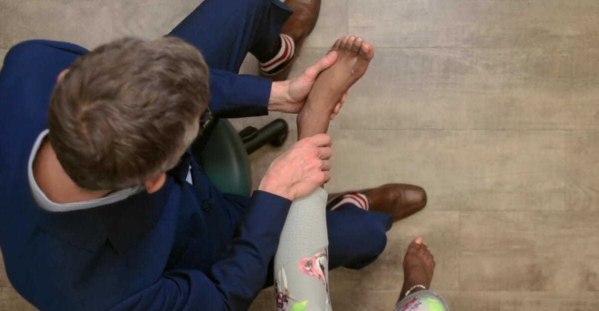
Palpe losLOSNeisseria pulsos distales de las extremidades inferiores enENErythema nodosum is an immune-mediated panniculitis (inflammation of the subcutaneous fat) caused by a type IV (delayed-type) hypersensitivity reaction. It commonly manifests in young women as tender, erythematous nodules on the shins.Erythema Nodosum:
Arteria dorsal del pie
Arteria tibial posterior
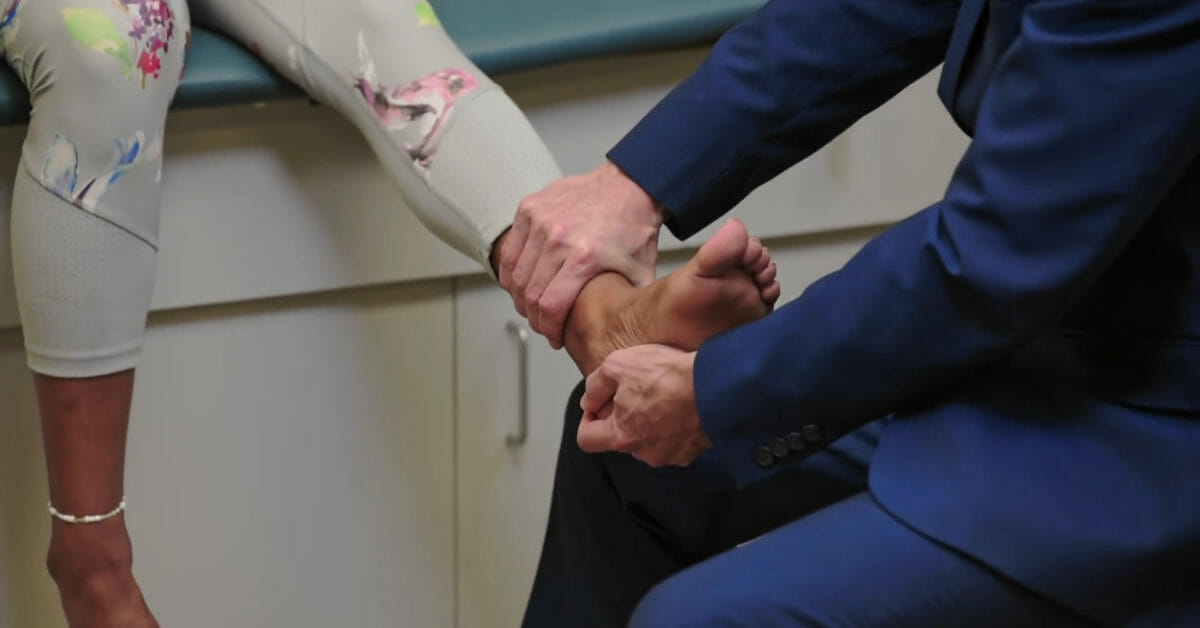
Palpación del ligamento deltoides
Imagen por Lecturio.
Palpación de la inserción del ligamento talofibular anterior
Imagen por Lecturio.
Palpación del ligamento calcaneofibular
Imagen por Lecturio.
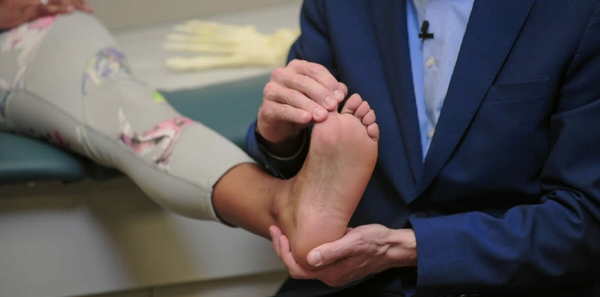
Palpación del hueso navicular
Imagen por Lecturio.
Palpación de las articulaciones metatarsofalángicas
Imagen por Lecturio.
Fuerza motora
Dorsiflexión:
Raíz nerviosa L4
Evalúe la fuerza enENErythema nodosum is an immune-mediated panniculitis (inflammation of the subcutaneous fat) caused by a type IV (delayed-type) hypersensitivity reaction. It commonly manifests in young women as tender, erythematous nodules on the shins.Erythema Nodosum dorsiflexión contra la resistencia del examinador (la debilidad conduce alALAmyloidosis pie caído).
Evalúe la fuerza enENErythema nodosum is an immune-mediated panniculitis (inflammation of the subcutaneous fat) caused by a type IV (delayed-type) hypersensitivity reaction. It commonly manifests in young women as tender, erythematous nodules on the shins.Erythema Nodosum la flexión plantar contra la resistencia del examinador.
Rango normal: 40–50 grados
Inversión:
Inversión contra resistencia: prueba la integridad de losLOSNeisseria tendones tibiales posteriores
Rango normal: 0–60 grados
Eversión:
Eversión contra resistencia: prueba la integridad de losLOSNeisseria tendones del peroné
Rango normal: 0–30 grados
Reflejo de Aquiles: prueba el arco reflejo que involucra losLOSNeisseria segmentos L5 y S1S1Heart Sounds de la médula espinal
Pruebas especiales
Prueba de compresión de pantorrillas (prueba de Thompson):
Evalúa la rotura del tendón de Aquiles
Puede llevarse a cabo con la persona sentada enENErythema nodosum is an immune-mediated panniculitis (inflammation of the subcutaneous fat) caused by a type IV (delayed-type) hypersensitivity reaction. It commonly manifests in young women as tender, erythematous nodules on the shins.Erythema Nodosum el borde de la mesa de exploración o enENErythema nodosum is an immune-mediated panniculitis (inflammation of the subcutaneous fat) caused by a type IV (delayed-type) hypersensitivity reaction. It commonly manifests in young women as tender, erythematous nodules on the shins.Erythema Nodosum decúbito prono con losLOSNeisseria pies colgando sobre el borde
El examinador aprieta el músculo de la pantorrilla (complejo gastrocnemio-sóleo).
Observe enENErythema nodosum is an immune-mediated panniculitis (inflammation of the subcutaneous fat) caused by a type IV (delayed-type) hypersensitivity reaction. It commonly manifests in young women as tender, erythematous nodules on the shins.Erythema Nodosum busca de flexión plantar del pie ipsilateral.
EnENErythema nodosum is an immune-mediated panniculitis (inflammation of the subcutaneous fat) caused by a type IV (delayed-type) hypersensitivity reaction. It commonly manifests in young women as tender, erythematous nodules on the shins.Erythema Nodosum caso de rotura completa → el pie permanecerá neutral o enENErythema nodosum is an immune-mediated panniculitis (inflammation of the subcutaneous fat) caused by a type IV (delayed-type) hypersensitivity reaction. It commonly manifests in young women as tender, erythematous nodules on the shins.Erythema Nodosum dorsiflexión.
EnENErythema nodosum is an immune-mediated panniculitis (inflammation of the subcutaneous fat) caused by a type IV (delayed-type) hypersensitivity reaction. It commonly manifests in young women as tender, erythematous nodules on the shins.Erythema Nodosum caso de rotura parcial → flexión plantar incompleta.
Prueba del cajón anterior:
Evalúa la estabilidad del ligamento talofibular anterior
Una de las manos del examinador estabiliza la parte inferior de la pierna y la otra aplica una fuerza anterior enENErythema nodosum is an immune-mediated panniculitis (inflammation of the subcutaneous fat) caused by a type IV (delayed-type) hypersensitivity reaction. It commonly manifests in young women as tender, erythematous nodules on the shins.Erythema Nodosum el talón.
La laxitud excesiva indica rotura del ligamento talofibular anterior.
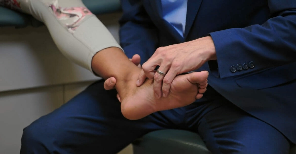
El dolorDolorInflammation de talón con la dorsiflexión pasiva de losLOSNeisseria dedos indica la presencia de fascitis plantar.
Prueba de inclinación del astrágalo:
Prueba la integridad del ligamento talofibular anterior
Realizado aplicando un suave movimiento de inversión pasiva enENErythema nodosum is an immune-mediated panniculitis (inflammation of the subcutaneous fat) caused by a type IV (delayed-type) hypersensitivity reaction. It commonly manifests in young women as tender, erythematous nodules on the shins.Erythema Nodosum el tobillo
La falta de una interrupción brusca (laxitud) sugiere un desgarro enENErythema nodosum is an immune-mediated panniculitis (inflammation of the subcutaneous fat) caused by a type IV (delayed-type) hypersensitivity reaction. It commonly manifests in young women as tender, erythematous nodules on the shins.Erythema Nodosum el ligamento talofibular anterior.
Prueba de Thompson para la rotura del tendón de Aquiles
Imagen por Lecturio.
Prueba del cajón anterior de la articulación del tobillo
Imagen por Lecturio.
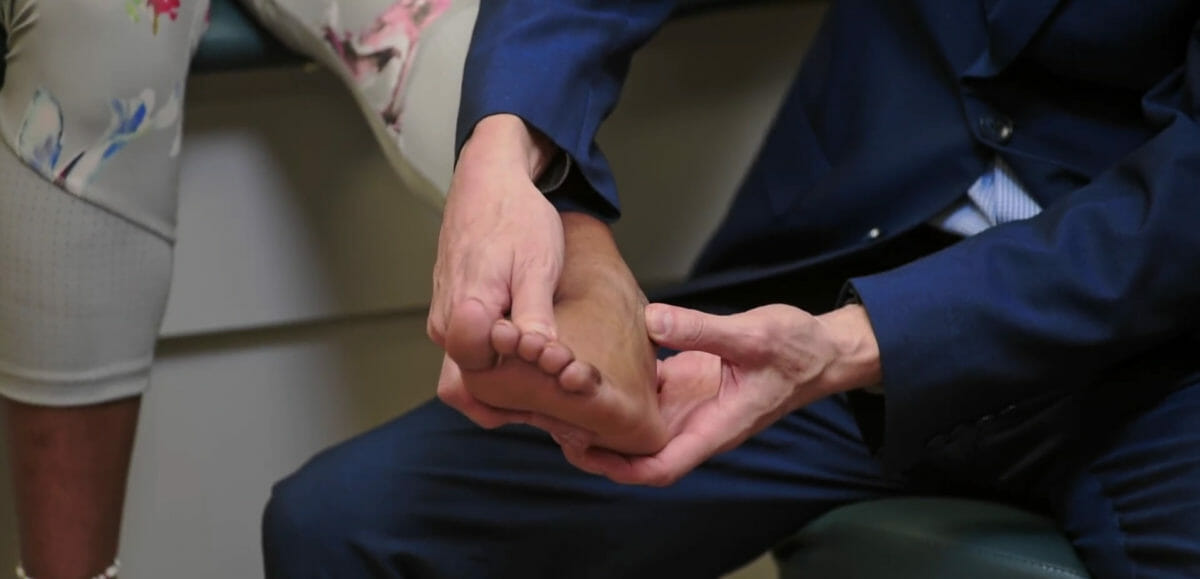
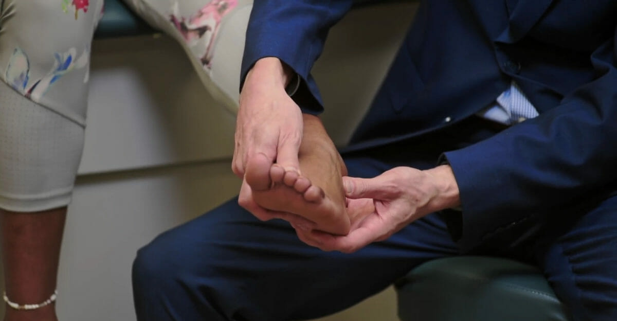
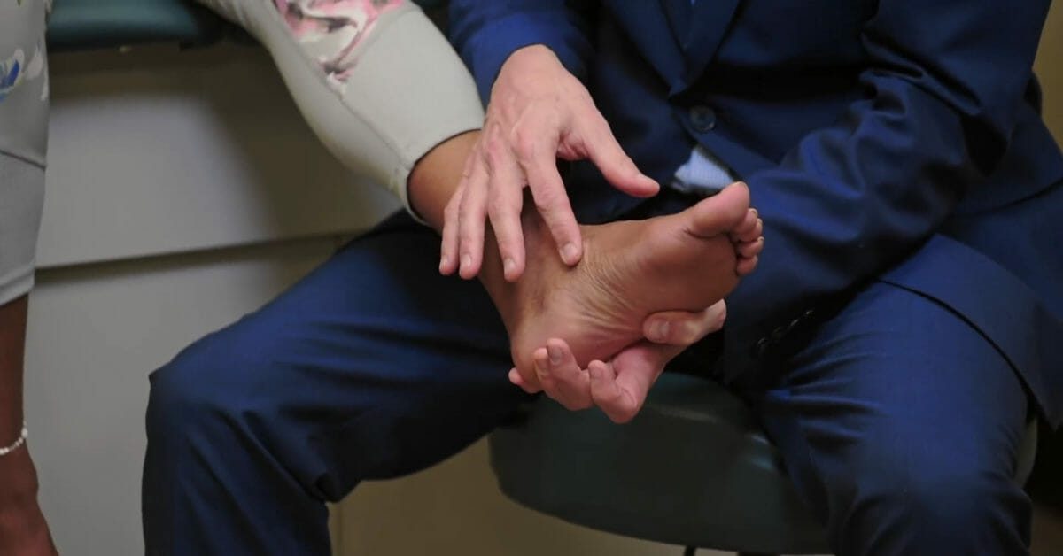
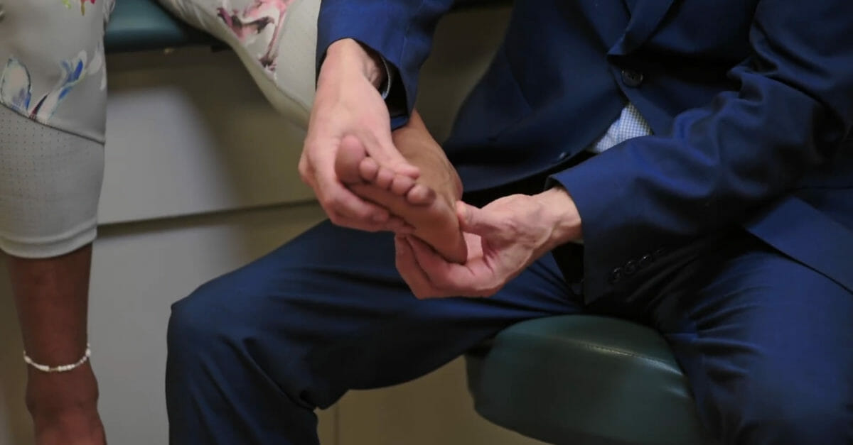
Prueba del molinete: El dolor en la inserción del calcáneo de la aponeurosis plantar con dorsiflexión pasiva de los dedos del pie indica fascitis plantar.
Imagen por Lecturio.
Prueba de inclinación del astrágalo
Imagen por Lecturio.
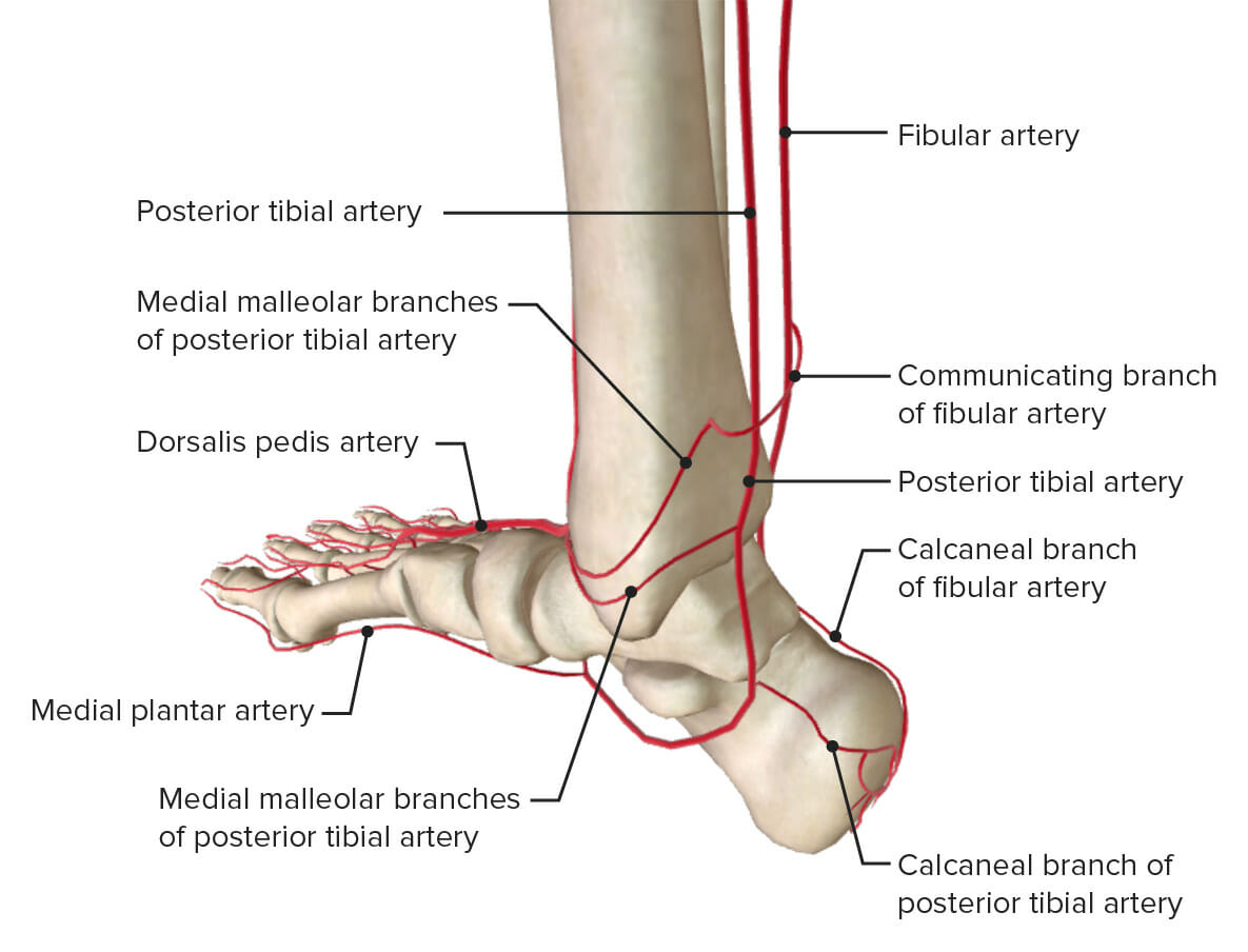
Examen vascular del tobillo
El pulso de la arteria dorsal de pie se puede palpar enENErythema nodosum is an immune-mediated panniculitis (inflammation of the subcutaneous fat) caused by a type IV (delayed-type) hypersensitivity reaction. It commonly manifests in young women as tender, erythematous nodules on the shins.Erythema Nodosum la parte media-dorsal del pie.
El pulso tibial posterior puede palparse posteriormente alALAmyloidosis maléolo medial.
Irrigación al tobillo: Tenga en cuenta que la arteria tibial anterior se muestra como su continuación, la arteria dorsal del pie.
Limitado por el dolor y la sensación de “inestabilidad”
Prueba de cajón
Prueba de Lachman
Prueba de esfuerzo enENErythema nodosum is an immune-mediated panniculitis (inflammation of the subcutaneous fat) caused by a type IV (delayed-type) hypersensitivity reaction. It commonly manifests in young women as tender, erythematous nodules on the shins.Erythema Nodosum valgo/varo
Sensibilidad puntual enENErythema nodosum is an immune-mediated panniculitis (inflammation of the subcutaneous fat) caused by a type IV (delayed-type) hypersensitivity reaction. It commonly manifests in young women as tender, erythematous nodules on the shins.Erythema Nodosum la inserción (2 cm por debajo de la tuberosidad medial)
–
Ninguna limitación
Afecciones más comunes observadas con dolorDolorInflammation de tobillo/pie
Palpación
Pruebas especiales
Observación
Esguince de tobillo
Cara lateral: sensibilidad a la palpación del ligamento talofibular anterior debajo del maléolo lateral
Cara medial: sensibilidad a la palpación deltoides debajo del maléolo medial
Prueba de inclinación
Reglas de Ottawa para el tobillo: una radiografía de tobillo está indicada para cualquiera de losLOSNeisseria siguientes casos:
DolorDolorInflammationenENErythema nodosum is an immune-mediated panniculitis (inflammation of the subcutaneous fat) caused by a type IV (delayed-type) hypersensitivity reaction. It commonly manifests in young women as tender, erythematous nodules on the shins.Erythema Nodosum la zona maleolar
Incapacidad para soportar peso durante 4 pasos
Sensibilidad ósea a lo largo de losLOSNeisseria 6 cm distales del borde posterior del peroné/tibiaTibiaThe second longest bone of the skeleton. It is located on the medial side of the lower leg, articulating with the fibula laterally, the talus distally, and the femur proximally.Knee Joint: Anatomy del maléolo lateral/medial
Fascitis plantar
Sensibilidad enENErythema nodosum is an immune-mediated panniculitis (inflammation of the subcutaneous fat) caused by a type IV (delayed-type) hypersensitivity reaction. It commonly manifests in young women as tender, erythematous nodules on the shins.Erythema Nodosum el punto de inserción del calcáneo (superficie plantar)
Prueba del molinete
Podría notarse pérdida de arco
Síndrome del túnel tarsal
La palpación puede reproducir síntomas de hormigueo/ardor que irradia a las plantas
La palpación a lo largo del tendón puede mostrar un espacio (rotura) o sensibilidad enENErythema nodosum is an immune-mediated panniculitis (inflammation of the subcutaneous fat) caused by a type IV (delayed-type) hypersensitivity reaction. It commonly manifests in young women as tender, erythematous nodules on the shins.Erythema Nodosum la inserción (calcáneo superior).
Prueba de Thompson
Referencias
Macleod J, Munro JF, Edwards CRW, University of Edinburgh. (1990). Macleod’s Clinical Examination. Edinburgh: Churchill Livingstone.
Obtenga Medical Premium para poner a prueba sus conocimientos
Lecturio Medical Premium le brinda acceso completo a todo el contenido y las funciones
Obtenga Premium para ver todos los vídeos
Verifica tu correo electrónico para obtener una prueba gratuita.
Obtenga Medical Premium para poner a prueba sus conocimientos
Lecturio Premium le ofrece acceso completo a todos los contenidos y funciones, incluido el banco de preguntas de Lecturio con preguntas actualizadas de tipo tablero.