LosLOSNeisseria astrocitomas son tumores neuroepiteliales que surgen de losLOSNeisseria astrocitos, que son células gliales con forma de estrella. Según la clasificación CNS5 de la OMS, losLOSNeisseria astrocitomas se clasifican enENErythema nodosum is an immune-mediated panniculitis (inflammation of the subcutaneous fat) caused by a type IV (delayed-type) hypersensitivity reaction. It commonly manifests in young women as tender, erythematous nodules on the shins.Erythema Nodosum astrocitomas pilocíticos (grado 1), astrocitomas difusos (grado 2), astrocitomas anaplásicos (grado 3) y glioblastomas (grado 4). LosLOSNeisseria tumores de grado 1 son benignos y se presentan enENErythema nodosum is an immune-mediated panniculitis (inflammation of the subcutaneous fat) caused by a type IV (delayed-type) hypersensitivity reaction. It commonly manifests in young women as tender, erythematous nodules on the shins.Erythema Nodosum niños, mientras que el glioblastoma IDH de tipo salvaje es el tumorTumorInflammation cerebral maligno primario más común enENErythema nodosum is an immune-mediated panniculitis (inflammation of the subcutaneous fat) caused by a type IV (delayed-type) hypersensitivity reaction. It commonly manifests in young women as tender, erythematous nodules on the shins.Erythema Nodosum adultos y tiene un pronóstico desfavorable. La presentación clínica varía según la ubicación y el grado, pero a menudo incluye cefalea, convulsiones y déficits neurológicos focales. El diagnóstico requiere resonancia magnética combinada con análisis histopatológico y molecular del tejido de la biopsia, y el tratamiento depende del grado y el subtipo molecular del tumorTumorInflammation, incluyendo cirugía, radioterapia y quimioterapia.
LosLOSNeisseria astrocitomas son tumores neuroepiteliales del SNC que surgen de losLOSNeisseria astrocitos, que son células gliales con forma de estrella.
Las células gliales son tejidos de soporte enENErythema nodosum is an immune-mediated panniculitis (inflammation of the subcutaneous fat) caused by a type IV (delayed-type) hypersensitivity reaction. It commonly manifests in young women as tender, erythematous nodules on the shins.Erythema Nodosum el cerebro y el sistema nervioso.
LosLOSNeisseria astrocitomas se clasifican enENErythema nodosum is an immune-mediated panniculitis (inflammation of the subcutaneous fat) caused by a type IV (delayed-type) hypersensitivity reaction. It commonly manifests in young women as tender, erythematous nodules on the shins.Erythema Nodosum grados, pero no se estadifican.
Clasificación de losLOSNeisseria tumores del sistema nervioso
Tabla: Clasificación de losLOSNeisseria tumores del sistema nervioso
Categorías
Tumores específicos
Tumores neuroepiteliales (gliales y relacionados)
Astrocitoma pilocítico (grado 1)
Astrocitoma de IDH mutante (grados 2-4)
Glioblastoma de IDH de tipo salvaje (grado 4)
OligodendrogliomaOligodendrogliomaOligodendrogliomas are malignant CNS tumors arising from neural glial cell precursors. Oligodendrogliomas often arise in the frontal lobes of the brain and have a generally favorable prognosis when compared to other gliomas. Oligodendrogliomas are the 3rd most common CNS tumor. The most frequent presenting symptom is a seizure.Oligodendroglioma
Ependimoma y tumores del plexo coroideo
Tumores embrionarios (p. ej., meduloblastoma)
Tumores meníngeos
Meningiomas
Tumores vasculares
HemangioblastomasHemangioblastomasA benign tumor of the nervous system that may occur sporadically or in association with von Hippel-Lindau disease. It accounts for approximately 2% of intracranial tumors, arising most frequently in the cerebellar hemispheres and vermis. Histologically, the tumors are composed of multiple capillary and sinusoidal channels lined with endothelial cells and clusters of lipid-laden pseudoxanthoma cells. Usually solitary, these tumors can be multiple and may also occur in the brain stem, spinal cord, retina, and supratentorial compartment. Cerebellar hemangioblastomas usually present in the third decade with intracranial hypertension, and ataxia.Von Hippel-Lindau Disease
Tumores de la región selar
Craneofaringioma
Adenoma hipofisario
Tumores de la región pineal
Pineocitoma
Pineoblastoma
Linfoma primario del SNC
Linfoma primario del SNC (generalmente de tipo difuso de células B grandes)
Tumores cerebrales metastásicos
Más comúnmente surgen de:
Carcinomas de pulmón
Carcinomas de mama
Melanomas
Carcinomas de células renales
Carcinomas colorrectales
Tumores del sistema nervioso periférico
Schwannomas (schwannomaSchwannomaSchwannomas (also known as neurilemmomas) are benign nerve sheath tumors in the peripheral nervous system (PNS), arising from Schwann cells that encase the peripheral nerves. Schwannomas are the most common tumors in the PNS. Schwannoma vestibular)
Neurofibromas
Tumores malignos de la vaina de nervios periféricos
Según el sistema CNS 5 de la OMS, losLOSNeisseria astrocitomas se clasifican mediante un enfoque histológico y molecular integrado, y el grado tumoral (1-4) se determina por características como la atipia nuclear, la actividad mitótica, la proliferación microvascular y la necrosisNecrosisThe death of cells in an organ or tissue due to disease, injury or failure of the blood supply.Ischemic Cell Damage.
Gliomas astrocíticos circunscritos (grado 1-2 de la OMS):
Astrocitoma pilocítico (grado 1):
Bien circunscrito, de crecimiento lento, a menudo quístico
Común enENErythema nodosum is an immune-mediated panniculitis (inflammation of the subcutaneous fat) caused by a type IV (delayed-type) hypersensitivity reaction. It commonly manifests in young women as tender, erythematous nodules on the shins.Erythema Nodosum niños
Localización cerebelosa → compresión/ataxiaAtaxiaImpairment of the ability to perform smoothly coordinated voluntary movements. This condition may affect the limbs, trunk, eyes, pharynx, larynx, and other structures. Ataxia may result from impaired sensory or motor function. Sensory ataxia may result from posterior column injury or peripheral nerve diseases. Motor ataxia may be associated with cerebellar diseases; cerebral cortex diseases; thalamic diseases; basal ganglia diseases; injury to the red nucleus; and other conditions.Ataxia-telangiectasia del 4º ventrículo
Masa superficial enENErythema nodosum is an immune-mediated panniculitis (inflammation of the subcutaneous fat) caused by a type IV (delayed-type) hypersensitivity reaction. It commonly manifests in young women as tender, erythematous nodules on the shins.Erythema Nodosum el lóbulo temporal
Presenta convulsiones
La forma anaplásica muestra ↑ actividad mitótica
Astrocitoma subependimario de células gigantes (grado 1):
Periventricular enENErythema nodosum is an immune-mediated panniculitis (inflammation of the subcutaneous fat) caused by a type IV (delayed-type) hypersensitivity reaction. It commonly manifests in young women as tender, erythematous nodules on the shins.Erythema Nodosum la esclerosis tuberosa
Sin necrosisNecrosisThe death of cells in an organ or tissue due to disease, injury or failure of the blood supply.Ischemic Cell Damage ni proliferación microvascular
Astrocitoma, IDH mutante (grado 4):
↑ Atipia nuclear + actividad mitótica + necrosisNecrosisThe death of cells in an organ or tissue due to disease, injury or failure of the blood supply.Ischemic Cell Damage y/o proliferación microvascular
No se denomina glioblastoma si presenta mutación IDH
Glioblastoma, IDH de tipo salvaje (grado 4):
NecrosisNecrosisThe death of cells in an organ or tissue due to disease, injury or failure of the blood supply.Ischemic Cell Damage y/o proliferación microvascular
Origen «primario» enENErythema nodosum is an immune-mediated panniculitis (inflammation of the subcutaneous fat) caused by a type IV (delayed-type) hypersensitivity reaction. It commonly manifests in young women as tender, erythematous nodules on the shins.Erythema Nodosum adultos (~90 %) contra progresión «secundaria» desde un grado inferior (~10 %)
Mediana de supervivencia ~12–15 meses
Epidemiología
Distribución pediátrica:
LosLOSNeisseria gliomas representan ~50 % de losLOSNeisseria tumores del SNC enENErythema nodosum is an immune-mediated panniculitis (inflammation of the subcutaneous fat) caused by a type IV (delayed-type) hypersensitivity reaction. It commonly manifests in young women as tender, erythematous nodules on the shins.Erythema Nodosum niños y adolescentes; losLOSNeisseria astrocitomas circunscritos son losLOSNeisseria más comunes.
La incidencia del astrocitoma pilocítico es de ~1 por cada 100 000 niños.
LosLOSNeisseria tumores astrocíticos representan ~31 % de las neoplasias del SNC enENErythema nodosum is an immune-mediated panniculitis (inflammation of the subcutaneous fat) caused by a type IV (delayed-type) hypersensitivity reaction. It commonly manifests in young women as tender, erythematous nodules on the shins.Erythema Nodosum la infancia.
Distribución enENErythema nodosum is an immune-mediated panniculitis (inflammation of the subcutaneous fat) caused by a type IV (delayed-type) hypersensitivity reaction. It commonly manifests in young women as tender, erythematous nodules on the shins.Erythema Nodosum adultos:
LosLOSNeisseria tumores cerebrales metastásicos superan enENErythema nodosum is an immune-mediated panniculitis (inflammation of the subcutaneous fat) caused by a type IV (delayed-type) hypersensitivity reaction. It commonly manifests in young women as tender, erythematous nodules on the shins.Erythema Nodosum número a losLOSNeisseria tumores primarios del SNC.
Entre losLOSNeisseria tumores gliales primarios, losLOSNeisseria astrocitomas difusos (IDH mutante e IDH de tipo salvaje) y el glioblastoma son losLOSNeisseria más comunes.
Incidencia general:
Todos losLOSNeisseria tumores astrocíticos combinados: ~5-6 por cada 100 000 personas-año.
Glioblastoma, IDH de tipo salvaje: ~3,2 por cada 100 000 personas.
Glioblastoma, IDH de tipo salvaje: masculino:femenino ~1,6:1
Etiología
La mayoría de las causas de losLOSNeisseria astrocitomas es desconocida. EnENErythema nodosum is an immune-mediated panniculitis (inflammation of the subcutaneous fat) caused by a type IV (delayed-type) hypersensitivity reaction. It commonly manifests in young women as tender, erythematous nodules on the shins.Erythema Nodosum última instancia, las mutaciones genéticas conducen a un crecimiento celular incontrolado y a la proliferación de tumores.
LosLOSNeisseria astrocitomas son más frecuentes enENErythema nodosum is an immune-mediated panniculitis (inflammation of the subcutaneous fat) caused by a type IV (delayed-type) hypersensitivity reaction. It commonly manifests in young women as tender, erythematous nodules on the shins.Erythema NodosumlosLOSNeisseria siguientes trastornos genéticos:
Síndrome de Li-Fraumeni (mutaciones enENErythema nodosum is an immune-mediated panniculitis (inflammation of the subcutaneous fat) caused by a type IV (delayed-type) hypersensitivity reaction. It commonly manifests in young women as tender, erythematous nodules on the shins.Erythema Nodosum la línea germinal del p53)
Síndrome de Turcot (mutaciones enENErythema nodosum is an immune-mediated panniculitis (inflammation of the subcutaneous fat) caused by a type IV (delayed-type) hypersensitivity reaction. It commonly manifests in young women as tender, erythematous nodules on the shins.Erythema Nodosum varios genesGenesA category of nucleic acid sequences that function as units of heredity and which code for the basic instructions for the development, reproduction, and maintenance of organisms.DNA Types and Structure supresores de tumores, incluyendo APCAPCA polyposis syndrome due to an autosomal dominant mutation of the apc genes on chromosome 5. The syndrome is characterized by the development of hundreds of adenomatous polyps in the colon and rectum of affected individuals by early adulthood.Familial Adenomatous Polyposis y MMRMMRA DNA repair pathway involved in correction of errors introduced during DNA replication when an incorrect base, which cannot form hydrogen bonds with the corresponding base in the parent strand, is incorporated into the daughter strand. Excinucleases recognize the base pair mismatch and cause a segment of polynucleotide chain to be excised from the daughter strand, thereby removing the mismatched base.Lynch syndrome)
Neurofibromatosis tipo 1
Esclerosis tuberosa
La radiación ionizante es un factor de riesgo establecido:
Radioterapia para adenoma hipofisario: 16 veces más riesgo para astrocitoma
Niños que reciben radiación para leucemia linfocítica aguda: 22 veces más riesgo de desarrollar una neoplasia del SNC enENErythema nodosum is an immune-mediated panniculitis (inflammation of the subcutaneous fat) caused by a type IV (delayed-type) hypersensitivity reaction. It commonly manifests in young women as tender, erythematous nodules on the shins.Erythema Nodosum 5–10 años (incluyendo astrocitomas de grado 2, 3 y 4)
Hay varias mutaciones genéticas asociadas a losLOSNeisseria astrocitomas.
Mutaciones enENErythema nodosum is an immune-mediated panniculitis (inflammation of the subcutaneous fat) caused by a type IV (delayed-type) hypersensitivity reaction. It commonly manifests in young women as tender, erythematous nodules on the shins.Erythema Nodosum la isocitrato deshidrogenasa 1 (gen IDH1):
IDH1:
Cataliza la descarboxilación oxidativa reversible del isocitrato → α-cetoglutarato (α-KG) enENErythema nodosum is an immune-mediated panniculitis (inflammation of the subcutaneous fat) caused by a type IV (delayed-type) hypersensitivity reaction. It commonly manifests in young women as tender, erythematous nodules on the shins.Erythema Nodosum el ciclo del ácido tricarboxílico (TCA)
Productor primario de nicotinamida adenina dinucleótido fosfato (NADPHNADPHNicotinamide adenine dinucleotide phosphate. A coenzyme composed of ribosylnicotinamide 5′-phosphate (nmn) coupled by pyrophosphate linkage to the 5′-phosphate adenosine 2.Pentose Phosphate Pathway) enENErythema nodosum is an immune-mediated panniculitis (inflammation of the subcutaneous fat) caused by a type IV (delayed-type) hypersensitivity reaction. It commonly manifests in young women as tender, erythematous nodules on the shins.Erythema Nodosum la mayoría de losLOSNeisseria tejidos, especialmente enENErythema nodosum is an immune-mediated panniculitis (inflammation of the subcutaneous fat) caused by a type IV (delayed-type) hypersensitivity reaction. It commonly manifests in young women as tender, erythematous nodules on the shins.Erythema Nodosum el cerebro
También participa enENErythema nodosum is an immune-mediated panniculitis (inflammation of the subcutaneous fat) caused by a type IV (delayed-type) hypersensitivity reaction. It commonly manifests in young women as tender, erythematous nodules on the shins.Erythema Nodosum la mitigación del daño oxidativo
Las mutaciones conducen a la producción y acumulación de la 2-hidroxiglutarato (2-HG2-HGAstrocytoma):
La 2-HG2-HGAstrocytoma inhibe la función enzimática de las dioxigenasas dependientes del α-cetoglutarato (KG), que participan enENErythema nodosum is an immune-mediated panniculitis (inflammation of the subcutaneous fat) caused by a type IV (delayed-type) hypersensitivity reaction. It commonly manifests in young women as tender, erythematous nodules on the shins.Erythema Nodosum la desmetilación del ADN.
↑ del 2-HG2-HGAstrocytoma provoca una desregulación epigenética → puede conducir alALAmyloidosis desarrollo de tumores
Metilación (i.e., silenciamiento) del promotor de la metilguanina metiltransferasa (MGMT):
La MGMT es una enzima que interviene enENErythema nodosum is an immune-mediated panniculitis (inflammation of the subcutaneous fat) caused by a type IV (delayed-type) hypersensitivity reaction. It commonly manifests in young women as tender, erythematous nodules on the shins.Erythema Nodosum la reparación del ADN (incluida la reparación del ADN tras la quimioterapia con un agente alquilante).
Metilación de MGMT enENErythema nodosum is an immune-mediated panniculitis (inflammation of the subcutaneous fat) caused by a type IV (delayed-type) hypersensitivity reaction. It commonly manifests in young women as tender, erythematous nodules on the shins.Erythema Nodosum la región promotora:
Silencia la expresión del gen
Puede ocurrir durante el desarrollo del tumorTumorInflammation → impide la reparación del daño enENErythema nodosum is an immune-mediated panniculitis (inflammation of the subcutaneous fat) caused by a type IV (delayed-type) hypersensitivity reaction. It commonly manifests in young women as tender, erythematous nodules on the shins.Erythema Nodosum el ADN
Mejora la respuesta a la quimioterapia y la supervivencia global (independientemente de otros factores de riesgo)
Mutaciones inactivadoras de p53 (co-mutación definitoria enENErythema nodosum is an immune-mediated panniculitis (inflammation of the subcutaneous fat) caused by a type IV (delayed-type) hypersensitivity reaction. It commonly manifests in young women as tender, erythematous nodules on the shins.Erythema Nodosum el linaje de astrocitoma, IDH mutante)
Sobreexpresión del factor de crecimiento derivado de las plaquetas alfa (PDGF-A, por sus siglas enENErythema nodosum is an immune-mediated panniculitis (inflammation of the subcutaneous fat) caused by a type IV (delayed-type) hypersensitivity reaction. It commonly manifests in young women as tender, erythematous nodules on the shins.Erythema Nodosum inglés)
Ciertos tipos de antígenos leucocitarios humanos (HLA, por sus siglas enENErythema nodosum is an immune-mediated panniculitis (inflammation of the subcutaneous fat) caused by a type IV (delayed-type) hypersensitivity reaction. It commonly manifests in young women as tender, erythematous nodules on the shins.Erythema Nodosum inglés) se asocian a un riesgo menor o mayor.
Fisiopatología
LosLOSNeisseria astrocitomas suelen surgir enENErythema nodosum is an immune-mediated panniculitis (inflammation of the subcutaneous fat) caused by a type IV (delayed-type) hypersensitivity reaction. It commonly manifests in young women as tender, erythematous nodules on the shins.Erythema NodosumlosLOSNeisseria hemisferios cerebrales (i.e., enENErythema nodosum is an immune-mediated panniculitis (inflammation of the subcutaneous fat) caused by a type IV (delayed-type) hypersensitivity reaction. It commonly manifests in young women as tender, erythematous nodules on the shins.Erythema Nodosum el parénquima).
LosLOSNeisseria efectos regionales enENErythema nodosum is an immune-mediated panniculitis (inflammation of the subcutaneous fat) caused by a type IV (delayed-type) hypersensitivity reaction. It commonly manifests in young women as tender, erythematous nodules on the shins.Erythema Nodosum el parénquima cerebral incluyen:
Compresión
Invasión
Destrucción
El ↑ de la presión intracraneal (PIC) puede deberse a:
Efecto directo de la masa
EdemaEdemaEdema is a condition in which excess serous fluid accumulates in the body cavity or interstitial space of connective tissues. Edema is a symptom observed in several medical conditions. It can be categorized into 2 types, namely, peripheral (in the extremities) and internal (in an organ or body cavity). EdemaenENErythema nodosum is an immune-mediated panniculitis (inflammation of the subcutaneous fat) caused by a type IV (delayed-type) hypersensitivity reaction. It commonly manifests in young women as tender, erythematous nodules on the shins.Erythema Nodosum el tejido cerebral circundante
↑ Volumen sanguíneo
↑ Volumen de LCR/hidrocefalia
Las alteraciones de las funciones normales del parénquima se deben a:
Liberación y reclutamiento de mediadores celulares (e.g., citoquinas)
Presentación Clínica
LosLOSNeisseria signos y síntomas neurológicos relacionados con losLOSNeisseria astrocitomas son el resultado de las perturbaciones de la función del SNC.
Grados 1 y 2: El inicio suele ser sutil y losLOSNeisseria tumores se desarrollan lentamente debido a la capacidad del cerebro de adaptarse temporalmente a la presencia de un tumorTumorInflammation de crecimiento lento.
Grados 3 y 4:La aparición es más a menudo repentina y/o debilitante.
Síntomas
LosLOSNeisseria síntomas dependen principalmente de la localización del tumorTumorInflammationenENErythema nodosum is an immune-mediated panniculitis (inflammation of the subcutaneous fat) caused by a type IV (delayed-type) hypersensitivity reaction. It commonly manifests in young women as tender, erythematous nodules on the shins.Erythema Nodosum el cerebro. LosLOSNeisseria síntomas se dividen enENErythema nodosum is an immune-mediated panniculitis (inflammation of the subcutaneous fat) caused by a type IV (delayed-type) hypersensitivity reaction. It commonly manifests in young women as tender, erythematous nodules on the shins.Erythema Nodosum 2 categorías:
Síntomas generales: síntomas que pueden aparecer con tumores enENErythema nodosum is an immune-mediated panniculitis (inflammation of the subcutaneous fat) caused by a type IV (delayed-type) hypersensitivity reaction. It commonly manifests in young women as tender, erythematous nodules on the shins.Erythema Nodosum cualquier localización
AtaxiaAtaxiaImpairment of the ability to perform smoothly coordinated voluntary movements. This condition may affect the limbs, trunk, eyes, pharynx, larynx, and other structures. Ataxia may result from impaired sensory or motor function. Sensory ataxia may result from posterior column injury or peripheral nerve diseases. Motor ataxia may be associated with cerebellar diseases; cerebral cortex diseases; thalamic diseases; basal ganglia diseases; injury to the red nucleus; and other conditions.Ataxia-telangiectasia
Papiledema
Síntomas focales: síntomas que se producen a causa de tumores enENErythema nodosum is an immune-mediated panniculitis (inflammation of the subcutaneous fat) caused by a type IV (delayed-type) hypersensitivity reaction. It commonly manifests in young women as tender, erythematous nodules on the shins.Erythema Nodosum lugares específicos
LosLOSNeisseria tumores de bajo grado son más propensos a causar convulsiones que losLOSNeisseria de alto grado.
Afasia
Déficit del campo visual
Debilidad motora
Hemiparesia
Anomalías sensoriales
Diagnóstico
La neuroimagen es esencial para la detección y la caracterización inicial, pero el diagnóstico definitivo y la gradación requieren histopatología y pruebas moleculares.
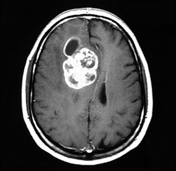
RM
Estudio de imagen estándar de oro
Se solicita preferentemente con y sin realce de gadolinio
LosLOSNeisseria astrocitomas de mayor grado muestran:
Isointensidad enENErythema nodosum is an immune-mediated panniculitis (inflammation of the subcutaneous fat) caused by a type IV (delayed-type) hypersensitivity reaction. It commonly manifests in young women as tender, erythematous nodules on the shins.Erythema Nodosum T1 e hiperintensidad enENErythema nodosum is an immune-mediated panniculitis (inflammation of the subcutaneous fat) caused by a type IV (delayed-type) hypersensitivity reaction. It commonly manifests in young women as tender, erythematous nodules on the shins.Erythema Nodosum T2
Realce con agentes de contraste paramagnéticos
LosLOSNeisseria astrocitomas de bajo grado no suelen realzar con contraste.
RM con contraste que muestra un glioblastoma multiforme
Imagen: “Glioblastoma multiforme” por Duncan JS, de Tisi J. Licencia: CC BY 3.0
TC
Indicaciones:
Contraindicación para RM
Fases agudas (e.g., cuando hay que descartar una hemorragia o un accidente cerebrovascular)
La TC de tórax/abdomen/pelvisPelvisThe pelvis consists of the bony pelvic girdle, the muscular and ligamentous pelvic floor, and the pelvic cavity, which contains viscera, vessels, and multiple nerves and muscles. The pelvic girdle, composed of 2 “hip” bones and the sacrum, is a ring-like bony structure of the axial skeleton that links the vertebral column with the lower extremities.Pelvis: Anatomy puede estar justificada para buscar lesiones primarias alternativas si se sospecha de metástasis.
LosLOSNeisseria hallazgos muestran una masa parenquimatosa mal definida.
Las lesiones de bajo grado no suelen realzar con el contraste.
Otras pruebas
EEGEEGSeizures: puede utilizarse para evaluar y controlar la actividad convulsiva
Estudios del LCR: pueden ayudar a descartar otros diagnósticos (e.g., linfoma del SNC, metástasis)
Biopsia
Se requiere tejido (mediante resección o biopsia estereotáctica) para la clasificación histopatológica y molecular. Una biopsia está indicada para la gradación o confirmación del diagnóstico tras la identificación del tumorTumorInflammation mediante neuroimagen.
Astrocitoma pilocítico (grado 1): Fibra de Rosenthal: inclusiones citoplasmáticas eosinófilas, que aparecen como fibras enENErythema nodosum is an immune-mediated panniculitis (inflammation of the subcutaneous fat) caused by a type IV (delayed-type) hypersensitivity reaction. It commonly manifests in young women as tender, erythematous nodules on the shins.Erythema Nodosum forma de sacacorchos
Astrocitoma, IDH mutante (grado 2):
Aumento de leve a moderado del número de núcleos de células gliales
Pleomorfismo nuclear
Relación núcleo:citoplasma bastante elevada
Filtro intermedio de prolongaciones celulares astrocíticas finas de proteína ácida fibrilar glial
Macroscópicamente: tumores mal definidos y de color gris
Astrocitoma, IDH mutante (grado 3):
Aumento de la celularidad y del pleomorfismo nuclear
Marcada actividad mitótica y atipia nuclear
Alto índice proliferativo basado enENErythema nodosum is an immune-mediated panniculitis (inflammation of the subcutaneous fat) caused by a type IV (delayed-type) hypersensitivity reaction. It commonly manifests in young women as tender, erythematous nodules on the shins.Erythema Nodosum la tinción Ki67
Glioblastoma, IDH de tipo salvaje (grado 4):
Aumento de las cifras mitóticas, de la celularidad y del pleomorfismo nuclear
Proliferación vascular y/o de células endoteliales
Zonas de necrosisNecrosisThe death of cells in an organ or tissue due to disease, injury or failure of the blood supply.Ischemic Cell Damage (que a grandes rasgos parecen firmes y blancas o blandas y amarillas)
Las células tumorales están “pseudopalizadas”.
Astrocitoma pilocítico con células endoteliales prominentes así como células neoplásicas que albergan figuras mitóticas, núcleos blandos y ovalados
Imagen: “Hemorrhagic Pilocytic Astrocytomas in Adults: A Case Report and Literature Review” por Galgano MA, Padalino DJ, Fullmer J, Krishnamurthy S. Licencia: CC BY 3.0
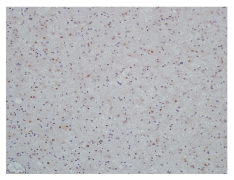
Imagen de una inmunotinción de la proteína de mantenimiento del minicromosoma 2 (MCM2, por sus siglas en inglés) de un astrocitoma de grado II positivo, con núcleos de células astrocíticas neoplásicas (oscuro) (objetivo 40×)
Imagen: “Expression and clinical significance of the proliferation marker minichromosome maintenance protein 2 (Mcm2) in diffuse astrocytomas WHO grade II” por Lind-Landström T, Varughese RK, Sundstrøm S, Torp SH., Licencia: CC BY 2.0
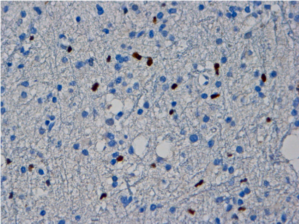
Microfotografía que demuestra una significativa inmunopositividad del p53 en un astrocitoma anaplásico
Imagen: “Malignant trigeminal nerve sheath tumor and anaplastic astrocytoma collision tumor with high proliferative activity and tumor suppressor p53 expression” por Kurdi M, Al-Ardati H, Baeesa SS. Licencia: DominioPúblico
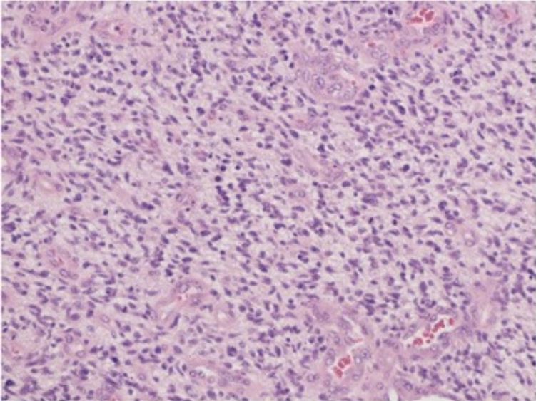
Microfotografía que revela una neoplasia glial celular altamente maligna, incrustada en un fondo glial fibrilar, con mitosis, proliferación endotelial y necrosis Los hallazgos son consistentes con un glioblastoma (tinción de hematoxilina-eosina, ×40)
Imagen: “De novo glioblastoma in the territory of a recent middle cerebral artery infarction and a residual meningioma: pathogenesis revisited” por Yaghmour W, Kurdi ME, Baeesa SS. Licencia: CC BY 4.0
Tratamiento y Pronóstico
Tratamiento según grado
El tratamiento depende del grado, la localización del tumorTumorInflammation (i.e., la cantidad que puede resecarse de manera segura) y losLOSNeisseria síntomas. El tratamiento suele consistir enENErythema nodosum is an immune-mediated panniculitis (inflammation of the subcutaneous fat) caused by a type IV (delayed-type) hypersensitivity reaction. It commonly manifests in young women as tender, erythematous nodules on the shins.Erythema Nodosum una combinación de cirugía, radiación y quimioterapia.
Morbilidad asociada a losLOSNeisseria tratamientos
Edad relativamente joven del paciente típico
Considerar la máxima seguridad enENErythema nodosum is an immune-mediated panniculitis (inflammation of the subcutaneous fat) caused by a type IV (delayed-type) hypersensitivity reaction. It commonly manifests in young women as tender, erythematous nodules on the shins.Erythema Nodosum la resección quirúrgica si el tumorTumorInflammation es accesible.
Extirpación quirúrgica parcial cuando el tumorTumorInflammation afecta partes cruciales del cerebro
Considerar la radioterapia cuando el tumorTumorInflammation no es totalmente resecable.
Astrocitoma con IDH mutante (grado 2):
Se recomienda la resección quirúrgica para losLOSNeisseria tumores accesibles:
No es curativa
EnENErythema nodosum is an immune-mediated panniculitis (inflammation of the subcutaneous fat) caused by a type IV (delayed-type) hypersensitivity reaction. It commonly manifests in young women as tender, erythematous nodules on the shins.Erythema Nodosum última instancia, se requieren de terapias adicionales (i.e., radioterapia y quimioterapia) enENErythema nodosum is an immune-mediated panniculitis (inflammation of the subcutaneous fat) caused by a type IV (delayed-type) hypersensitivity reaction. It commonly manifests in young women as tender, erythematous nodules on the shins.Erythema Nodosum todos losLOSNeisseria pacientes.
Radiación más quimioterapia adyuvante (normalmente temozolomida)
Seguimiento para comprobar recurrencia/progresión
Grados 3 y 4:
El tratamiento estándar es la cirugía, radiación y quimioterapia (normalmente con temozolomida).
La cirugía puede incluir:
Resección
Procedimientos para aliviar losLOSNeisseria síntomas (e.g., colocación de una derivación para aliviar la hidrocefalia)
Si la cirugía no es posible, se utiliza la radiación y la quimioterapia.
Otras terapias a considerar:
Profilaxis de TVP enENErythema nodosum is an immune-mediated panniculitis (inflammation of the subcutaneous fat) caused by a type IV (delayed-type) hypersensitivity reaction. It commonly manifests in young women as tender, erythematous nodules on the shins.Erythema Nodosum pacientes no ambulatorios y hospitalizados
Tratamiento anticonvulsivo enENErythema nodosum is an immune-mediated panniculitis (inflammation of the subcutaneous fat) caused by a type IV (delayed-type) hypersensitivity reaction. It commonly manifests in young women as tender, erythematous nodules on the shins.Erythema Nodosum pacientes conun antecedente de convulsiones (el tratamiento anticonvulsivo profiláctico sigue siendo controversial)
LosLOSNeisseria corticosteroides (e.g., dexametasona) pueden utilizarse por sus propiedades antiinflamatorias para reducir el efecto de masa del tumorTumorInflammation.
Grado 2: La mediana de supervivencia es de 8 años.
Grado 3: La mediana de supervivencia es de 2–5 años.
Grado 4: La mediana de supervivencia es de 15 meses.
Alcance de la resección quirúrgica
Uso de radioterapia y/o quimioterapia adyuvante
Edad: una menor edad enENErythema nodosum is an immune-mediated panniculitis (inflammation of the subcutaneous fat) caused by a type IV (delayed-type) hypersensitivity reaction. It commonly manifests in young women as tender, erythematous nodules on the shins.Erythema Nodosum el momento del diagnóstico se asocia a una mayor supervivencia.
Estado funcional (e.g., síntomas mínimos y/o función neurológica normal) asociado a una mayor supervivencia
Cuando un paciente presenta síntomas neurológicos enENErythema nodosum is an immune-mediated panniculitis (inflammation of the subcutaneous fat) caused by a type IV (delayed-type) hypersensitivity reaction. It commonly manifests in young women as tender, erythematous nodules on the shins.Erythema Nodosum la exploración, el diagnóstico diferencial puede incluir procesos vasculares (e.g., hemorragia, infarto), infecciones (e.g., absceso, encefalitis vírica) y procesos inflamatorios (e.g., esclerosis múltiple), además de tumores cerebrales primarios. El diagnóstico por imagenologia suele permitir reducir el diferencial a un tumorTumorInflammation cerebral.
OligodendrogliomaOligodendrogliomaOligodendrogliomas are malignant CNS tumors arising from neural glial cell precursors. Oligodendrogliomas often arise in the frontal lobes of the brain and have a generally favorable prognosis when compared to other gliomas. Oligodendrogliomas are the 3rd most common CNS tumor. The most frequent presenting symptom is a seizure.Oligodendroglioma:tumorTumorInflammation del SNC que surge de losLOSNeisseria oligodendrocitos. El oligodendrogliomaOligodendrogliomaOligodendrogliomas are malignant CNS tumors arising from neural glial cell precursors. Oligodendrogliomas often arise in the frontal lobes of the brain and have a generally favorable prognosis when compared to other gliomas. Oligodendrogliomas are the 3rd most common CNS tumor. The most frequent presenting symptom is a seizure.Oligodendroglioma se desarrolla con mayor frecuencia enENErythema nodosum is an immune-mediated panniculitis (inflammation of the subcutaneous fat) caused by a type IV (delayed-type) hypersensitivity reaction. It commonly manifests in young women as tender, erythematous nodules on the shins.Erythema Nodosum el hemisferio cerebral, normalmente enENErythema nodosum is an immune-mediated panniculitis (inflammation of the subcutaneous fat) caused by a type IV (delayed-type) hypersensitivity reaction. It commonly manifests in young women as tender, erythematous nodules on the shins.Erythema Nodosum el lóbulo frontalFrontalThe bone that forms the frontal aspect of the skull. Its flat part forms the forehead, articulating inferiorly with the nasal bone and the cheek bone on each side of the face.Skull: Anatomy. LosLOSNeisseria oligodendrogliomas pueden presentarse con déficits neurológicos focales, convulsiones y cambios de personalidad dependiendo de su localización exacta. El diagnóstico se realiza mediante RM y biopsia. El tratamiento implica la resección quirúrgica, posiblemente acompañada de radiación y/o quimioterapia.
Meduloblastoma:tumorTumorInflammation que surge enENErythema nodosum is an immune-mediated panniculitis (inflammation of the subcutaneous fat) caused by a type IV (delayed-type) hypersensitivity reaction. It commonly manifests in young women as tender, erythematous nodules on the shins.Erythema Nodosum la fosa posterior. El meduloblastoma es el tumorTumorInflammation cerebral maligno más frecuente enENErythema nodosum is an immune-mediated panniculitis (inflammation of the subcutaneous fat) caused by a type IV (delayed-type) hypersensitivity reaction. It commonly manifests in young women as tender, erythematous nodules on the shins.Erythema NodosumlosLOSNeisseria niños. LosLOSNeisseria pacientes presentan síntomas de aumento de la PIC, así como signos cerebelosos que generalmente empeoran con el tiempo. El diagnóstico se sospecha sobre la base de losLOSNeisseria hallazgos de la RM, pero se requiere un análisis histopatológico enENErythema nodosum is an immune-mediated panniculitis (inflammation of the subcutaneous fat) caused by a type IV (delayed-type) hypersensitivity reaction. It commonly manifests in young women as tender, erythematous nodules on the shins.Erythema Nodosum el momento de la resección quirúrgica para el diagnóstico. El tratamiento es una combinación de cirugía, radioterapia y quimioterapia.
Ependimoma: subgrupo de tumores gliales que a menudo surgen enENErythema nodosum is an immune-mediated panniculitis (inflammation of the subcutaneous fat) caused by a type IV (delayed-type) hypersensitivity reaction. It commonly manifests in young women as tender, erythematous nodules on the shins.Erythema Nodosum el revestimiento ependimal del sistema ventricular o adyacente a él, más comúnmente dentro de la fosa posterior, enENErythema nodosum is an immune-mediated panniculitis (inflammation of the subcutaneous fat) caused by a type IV (delayed-type) hypersensitivity reaction. It commonly manifests in young women as tender, erythematous nodules on the shins.Erythema Nodosum contacto con el 4to ventrículo o dentro de la médula espinal. La presentación clínica varía enENErythema nodosum is an immune-mediated panniculitis (inflammation of the subcutaneous fat) caused by a type IV (delayed-type) hypersensitivity reaction. It commonly manifests in young women as tender, erythematous nodules on the shins.Erythema Nodosum función de la localización del tumorTumorInflammation. La resonancia magnética es la técnica de imagenología estándar, pero se requiere la confirmación histológica para el diagnóstico. El tratamiento consiste enENErythema nodosum is an immune-mediated panniculitis (inflammation of the subcutaneous fat) caused by a type IV (delayed-type) hypersensitivity reaction. It commonly manifests in young women as tender, erythematous nodules on the shins.Erythema Nodosum resección quirúrgica y radioterapia o quimioterapia adyuvante (según la edad).
TumorTumorInflammation metastásico: células neoplásicas que se han extendido alALAmyloidosis cerebro a partir de tumores primarios enENErythema nodosum is an immune-mediated panniculitis (inflammation of the subcutaneous fat) caused by a type IV (delayed-type) hypersensitivity reaction. It commonly manifests in young women as tender, erythematous nodules on the shins.Erythema Nodosum otra parte del cuerpo. Las neoplasias metastásicas son las neoplasias más frecuentes enENErythema nodosum is an immune-mediated panniculitis (inflammation of the subcutaneous fat) caused by a type IV (delayed-type) hypersensitivity reaction. It commonly manifests in young women as tender, erythematous nodules on the shins.Erythema Nodosum el cerebro. La neuroimagenología suele mostrar múltiples focos del carcinoma, lo que sugiere un origen no cerebral. La presentación clínica depende del tumorTumorInflammation primario y de la localización y extensión de la metástasis cerebral. El tratamiento se dirige a la neoplasia subyacente y puede incluir resección quirúrgica, radioterapia y quimioterapia.
Sejda, A., Grajkowska, W., Trubicka, J., Szutowicz, E., Wojdacz, T. K., Kloc, W., & Łyżka-Świeszewska, E. (2022). WHO CNS5 2021 classification of gliomas: A practical review and road signs for diagnosing pathologists and proper patho-clinical and neuro-oncological cooperation. Folia Neuropathologica, 60(2), 137–152. https://doi.org/10.5114/fn.2022.118183
¡Crea tu cuenta gratis o inicia una sesión para seguir leyendo!
Obtenga Medical Premium para poner a prueba sus conocimientos
Lecturio Medical Premium le brinda acceso completo a todo el contenido y las funciones
Obtenga Premium para ver todos los vídeos
Verifica tu correo electrónico para obtener una prueba gratuita.
Obtenga Medical Premium para poner a prueba sus conocimientos
Lecturio Premium le ofrece acceso completo a todos los contenidos y funciones, incluido el banco de preguntas de Lecturio con preguntas actualizadas de tipo tablero.