A nursing skin assessment is a foundational and comprehensive examination of a patient’s integumentary system. It involves inspecting and palpating the skin and its appendages to identify any abnormalities. A skin assessment is a valuable clinical tool, as the skin as the largest organ often mirrors underlying health conditions and offers insights into a patient’s overall well-being.
Hair, nails: texture, distribution, pests like lice, nail clubbing/ridges
What is skin turgor?
Skin turgor refers to the skin’s elasticity and how well it returns to its original shape. It gives insight into the state of the skin’s hydration. It can be assessed by pinching a fold of skin, as on the back of the hand, and then releasing it. If skin turgor is good, the skin quickly returns to its original position. If skin turgor is poor, the skin takes longer to reshape or remains elevated.
Skin assessment tools
Standardized tools or instruments can help guide skin assessment procedures, for example:
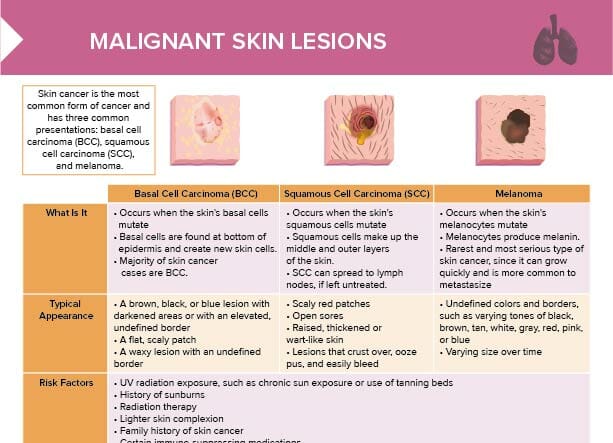
ABCDE is a skin assessment tool and mnemonic used to evaluate skin lesions for skin cancer:
Asymmetry (of moles/lesions)
Border (irregular, ragged, blurred edges)
Color (inconsistent)
Diameter (larger than 6 mm, although melanomas can be smaller)
Evolving (changes over time)
Guided checklists
Scoring systems for pressure injury stages
Braden scale (predictive tool for pressure injury risk)
Skin assessment documentation
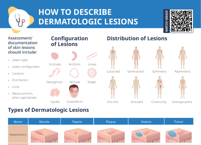
The documentation of skin lesions should include:
Lesion type
Lesion configuration
Location
Distribution
Color
Measurements
A skin assessment form may be used to facilitate documentation.
Using standardized terminology when describing skin lesions helps communication and efficiency in the healthcare team, and minimizes the risk of misinterpretations.
The configuration of lesions can be described as follows:
Circinate
Arciform
Linear
Serpiginous
Annular
Target
Gyrate
Zosteriform
The distribution of lesions can be categorized as follows:
Localized
Generalized
Symmetric
Asymmetric
Discrete
Grouped
Coalescing
Cleavage plane
Common skin lesions
Macule: localized change in skin color, < 1 cm in diameter
Papule: solid, elevated lesion, < 0.5 cm in diameter
Plaque: solid, elevated lesion, > 0.5 cm in diameter
Nodule: solid, elevated, extends into dermis or subcutaneous tissue, 0.5–2 cm in diameter
Tumor: same as nodule, but > 2 cm in diameter
Wheal: localized edema of epidermis causing irregular elevation
Vesicle: elevated mass containing serous fluid, < 0.5 cm
Bullae: same as vesicle, only > 0.5 cm
Pustule: vesicle or bullae that become filled with pus
Cyst: encapsulated fluid-filled or semi-solid mass
Nursing skin assessment examples
Lesion
Examples
Macule
Freckle
Papule
Elevated nevi, seborrheic keratosis
Plaque
Psoriasis, eczema
Nodule
Lipoma, melanoma
Tumor
Breast carcinoma
Wheal
Insect bite, hive, angioedema
Vesicle
Herpes simplex, chicken pox
Bullae
Contact dermatitis, second-degree burns
Pustule
Acne, impetigo, furuncle, folliculitis
Cyst
Sebaceous cyst, epidermoid cyst
Important skin assessment findings
The following are important skin assessment findings that are examples of findings that require monitoring for other conditions or diagnoses:
Very pale skin may suggest anemia
Cyanosis may suggest poor oxygenation
Jaundice may suggest liver dysfunction
Erythema can be from inflammation or infection
Moles can indicate melanoma
Rashes can be indicative of allergies
Heat can be indicative of inflammation or infection
Poor skin turgor can hint to dehydration
Bruising can suggest clotting disorders or physical abuse