Domina Conceptos Médicos

Estudia para la escuela de medicina y tus examenes con Lecturio.

Esofagitis

La esofagitis es la inflamación o irritación del esófago. Los LOS Neisseria principales tipos de esofagitis son la inducida por medicamentos, la infecciosa, la eosinofílica, la corrosiva y por reflujo ácido del estómago. Los LOS Neisseria pacientes suelen presentar odinofagia, disfagia y dolor Dolor Inflammation torácico retroesternal. El diagnóstico se realiza mediante endoscopia y biopsia. Se obtienen pruebas de laboratorio y de imagenología, dependiendo del grado de daño y de la afectación de otros sistemas. El tratamiento de la esofagitis depende de la etiología subyacente e incluye cambios en EN Erythema nodosum is an immune-mediated panniculitis (inflammation of the subcutaneous fat) caused by a type IV (delayed-type) hypersensitivity reaction. It commonly manifests in young women as tender, erythematous nodules on the shins. Erythema Nodosum la dieta, evitar los LOS Neisseria agentes causantes, terapia con antibióticos o uso de inhibidores de la bomba de protones. En EN Erythema nodosum is an immune-mediated panniculitis (inflammation of the subcutaneous fat) caused by a type IV (delayed-type) hypersensitivity reaction. It commonly manifests in young women as tender, erythematous nodules on the shins. Erythema Nodosum los LOS Neisseria casos graves, como en EN Erythema nodosum is an immune-mediated panniculitis (inflammation of the subcutaneous fat) caused by a type IV (delayed-type) hypersensitivity reaction. It commonly manifests in young women as tender, erythematous nodules on the shins. Erythema Nodosum las lesiones por corrosión, la cirugía puede ser necesaria. Si no se trata, la esofagitis puede dar lugar a complicaciones como estenosis, metaplasia Metaplasia A condition in which there is a change of one adult cell type to another similar adult cell type. Cellular Adaptation del esófago y desarrollo de malignidad.

Last updated: Dec 15, 2025

Editorial responsibility: Stanley Oiseth, Lindsay Jones, Evelin Maza

Descripción General

  • Esofagitis: inflamación o lesión de la mucosa esofágica
  • Principales etiologías:
    • Esofagitis inducida por medicamentos/pastillas
    • Esofagitis infecciosa
    • Esofagitis eosinofílica
    • Esofagitis corrosiva
    • Esofagitis por reflujo
  • El diagnóstico se realiza mediante endoscopia superior; los LOS Neisseria hallazgos varían según la etiología.

Mnemotecnia

Para recordar las causas comunes de la esofagitis: “PIECE” ( en EN Erythema nodosum is an immune-mediated panniculitis (inflammation of the subcutaneous fat) caused by a type IV (delayed-type) hypersensitivity reaction. It commonly manifests in young women as tender, erythematous nodules on the shins. Erythema Nodosum inglés)

  • P – Pill-induced esophagitis Esophagitis Esophagitis is the inflammation or irritation of the esophagus. The major types of esophagitis are medication-induced, infectious, eosinophilic, corrosive, and acid reflux. Patients typically present with odynophagia, dysphagia, and retrosternal chest pain. Esophagitis (esofagitis inducida por pastillas)
  • I – Infectious esophagitis Esophagitis Esophagitis is the inflammation or irritation of the esophagus. The major types of esophagitis are medication-induced, infectious, eosinophilic, corrosive, and acid reflux. Patients typically present with odynophagia, dysphagia, and retrosternal chest pain. Esophagitis (esofagitis infecciosa)
  • E Eosinophilic esophagitis Eosinophilic esophagitis Chronic esophagitis characterized by esophageal mucosal eosinophilia. It is diagnosed when an increase in eosinophils are present over the entire esophagus. The reflux symptoms fail to respond to proton pump inhibitors treatment, unlike in gastroesophageal reflux disease. The symptoms are associated with ige-mediated hypersensitivity to food or inhalant allergens. Esophagitis (esofagitis eosinofílica)
  • C – Corrosive esophagitis Esophagitis Esophagitis is the inflammation or irritation of the esophagus. The major types of esophagitis are medication-induced, infectious, eosinophilic, corrosive, and acid reflux. Patients typically present with odynophagia, dysphagia, and retrosternal chest pain. Esophagitis (esofagitis corrosiva)
  • E Etc ETC The electron transport chain (ETC) sends electrons through a series of proteins, which generate an electrochemical proton gradient that produces energy in the form of adenosine triphosphate (ATP). Electron Transport Chain (ETC). (esofagitis por reflujo)

Related videos

Esofagitis Inducida por Medicamentos

Etiología

  • Antibióticos: tetraciclinas, doxiciclina, clindamicina
  • Antiinflamatorios: Antiinflamatorios no esteroideos (AINE), aspirina
  • Bifosfonatos: alendronato, risedronato
  • Otros: cloruro de potasio, quinidina, suplementos de hierro, ácido ascórbico

Fisiopatología

  • El contacto directo y prolongado del medicamento o los LOS Neisseria medicamentos con la mucosa provoca irritación, erosiones y ulceraciones
  • Afecta con frecuencia a la(s) zona(s) de estrechamiento anatómico (parte media del esófago, cerca del nivel del arco aórtico)
  • Mecanismos:
    • Alteración de la barrera protectora normal de la mucosa: aspirina, AINE
    • Daño cáustico local (e.g., la doxiciclina es ácida una vez disuelta)
    • Las soluciones hiperosmóticas contribuyen a las lesiones tisulares y vasculares (e.g., el cloruro de potasio).

Factores de riesgo

  • Ocurre con más frecuencia en EN Erythema nodosum is an immune-mediated panniculitis (inflammation of the subcutaneous fat) caused by a type IV (delayed-type) hypersensitivity reaction. It commonly manifests in young women as tender, erythematous nodules on the shins. Erythema Nodosum pacientes de edad avanzada
  • Posición del paciente (supina > vertical)
  • Tamaño del medicamento (tránsito retardado con pastillas grandes)
  • Cantidad de líquido ingerido con el medicamento (se aconseja la ingesta de al AL Amyloidosis menos > 100 mL)

Presentación clínica

Diagnóstico

  • Diagnóstico clínico basado en EN Erythema nodosum is an immune-mediated panniculitis (inflammation of the subcutaneous fat) caused by a type IV (delayed-type) hypersensitivity reaction. It commonly manifests in young women as tender, erythematous nodules on the shins. Erythema Nodosum el antecedente clínico
  • Endoscopia alta:
    • Realizar si hay síntomas severos o persistentes (a pesar de la interrupción del medicamento causante)
    • Hallazgos: úlcera discreta, con mucosa limítrofe normal
Esofagitis inducida por cloxacilina

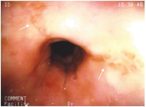
Erosiones estrelladas en el esófago medial (flechas blancas) en un paciente con esofagitis inducida por cloxacilina

Imagen: “Cloxacillin: A New Cause of Pill-Induced Esophagitis” por Zezos P, Harel Z, Saibil F. Licencia: CC BY 4.0

Prevención y tratamiento

  • Evite el medicamento.
  • Resolución esperada en EN Erythema nodosum is an immune-mediated panniculitis (inflammation of the subcutaneous fat) caused by a type IV (delayed-type) hypersensitivity reaction. It commonly manifests in young women as tender, erythematous nodules on the shins. Erythema Nodosum < 2 semanas después de la interrupción.
  • Si el medicamento es necesario, se debe tomar con suficiente agua y mantener una posición vertical durante al AL Amyloidosis menos 30 minutos.

Esofagitis Infecciosa: Citomegalovirus (CMV)

Etiología

  • Infección previa por CMV + recuento de células CD4 < 50 células/microL → ↑ riesgo de reactivación → infección de órganos como el esófago
  • La enfermedad gastrointestinal por CMV no es frecuente, pero es una complicación grave del SIDA (síndrome de inmunodeficiencia adquirida).

Factores de riesgo

  • Pacientes con SIDA (la incidencia ha HA Hemolytic anemia (HA) is the term given to a large group of anemias that are caused by the premature destruction/hemolysis of circulating red blood cells (RBCs). Hemolysis can occur within (intravascular hemolysis) or outside the blood vessels (extravascular hemolysis). Hemolytic Anemia disminuido desde la disponibilidad de la terapia antirretroviral)
  • Receptores de trasplantes de órganos sólidos y de médula ósea
  • Pacientes que reciben terapia inmunosupresora y quimioterapia

Presentación clínica

  • Odinofagia con disfagia
  • Náuseas, fiebre, dolor Dolor Inflammation ardiente subesternal
  • Puede tener infección concurrente en EN Erythema nodosum is an immune-mediated panniculitis (inflammation of the subcutaneous fat) caused by a type IV (delayed-type) hypersensitivity reaction. It commonly manifests in young women as tender, erythematous nodules on the shins. Erythema Nodosum retina Retina The ten-layered nervous tissue membrane of the eye. It is continuous with the optic nerve and receives images of external objects and transmits visual impulses to the brain. Its outer surface is in contact with the choroid and the inner surface with the vitreous body. The outermost layer is pigmented, whereas the inner nine layers are transparent. Eye: Anatomy, pulmón, hígado, colon Colon The large intestines constitute the last portion of the digestive system. The large intestine consists of the cecum, appendix, colon (with ascending, transverse, descending, and sigmoid segments), rectum, and anal canal. The primary function of the colon is to remove water and compact the stool prior to expulsion from the body via the rectum and anal canal. Colon, Cecum, and Appendix: Anatomy

Diagnóstico

El diagnóstico se realiza mediante una endoscopia superior.

  • Ulceraciones profundas lineales o longitudinales, erosiones
  • Biopsia: células citomegálicas (con inclusiones intracitoplasmáticas o intranucleares)
Esofagitis por citomegalovirus

Hallazgos de una endoscopia gastrointestinal superior en un paciente con esofagitis por citomegalovirus. Se observa una úlcera con una base blanca en el esófago.

Imagen: “Cytomegalovirus esophagitis developing during chemoradiotherapy for esophageal cancer” por Journal of Medical Case Reports. Licencia: CC BY 4.0

Tratamiento

  • Ganciclovir Ganciclovir An acyclovir analog that is a potent inhibitor of the herpesvirus family including cytomegalovirus. Ganciclovir is used to treat complications from aids-associated cytomegalovirus infections. Antivirals for Herpes Virus
  • Con VIH/SIDA: terapia antirretroviral

Esofagitis Infecciosa: Virus del Herpes Simple (VHS)

Etiología

  • La mayoría son VHS tipo 1.
  • Puede ser una infección primaria o una reactivación del VHS

Factores de riesgo

  • SIDA
  • Receptores de trasplantes de órganos o de médula ósea
  • Pacientes en EN Erythema nodosum is an immune-mediated panniculitis (inflammation of the subcutaneous fat) caused by a type IV (delayed-type) hypersensitivity reaction. It commonly manifests in young women as tender, erythematous nodules on the shins. Erythema Nodosum tratamiento inmunosupresor

Presentación clínica

  • Odinofagia con disfagia
  • Dolor Dolor Inflammation torácico retroesternal, +/- fiebre, úlceras orofaríngeas recurrentes

Diagnóstico

El diagnóstico se realiza mediante una endoscopia superior.

  • Lesión temprana: vesículas (raramente se observan)
  • Úlceras perforadas, +/- placas
  • Biopsia: células infectadas por el VHS con cuerpos de inclusión de tipo Cowdry A
  • Cultivo viral si no hay respuesta al AL Amyloidosis tratamiento
Esofagitis herpética

Endoscopia digestiva alta de un paciente que muestra numerosas lesiones en la superficie esofágica de color amarillo-blanquecino, pleomórficas y pequeñas placas circulares aisladas con erosiones centrales.

Imagen: “Herpetic esophagitis in immunocompetent medical student” por Marinho AV, Bonfim VM, de Alencar LR, Pinto SA, de Araújo Filho JA. Licencia: CC BY 3.0

Tratamiento

  • Aciclovir
  • Con VIH/SIDA: terapia antirretroviral

Esofagitis Infecciosa: Candidiasis

Etiología

  • Candida albicans Candida albicans A unicellular budding fungus which is the principal pathogenic species causing candidiasis (moniliasis). Candida/Candidiasis: la más frecuente
  • Candida Candida Candida is a genus of dimorphic, opportunistic fungi. Candida albicans is part of the normal human flora and is the most common cause of candidiasis. The clinical presentation varies and can include localized mucocutaneous infections (e.g., oropharyngeal, esophageal, intertriginous, and vulvovaginal candidiasis) and invasive disease (e.g., candidemia, intraabdominal abscess, pericarditis, and meningitis). Candida/Candidiasis glabrata, Candida Candida Candida is a genus of dimorphic, opportunistic fungi. Candida albicans is part of the normal human flora and is the most common cause of candidiasis. The clinical presentation varies and can include localized mucocutaneous infections (e.g., oropharyngeal, esophageal, intertriginous, and vulvovaginal candidiasis) and invasive disease (e.g., candidemia, intraabdominal abscess, pericarditis, and meningitis). Candida/Candidiasis krusei: menos comunes

Factores de riesgo

  • Virus Virus Viruses are infectious, obligate intracellular parasites composed of a nucleic acid core surrounded by a protein capsid. Viruses can be either naked (non-enveloped) or enveloped. The classification of viruses is complex and based on many factors, including type and structure of the nucleoid and capsid, the presence of an envelope, the replication cycle, and the host range. Virology de la inmunodeficiencia humana (VIH) con recuento de células CD4 < 200 células/μL
  • Malignidad hematológica
  • Receptor Receptor Receptors are proteins located either on the surface of or within a cell that can bind to signaling molecules known as ligands (e.g., hormones) and cause some type of response within the cell. Receptors de un trasplante de células hematopoyéticas
  • Pacientes bajo quimioterapia citotóxica
  • Posible en EN Erythema nodosum is an immune-mediated panniculitis (inflammation of the subcutaneous fat) caused by a type IV (delayed-type) hypersensitivity reaction. It commonly manifests in young women as tender, erythematous nodules on the shins. Erythema Nodosum pacientes con inhalación crónica de corticosteroides

Presentación clínica

  • Odinofagia con disfagia
  • Puede o no tener aftas orales

Diagnóstico

El diagnóstico se realiza mediante una endoscopia superior.

  • Pseudomembrana blanca o lesiones en EN Erythema nodosum is an immune-mediated panniculitis (inflammation of the subcutaneous fat) caused by a type IV (delayed-type) hypersensitivity reaction. It commonly manifests in young women as tender, erythematous nodules on the shins. Erythema Nodosum placa
  • Biopsia: levaduras e hifas en EN Erythema nodosum is an immune-mediated panniculitis (inflammation of the subcutaneous fat) caused by a type IV (delayed-type) hypersensitivity reaction. It commonly manifests in young women as tender, erythematous nodules on the shins. Erythema Nodosum las células de la mucosa
  • El cultivo muestra a Candida Candida Candida is a genus of dimorphic, opportunistic fungi. Candida albicans is part of the normal human flora and is the most common cause of candidiasis. The clinical presentation varies and can include localized mucocutaneous infections (e.g., oropharyngeal, esophageal, intertriginous, and vulvovaginal candidiasis) and invasive disease (e.g., candidemia, intraabdominal abscess, pericarditis, and meningitis). Candida/Candidiasis
Malestar epigástrico causado por la candidiasis esofágica

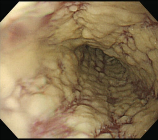
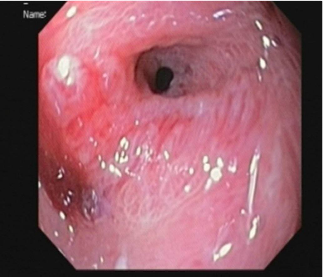
La esofagogastroduodenoscopia ilustra lesiones blancas difusas en el esófago características de la esofagitis por Candida.

Imagen: “Epigastric Distress Caused by Esophageal Candidiasis” por Chen KH, Weng MT, Chou YH, Lu YF, Hsieh CH. Licencia: CC BY 4.0

Tratamiento

  • Cándida: fluconazol oral (nistatina para la candidiasis Candidiasis Candida is a genus of dimorphic, opportunistic fungi. Candida albicans is part of the normal human flora and is the most common cause of candidiasis. The clinical presentation varies and can include localized mucocutaneous infections (e.g., oropharyngeal, esophageal, intertriginous, and vulvovaginal candidiasis) and invasive disease (e.g., candidemia, intraabdominal abscess, pericarditis, and meningitis). Candida/Candidiasis oral recurrente)
  • Con VIH/SIDA: terapia antirretroviral

Esofagitis Eosinofílica

Etiología

  • Trastorno alérgico asociado a:
    • Sensibilización a antígenos a través de alimentos o aeroalérgenos
    • Eosinofilia y disfunción esofágica
  • Nota: Los LOS Neisseria eosinófilos no se encuentran normalmente en EN Erythema nodosum is an immune-mediated panniculitis (inflammation of the subcutaneous fat) caused by a type IV (delayed-type) hypersensitivity reaction. It commonly manifests in young women as tender, erythematous nodules on the shins. Erythema Nodosum el esófago.

Factores de riesgo

Epidemiología

  • Hombres > mujeres
  • Edad común de presentación: 20–30 años
  • Los LOS Neisseria caucásicos son los LOS Neisseria más afectados.

Fisiopatología

  • Los LOS Neisseria eosinófilos se reúnen al AL Amyloidosis exponerse a alérgenos inhalados o congestionados.
  • Se produce una infiltración eosinofílica → los LOS Neisseria eosinófilos liberan interleucinas → respuesta inflamatoria

Presentación clínica

  • Disfagia (a los LOS Neisseria alimentos sólidos)
  • Impactación de alimentos
  • Odinofagia

Diagnóstico

El diagnóstico se realiza mediante una endoscopia superior.

  • Anillos mucosos en EN Erythema nodosum is an immune-mediated panniculitis (inflammation of the subcutaneous fat) caused by a type IV (delayed-type) hypersensitivity reaction. It commonly manifests in young women as tender, erythematous nodules on the shins. Erythema Nodosum el esófago
  • Biopsia que muestra eosinófilos (> 15/campos de alta potencia (HPF, por sus siglas en EN Erythema nodosum is an immune-mediated panniculitis (inflammation of the subcutaneous fat) caused by a type IV (delayed-type) hypersensitivity reaction. It commonly manifests in young women as tender, erythematous nodules on the shins. Erythema Nodosum inglés))
  • Otras características:
    • La eosinofilia esofágica persiste incluso con la ingesta de inhibidores de la bomba de protones (IBP).
    • Monitoreo normal del pH pH The quantitative measurement of the acidity or basicity of a solution. Acid-Base Balance
Esofagitis eosinofílica

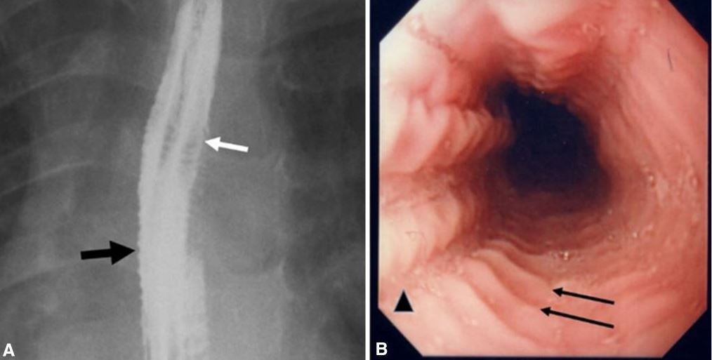
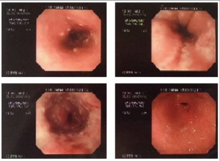
Imágenes de la esofagitis eosinofílica:
A. Esofagograma en un paciente con antecedente de impactaciones alimentarias recurrentes y disfagia que muestra múltiples anillos esofágicos (flecha blanca), dando la apariencia de un esófago corrugado o anillado e irregularidad de la mucosa (flecha negra).
B. La endoscopia muestra múltiples anillos transversales (flechas) y surcos en la mucosa (cabeza de flecha).

Imagen: “Eosinophilic esophagitis” por Al-Hussaini A, AboZeid A, Hai A. Licencia: CC BY 4.0

Tratamiento

  • Remisión a un alergólogo para orientación (terapia dietética)
  • Evite los LOS Neisseria alérgenos alimentarios conocidos (e.g., leche de vaca)
  • Terapia con medicamentos:
    • Glucocorticoides tópicos (e.g., fluticasona ingerida)
    • Glucocorticoides sistémicos si hay disfagia significativa, pérdida de peso, deshidratación
    • IBP para los LOS Neisseria síntomas del reflujo
    • Los LOS Neisseria antagonistas de los LOS Neisseria receptores de IL-4 (e.g., dupilumab Dupilumab Asthma Drugs) pueden considerarse para la enfermedad refractaria.

Esofagitis Corrosiva

Etiología

  • Álcali:
    • Limpiadores de desagües, productos de limpieza del hogar, pilas, lejías
    • Viscoso, insípido, incoloro
  • Ácidos:
    • Líquido para baterías, limpiadores de inodoros, líquidos para la limpieza de metales, soluciones antioxidantes
    • Sabor desagradable, maloliente

Factores de riesgo

  • Intencional (suicidio, enfermedad psiquiátrica subyacente)
  • Accidental (generalmente en EN Erythema nodosum is an immune-mediated panniculitis (inflammation of the subcutaneous fat) caused by a type IV (delayed-type) hypersensitivity reaction. It commonly manifests in young women as tender, erythematous nodules on the shins. Erythema Nodosum niños)

Fisiopatología

  • Lesión inducida por álcali:
    • Daño rápido, que afecta más al AL Amyloidosis esófago que al AL Amyloidosis estómago
    • Sin embargo, grandes cantidades provocan lesiones gástricas.
    • Proceso: necrosis Necrosis The death of cells in an organ or tissue due to disease, injury or failure of the blood supply. Ischemic Cell Damage licuefactiva en EN Erythema nodosum is an immune-mediated panniculitis (inflammation of the subcutaneous fat) caused by a type IV (delayed-type) hypersensitivity reaction. It commonly manifests in young women as tender, erythematous nodules on the shins. Erythema Nodosum el esófago
  • Lesión inducida por el ácido:
    • El dolor Dolor Inflammation al AL Amyloidosis contacto con la orofaringe limita la cantidad ingerida.
    • Mayor daño orofaríngeo y de las vías respiratorias que con las soluciones alcalinas
    • El ácido baja más rápido, causando más daño al AL Amyloidosis estómago.
    • Proceso: necrosis Necrosis The death of cells in an organ or tissue due to disease, injury or failure of the blood supply. Ischemic Cell Damage y coagulación superficial en EN Erythema nodosum is an immune-mediated panniculitis (inflammation of the subcutaneous fat) caused by a type IV (delayed-type) hypersensitivity reaction. It commonly manifests in young women as tender, erythematous nodules on the shins. Erythema Nodosum el esófago

Complicaciones en EN Erythema nodosum is an immune-mediated panniculitis (inflammation of the subcutaneous fat) caused by a type IV (delayed-type) hypersensitivity reaction. It commonly manifests in young women as tender, erythematous nodules on the shins. Erythema Nodosum daños graves

  • Perforación
  • Sangrado
  • Mediastinitis Mediastinitis Mediastinitis refers to an infection or inflammation involving the mediastinum (a region in the thoracic cavity containing the heart, thymus gland, portions of the esophagus, and trachea). Acute mediastinitis can be caused by bacterial infection due to direct contamination, hematogenous or lymphatic spread, or extension of infection from nearby structures. Mediastinitis
  • Obstrucción
  • Estenosis
  • Fístulas
  • Daño laríngeo y traqueobronquial por aspiración
  • Aumenta el riesgo para cáncer de células escamosas (SCC, por sus siglas en EN Erythema nodosum is an immune-mediated panniculitis (inflammation of the subcutaneous fat) caused by a type IV (delayed-type) hypersensitivity reaction. It commonly manifests in young women as tender, erythematous nodules on the shins. Erythema Nodosum inglés) del esófago

Presentación clínica

  • Dolor Dolor Inflammation orofaríngeo, retroesternal o epigástrico
  • Odinofagia y/o disfagia
  • Hipersalivación
  • Estridor o sibilancias por quemaduras de la laringe
  • Disnea
  • Náuseas/vómitos
  • Dolor Dolor Inflammation retroesternal severo (perforación y mediastinitis Mediastinitis Mediastinitis refers to an infection or inflammation involving the mediastinum (a region in the thoracic cavity containing the heart, thymus gland, portions of the esophagus, and trachea). Acute mediastinitis can be caused by bacterial infection due to direct contamination, hematogenous or lymphatic spread, or extension of infection from nearby structures. Mediastinitis)
  • Sensibilidad y rigidez abdominal (perforación y peritonitis Peritonitis Inflammation of the peritoneum lining the abdominal cavity as the result of infectious, autoimmune, or chemical processes. Primary peritonitis is due to infection of the peritoneal cavity via hematogenous or lymphatic spread and without intra-abdominal source. Secondary peritonitis arises from the abdominal cavity itself through rupture or abscess of intra-abdominal organs. Penetrating Abdominal Injury)

Diagnóstico

  • Antecedentes: Anote el tipo y la cantidad de agente ingerido.
  • Rayos X:
    • Tórax: comprobar si hay neumomediastino, neumonía por aspiración, cuerpo extraño (e.g., una pila)
    • Abdominal: comprobar si hay neumoperitoneo, cuerpo extraño
  • Tomografía computarizada (TC): comprobar la profundidad de la necrosis Necrosis The death of cells in an organ or tissue due to disease, injury or failure of the blood supply. Ischemic Cell Damage y ayuda a evaluar la necesidad de cirugía de urgencia
  • Endoscopia superior:
    • En EN Erythema nodosum is an immune-mediated panniculitis (inflammation of the subcutaneous fat) caused by a type IV (delayed-type) hypersensitivity reaction. It commonly manifests in young women as tender, erythematous nodules on the shins. Erythema Nodosum 24 horas si no hay contraindicaciones
    • Contraindicado en EN Erythema nodosum is an immune-mediated panniculitis (inflammation of the subcutaneous fat) caused by a type IV (delayed-type) hypersensitivity reaction. It commonly manifests in young women as tender, erythematous nodules on the shins. Erythema Nodosum inestabilidad hemodinámica, perforación gastrointestinal
Estenosis pilórica Esofagitis

Hallazgo en la endoscopia superior de un niño con ingestión de álcali: Estenosis pilórica desarrollada 2 meses después de la lesión.

Imagen: “Pyloric stenosis after 2 months” por Dehghani SM, Aldaghi M, Javaherizadeh H. Licencia: CC BY 3.0

Clasificación de las lesiones

  • Lesión de 1er grado: mucosa superficial afectada; eritema, edema Edema Edema is a condition in which excess serous fluid accumulates in the body cavity or interstitial space of connective tissues. Edema is a symptom observed in several medical conditions. It can be categorized into 2 types, namely, peripheral (in the extremities) and internal (in an organ or body cavity). Edema, hemorragia, con una curación esperada
  • Lesión de 2do grado: las úlceras y los LOS Neisseria exudados afectan hasta la capa submucosa; es posible que se produzcan cicatrices y estenosis
  • Lesión de 3er grado: profundidad transmural, con úlceras profundas y perforación de la pared

Tratamiento

  • Protección de la vía aérea; evaluar la necesidad de intubación
  • Reanimación con líquidos
  • Nada por vía oral
  • No usar sonda nasogástrica, ni eméticos
  • IBP
  • Antibióticos para la sospecha de perforación
  • Evaluar las indicaciones quirúrgicas
  • Vigilancia: endoscopia superior 15–20 años después para realizar tamizaje de carcinoma de células escamosas

Esofagitis por Reflujo

La esofagitis por reflujo también se conoce como enfermedad por reflujo gastroesofágico (ERGE).

Etiología y fisiopatología

  • Flujo retrógrado de ácido estomacal hacia el esófago debido a la relajación transitoria del esfínter esofágico inferior (EEI)
  • La mayoría de los LOS Neisseria episodios de relajación son desencadenados por la distensión gástrica.
  • Algunos tienen un esfínter esofágico inferior incompetente.
  • Las sustancias tóxicas (ácido gástrico, pepsina, sales biliares) provocan daños en EN Erythema nodosum is an immune-mediated panniculitis (inflammation of the subcutaneous fat) caused by a type IV (delayed-type) hypersensitivity reaction. It commonly manifests in young women as tender, erythematous nodules on the shins. Erythema Nodosum la mucosa esofágica distal.

Factores de riesgo

  • Tabaquismo
  • Consumo de alcohol
  • Obesidad
  • Hernia Hernia Protrusion of tissue, structure, or part of an organ through the bone, muscular tissue, or the membrane by which it is normally contained. Hernia may involve tissues such as the abdominal wall or the respiratory diaphragm. Hernias may be internal, external, congenital, or acquired. Abdominal Hernias hiatal
  • Estrés
  • Esclerodermia
  • Síndrome de Zollinger-Ellison que provoca un aumento de la secreción de ácido gástrico

Presentación clínica

  • Acidez retroesternal o pirosis
    • Empeora cuando es postprandial o al AL Amyloidosis acostarse
    • Puede interrumpir el sueño
    • Puede verse exacerbado por el estrés emocional
  • Regurgitación
  • Disfonía
  • Sabor “salobre” en EN Erythema nodosum is an immune-mediated panniculitis (inflammation of the subcutaneous fat) caused by a type IV (delayed-type) hypersensitivity reaction. It commonly manifests in young women as tender, erythematous nodules on the shins. Erythema Nodosum la boca
  • La sensación de nudo en EN Erythema nodosum is an immune-mediated panniculitis (inflammation of the subcutaneous fat) caused by a type IV (delayed-type) hypersensitivity reaction. It commonly manifests in young women as tender, erythematous nodules on the shins. Erythema Nodosum la garganta
  • Tos TOS Thoracic outlet syndrome (TOS) is a broad term used for a spectrum of syndromes related to the general region of the thoracic outlet, which involves the compression or irritation of elements of the brachial plexus, subclavian artery, or subclavian vein. Thoracic Outlet Syndrome crónica no productiva

Complicaciones

  • Esofagitis erosiva
  • Esófago de Barrett (con riesgo de adenocarcinoma)
  • Estenosis esofágica

Diagnóstico

  • Clínico (signos clásicos de acidez y regurgitación)
  • Se confirma con la monitorización del pH pH The quantitative measurement of the acidity or basicity of a solution. Acid-Base Balance esofágico si:
    • Existe un tratamiento insatisfactorio
    • Existen síntomas atípicos
    • Se está considerando la cirugía antirreflujo
  • La endoscopia superior se realiza si existe:
    • Disfagia
    • Pérdida de peso inexplicable
    • Anemia Anemia Anemia is a condition in which individuals have low Hb levels, which can arise from various causes. Anemia is accompanied by a reduced number of RBCs and may manifest with fatigue, shortness of breath, pallor, and weakness. Subtypes are classified by the size of RBCs, chronicity, and etiology. Anemia: Overview and Types o evidencia de hemorragia digestiva
    • Síntomas refractarios
    • ERGE de nueva aparición > 60 años
    • Odinofagia
    • Alto riesgo de esófago de Barrett
    • Familiar de primer grado con cáncer gastrointestinal
Esofagitis por reflujo

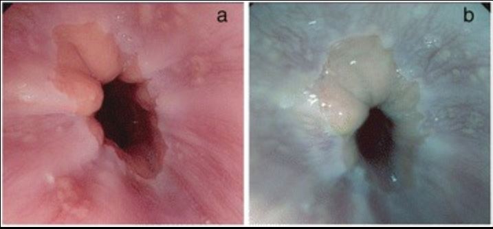
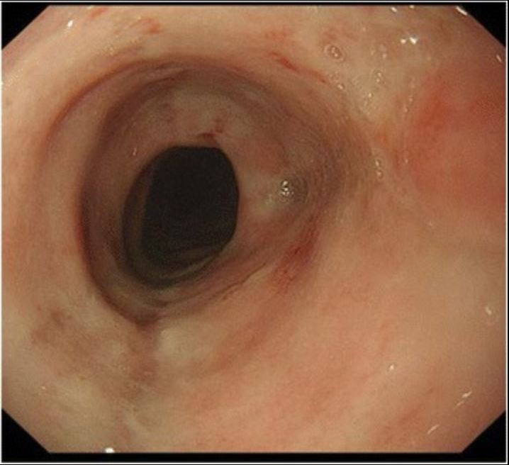
Esofagitis por reflujo en la endoscopia superior:
A: imagen de alta definición de los cambios esofágicos observados en la ERGE
B: imagen con realce de tono de la ERGE

Imagen: “Reflux esophagitis” por Netinatsunton N, Sottisuporn J, Attasaranya S, Witeerungrot T, Chamroonkul N, Jongboonyanuparp T, Geater A, Ovartlarnporn B. Licencia: CC BY 4.0

Tratamiento

  • Modificación del estilo de vida:
    • Pérdida de peso
    • Elevación de la cabecera de la cama
    • Eliminación de los LOS Neisseria desencadenantes dietéticos
    • Cese en EN Erythema nodosum is an immune-mediated panniculitis (inflammation of the subcutaneous fat) caused by a type IV (delayed-type) hypersensitivity reaction. It commonly manifests in young women as tender, erythematous nodules on the shins. Erythema Nodosum el consumo de tabaco/alcohol
  • Terapia con medicamentos: IBP
  • Opciones quirúrgicas incluyen la fundoplicación, que está indicada para los LOS Neisseria síntomas refractarios.

Referencias

  1. Azer, S., Reddivari, A. (2024). Reflux esophagitis. Retrieved July 19, 2025, from https://www.ncbi.nlm.nih.gov/books/NBK554462
  2. Bonnis, P., Kotton, C., Hirsch, M., Mitty, J. (2025). Herpes simplex virus infection of the esophagus. UpToDate. Retrieved July 19, 2025, from https://www.uptodate.com/contents/herpes-simplex-virus-infection-of-the-esophagus
  3. Kahrilas PJ, Lamont JT, Meyer C, et al. (2024). Pill esophagitis. UpToDate. Retrieved July 19, 2025, from https://www.uptodate.com/contents/pill-esophagitis
  4. De Lusong, M., Timbol, A., Tuazon, D. (2017). Management of esophageal caustic injury. World Journal of Gastrointestinal Pharmacology and Therapeutics 6; 8(2): 90–98.
  5. Greenberger, N.J. (2016). Eosinophilic esophagitis. Greenberger N.J., Blumberg R.S., Burakoff, R. (Eds.) Current Diagnosis & Treatment: Gastroenterology, Hepatology, & Endoscopy, 3rd ed. McGraw-Hill.
  6. Jacobson, M., Bartlett, J., Mitty, J. (2024). AIDS-related cytomegalovirus gastrointestinal disease. UpToDate. Retrieved July 19, 2025, from https://www.uptodate.com/contents/aids-related-cytomegalovirus-gastrointestinal-disease
  7. Kardon, E., Vearrier, D. (2024). Caustic ingestions. Medscape. Retrieved July 19, 2025, from https://emedicine.medscape.com/article/813772-overview
  8. McQuaid K.R. (2021). Caustic esophageal injury. Papadakis M.A., McPhee S.J., Rabow M.W. (Eds.) Current Medical Diagnosis & Treatment 2021. McGraw-Hill.
  9. McQuaid K.R. (2021). Gastroesophageal reflux disease. Papadakis, M.A., McPhee S.J., Rabow M.W. (Eds.) Current Medical Diagnosis & Treatment 2021. McGraw-Hill.
  10. Triadafilopoulos, G., Saltzman, J., Grover, S., Chen, W. (2025). Caustic esophageal injury in adults. UpToDate. Retrieved July 19, 2025, from https://www.uptodate.com/contents/caustic-esophageal-injury-in-adults
  11. Anwar KP, Malik A, et al. Profile of candidiasis in HIV infected patients. Iran J Microbiol. 2012 Dec;4(4):204–209. PMID: 23205253. PMCID: PMC3507311.

¡Crea tu cuenta gratis o inicia una sesión para seguir leyendo!

Regístrate ahora y obtén acceso gratuito a Lecturio con páginas de concepto, videos médicos y cuestionarios para tu educación médica.

User Reviews

Que tengas una sesión de estudio alegre y navideña 🎁 Ahorra 50% en todos los planes >>

Details