La ruptura o perforación esofágica es un defecto transmural que se produce en EN Erythema nodosum is an immune-mediated panniculitis (inflammation of the subcutaneous fat) caused by a type IV (delayed-type) hypersensitivity reaction. It commonly manifests in young women as tender, erythematous nodules on the shins. Erythema Nodosum el esófago, exponiendo el mediastino al AL Amyloidosis contenido gastrointestinal. La causa más común de perforación esofágica es el traumatismo iatrogénico por instrumentación o procedimientos quirúrgicos. La perforación también puede deberse a la ingesta de un cuerpo extraño o a un traumatismo no iatrogénico producido por vómitos excesivos. La perforación esofágica se presenta con dolor Dolor Inflammation torácico subesternal que puede tener un inicio repentino o insidioso. El diagnóstico puede realizarse mediante una tomografía computarizada de tórax y cuello, una radiografía de tórax o un esofagograma. El tratamiento suele incluir la reparación quirúrgica del defecto esofágico transmural. Sin embargo, el tratamiento conservador también puede considerarse para un paciente hemodinámicamente estable con un defecto pequeño. La principal complicación de la perforación esofágica es la mediastinitis Mediastinitis Mediastinitis refers to an infection or inflammation involving the mediastinum (a region in the thoracic cavity containing the heart, thymus gland, portions of the esophagus, and trachea). Acute mediastinitis can be caused by bacterial infection due to direct contamination, hematogenous or lymphatic spread, or extension of infection from nearby structures. Mediastinitis aguda. La tasa de mortalidad puede oscilar entre el 10%–50%.
Last updated: Dec 15, 2025
La perforación esofágica, también llamada ruptura esofágica, es un defecto transmural de la pared esofágica que expone el mediastino al AL Amyloidosis contenido gastrointestinal.
La patogénesis de la perforación esofágica depende de la causa.
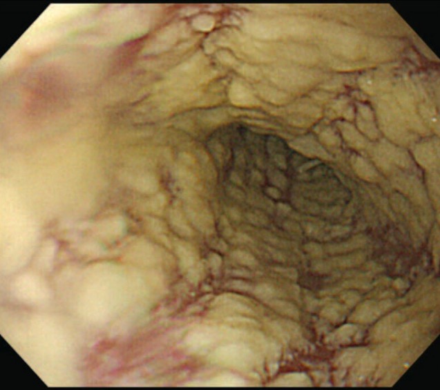
Esofagitis por Candida con placas blanco-amarillentas
Imagen: “Esophagogastroduodenoscopy” por Department of Medicine (C-HH), and Institute of Traditional Medicine, School of Medicine, National Yang-Ming University, Taipei, Taiwan. Licencia: CC BY 4.0Antecedentes:
Manifestaciones:
Hallazgos al AL Amyloidosis examen físico:
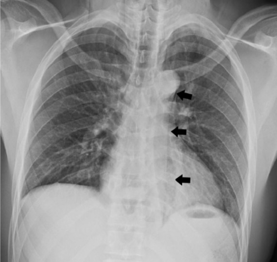
Radiografía de tórax posteroanterior que muestra un neumomediastino (flechas negras) debido a una ruptura esofágica
Imagen: “Chest X-ray displaying pneumomediastinum” por Faculty of Medicine, University of Toronto, Toronto, ON, Canada. Licencia: CC BY 4.0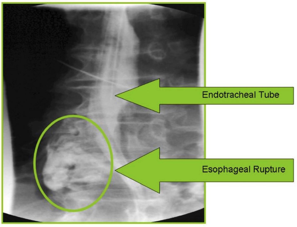
Esofagografía con gastrografin que muestra una ruptura esofágica distal con fuga de contraste (círculo verde)
Imagen: “Intubated patient showing esophageal rupture” por Rockyview General Hospital, Alberta Health Services, 7007-14th Street SW, Calgary, AB, T2P 1P9, Canada. Licencia: CC BY 2.0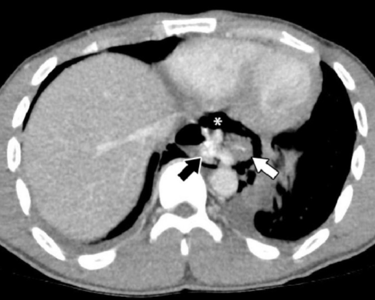
TC de tórax que muestra la ruptura distal del esófago (flecha blanca) con fuga de contraste (flecha negra) y neumomediastino (asterisco)
Imagen: “Chest computed tomography” por Faculty of Medicine, University of Toronto, Toronto, ON, Canada. Licencia: CC BY 4.0Abordaje inicial:
Obtenga una interconsulta quirúrgica (incluyendo cirugía cardiotorácica), ya que incluso los LOS Neisseria pacientes estables pueden deteriorarse y requerir cirugía.
Intervención adicional determinada por:
Indicaciones:
Procedimiento:
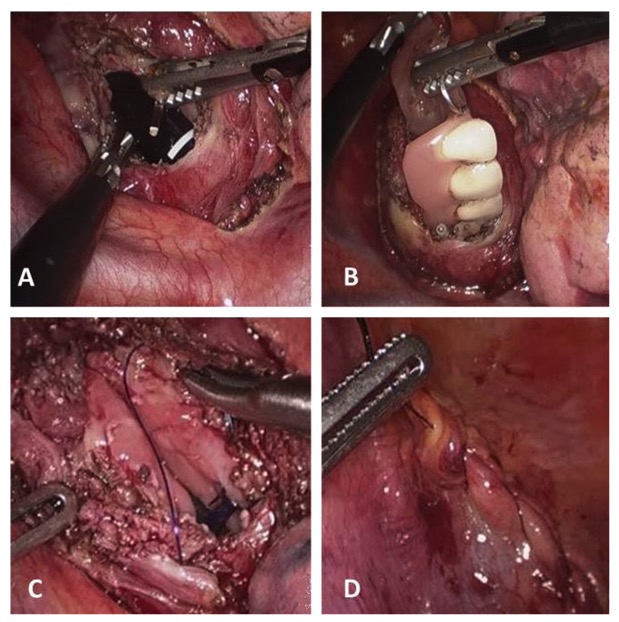
Perforación esofágica por cuerpo extraño (prótesis dental):
A: esofagotomía
B: extracción de la prótesis dental
C y D: sutura de la pared esofágica y de la pleura mediastínica