La paracoccidioidomicosis es una infección fúngica endémica causada por el complejo de Paracoccidioides Paracoccidioides Paracoccidioidomycosis (PCM) is an endemic fungal infection caused by Paracoccidioides brasiliensis and P. lutzii. The fungus is geographically distributed across Mexico and South and Central America. Transmission is by inhalation, and most infections are asymptomatic. Paracoccidioides/Paracoccidioidomycosis brasiliensis y P. lutzii P. lutzii Paracoccidioides/Paracoccidioidomycosis. El hongo se distribuye geográficamente en EN Erythema nodosum is an immune-mediated panniculitis (inflammation of the subcutaneous fat) caused by a type IV (delayed-type) hypersensitivity reaction. It commonly manifests in young women as tender, erythematous nodules on the shins. Erythema Nodosum México y América del Sur y Central. La transmisión es por inhalación y la mayoría de las infecciones son asintomáticas. El hongo infecta los LOS Neisseria pulmones y luego se propaga a la piel, las membranas mucosas y otras partes del cuerpo. La infección primaria suele ser autolimitada. Sin embargo, si el huésped no contiene la infección y/o el paciente no recibe tratamiento, pueden producirse complicaciones potencialmente mortales. Las infecciones agudas suelen afectar a poblaciones más jóvenes, con hallazgos de linfadenopatía, hepatoesplenomegalia, lesiones cutáneas y signos de disfunción de la médula ósea. Más comúnmente, se observa paracoccidioidomicosis crónica, especialmente en EN Erythema nodosum is an immune-mediated panniculitis (inflammation of the subcutaneous fat) caused by a type IV (delayed-type) hypersensitivity reaction. It commonly manifests in young women as tender, erythematous nodules on the shins. Erythema Nodosum hombres. La paracoccidioidomicosis crónica representa la reactivación de la infección, presentándose como enfermedad pulmonar o infección diseminada. El diagnóstico se realiza por microscopía, histopatología, cultivo y serología. El itraconazol se usa típicamente para la enfermedad leve a moderada y la anfotericina B para la enfermedad grave y/o del sistema nervioso central (SNC).
Last updated: Dec 15, 2025
Morfología:
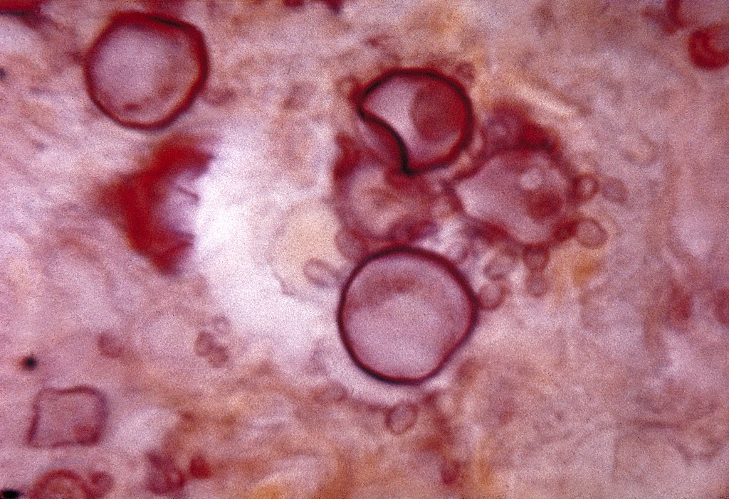
Histopatología del Paracoccidioides:
Células con células hijas en brotes (blastoconidios) que se ven en el tejido, como el timón de un barco (el timón del capitán o del piloto)
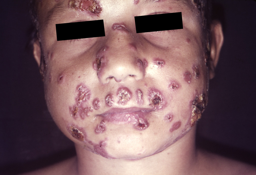
Lesiones faciales causadas por paracoccidioidomicosis en un niño brasileño.
Imagen: “Paracoccidioidomycosis lesions” por CDC/Dr. Martín Castro. Licencia: Dominio Público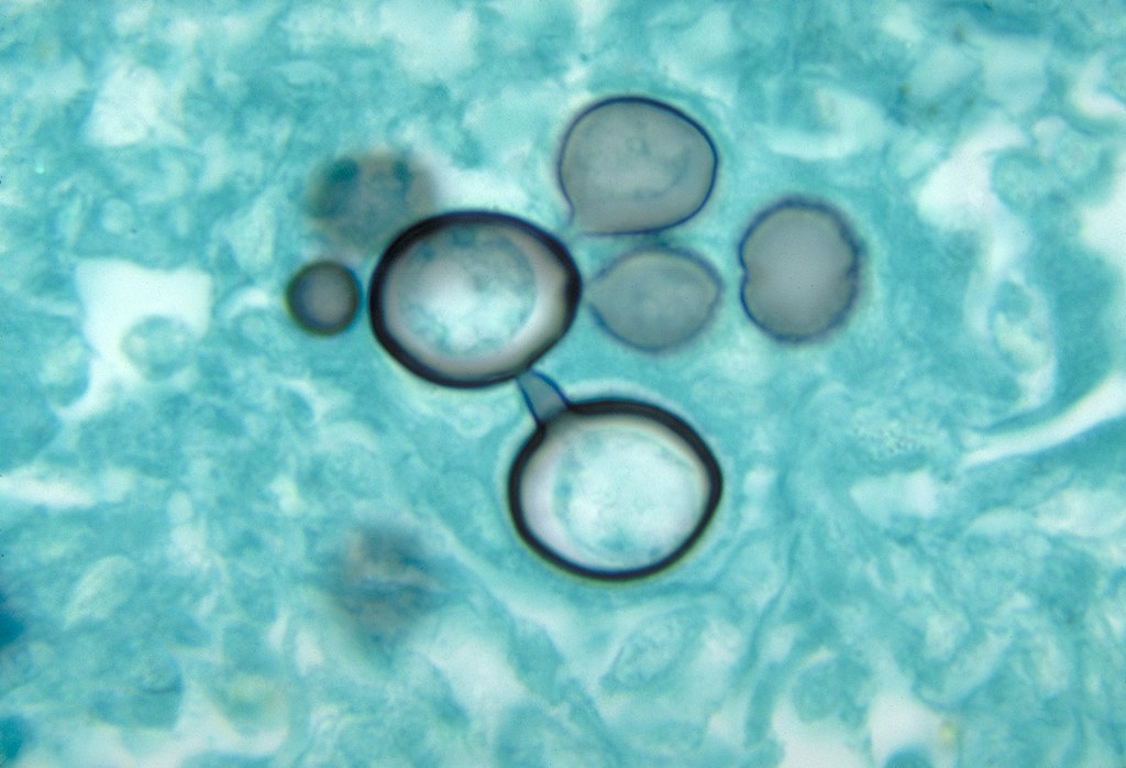
Histopatología de la paracoccidioidomicosis
Imagen: “Histopathology of paracoccidiodomycoisis” por CDC. Licencia: Dominio Público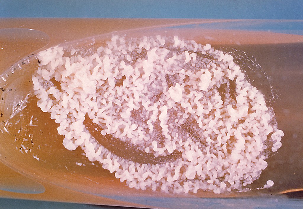
Un cultivo de Paracoccidioides brasiliensis durante su fase de levadura
Imagen: “A culture of Paracoccidioides brasiliensis during its yeast phase” por CDC. Licencia: Dominio Público