El linfoma extraganglionar de la zona marginal del tejido linfoide asociado a las mucosas (también llamado MALToma MALToma Extranodal marginal zone lymphoma (EMZL) of mucosa-associated lymphoid tissue (also called MALToma, MALT lymphoma, and pseudolymphoma) is a group of non-Hodgkin's lymphomas that have historically been grouped together because they appear to arise from postgerminal center marginal zone B cells and share a similar immunophenotype. MALT Lymphoma, linfoma MALT MALT Colon, Cecum, and Appendix: Anatomy y pseudolinfoma) es un grupo de linfomas no Hodgkin que históricamente se han agrupado porque parecen surgir de las células B de la zona marginal del centro postgerminal y comparten un inmunofenotipo similar. Se cree que el linfoma MALT MALT Colon, Cecum, and Appendix: Anatomy surge en EN Erythema nodosum is an immune-mediated panniculitis (inflammation of the subcutaneous fat) caused by a type IV (delayed-type) hypersensitivity reaction. It commonly manifests in young women as tender, erythematous nodules on the shins. Erythema Nodosum el marco de una estimulación inmunitaria crónica, que suele deberse a estímulos bacterianos, víricos o autoinmunes. Los LOS Neisseria linfomas MALT MALT Colon, Cecum, and Appendix: Anatomy se presentan con síntomas debido a la afectación localizada de los LOS Neisseria tejidos epiteliales glandulares en EN Erythema nodosum is an immune-mediated panniculitis (inflammation of the subcutaneous fat) caused by a type IV (delayed-type) hypersensitivity reaction. It commonly manifests in young women as tender, erythematous nodules on the shins. Erythema Nodosum el lugar específico donde se desarrollan. El diagnóstico del linfoma MALT MALT Colon, Cecum, and Appendix: Anatomy se realiza mediante el análisis morfológico, inmunofenotípico y genético de las muestras de biopsia. El linfoma MALT MALT Colon, Cecum, and Appendix: Anatomy gástrico positivo para Helicobacter pylori Helicobacter pylori A spiral bacterium active as a human gastric pathogen. It is a gram-negative, urease-positive, curved or slightly spiral organism initially isolated in 1982 from patients with lesions of gastritis or peptic ulcers in Western Australia. Helicobacter pylori was originally classified in the genus campylobacter, but RNA sequencing, cellular fatty acid profiles, growth patterns, and other taxonomic characteristics indicate that the micro-organism should be included in the genus Helicobacter. It has been officially transferred to Helicobacter gen. Helicobacter se trata con terapia de erradicación de H. pylori H. pylori A spiral bacterium active as a human gastric pathogen. It is a gram-negative, urease-positive, curved or slightly spiral organism initially isolated in 1982 from patients with lesions of gastritis or peptic ulcers in Western Australia. Helicobacter pylori was originally classified in the genus campylobacter, but RNA sequencing, cellular fatty acid profiles, growth patterns, and other taxonomic characteristics indicate that the micro-organism should be included in the genus Helicobacter. It has been officially transferred to Helicobacter gen. Helicobacter, y el linfoma MALT MALT Colon, Cecum, and Appendix: Anatomy gástrico negativo para H. pylori H. pylori A spiral bacterium active as a human gastric pathogen. It is a gram-negative, urease-positive, curved or slightly spiral organism initially isolated in 1982 from patients with lesions of gastritis or peptic ulcers in Western Australia. Helicobacter pylori was originally classified in the genus campylobacter, but RNA sequencing, cellular fatty acid profiles, growth patterns, and other taxonomic characteristics indicate that the micro-organism should be included in the genus Helicobacter. It has been officially transferred to Helicobacter gen. Helicobacter se trata con radioterapia. El linfoma MALT MALT Colon, Cecum, and Appendix: Anatomy no gástrico se trata en EN Erythema nodosum is an immune-mediated panniculitis (inflammation of the subcutaneous fat) caused by a type IV (delayed-type) hypersensitivity reaction. It commonly manifests in young women as tender, erythematous nodules on the shins. Erythema Nodosum función de la zona afectada y la extensión de la enfermedad. Los LOS Neisseria pacientes con linfoma MALT MALT Colon, Cecum, and Appendix: Anatomy tienen un buen pronóstico, con una supervivencia media de > 10 años.
Last updated: Dec 15, 2025
El linfoma del tejido linfoide asociado a mucosas ( MALT MALT Colon, Cecum, and Appendix: Anatomy, por sus siglas en EN Erythema nodosum is an immune-mediated panniculitis (inflammation of the subcutaneous fat) caused by a type IV (delayed-type) hypersensitivity reaction. It commonly manifests in young women as tender, erythematous nodules on the shins. Erythema Nodosum inglés) es un linfoma no Hodgkin clínicamente indolente que se postula que surge de las células B de memoria del centro postgerminal con capacidad para diferenciarse en EN Erythema nodosum is an immune-mediated panniculitis (inflammation of the subcutaneous fat) caused by a type IV (delayed-type) hypersensitivity reaction. It commonly manifests in young women as tender, erythematous nodules on the shins. Erythema Nodosum células de la zona marginal y células plasmáticas.
La presentación clínica varía en EN Erythema nodosum is an immune-mediated panniculitis (inflammation of the subcutaneous fat) caused by a type IV (delayed-type) hypersensitivity reaction. It commonly manifests in young women as tender, erythematous nodules on the shins. Erythema Nodosum función de la localización en EN Erythema nodosum is an immune-mediated panniculitis (inflammation of the subcutaneous fat) caused by a type IV (delayed-type) hypersensitivity reaction. It commonly manifests in young women as tender, erythematous nodules on the shins. Erythema Nodosum la que se desarrolla el linfoma MALT MALT Colon, Cecum, and Appendix: Anatomy.
Diseminación a otros sitios del tejido MALT MALT Colon, Cecum, and Appendix: Anatomy:
La sangre periférica no suele estar implicada.
Los LOS Neisseria síntomas sistémicos B son poco frecuentes:
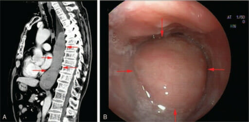
Linfoma del tejido linfoide asociado a la mucosa del esófago:
La tomografía computarizada (imagen A) y la endoscopia (imagen B) muestran una neoplasia del esófago medio e inferior.
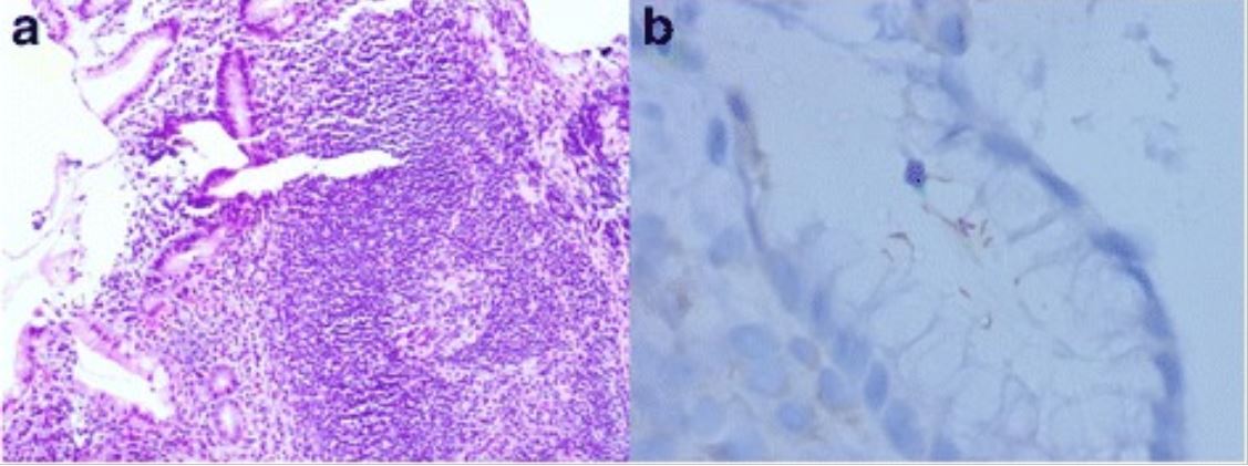
Linfoma MALT gástrico asociado a H. pylori:
a: Folículo linfoide con un centro germinal reactivo y una zona marginal ligeramente expandida (H&E, aumento original, 100X)
b: La inmunotinción de H. pylori muestra bacterias intrafoveolares con una configuración curva, característica de H. pylori.
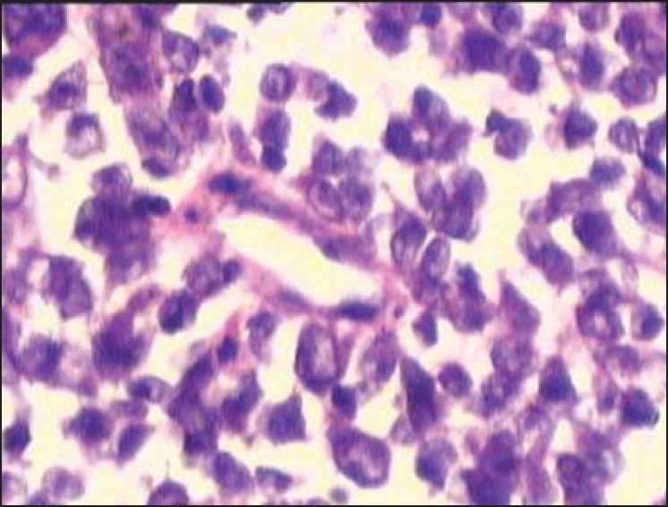
Linfoma MALT de colon:
Infiltración de células linfoides de aspecto maligno en la lámina propia que sugiere un linfoma de malta