Domina Conceptos Médicos

Estudia para la escuela de medicina y tus examenes con Lecturio.

Ependimoma

Los LOS Neisseria ependimomas son tumores de las células gliales que surgen de las células ependimarias productoras del LCR las cuales recubren el sistema ventricular. Los LOS Neisseria ependimomas suelen aparecer en EN Erythema nodosum is an immune-mediated panniculitis (inflammation of the subcutaneous fat) caused by a type IV (delayed-type) hypersensitivity reaction. It commonly manifests in young women as tender, erythematous nodules on the shins. Erythema Nodosum la fosa posterior, en EN Erythema nodosum is an immune-mediated panniculitis (inflammation of the subcutaneous fat) caused by a type IV (delayed-type) hypersensitivity reaction. It commonly manifests in young women as tender, erythematous nodules on the shins. Erythema Nodosum contacto con el 4to ventrículo o en EN Erythema nodosum is an immune-mediated panniculitis (inflammation of the subcutaneous fat) caused by a type IV (delayed-type) hypersensitivity reaction. It commonly manifests in young women as tender, erythematous nodules on the shins. Erythema Nodosum la médula espinal intramedular. La presentación clínica de los LOS Neisseria ependimomas varía en EN Erythema nodosum is an immune-mediated panniculitis (inflammation of the subcutaneous fat) caused by a type IV (delayed-type) hypersensitivity reaction. It commonly manifests in young women as tender, erythematous nodules on the shins. Erythema Nodosum función de la localización del tumor Tumor Inflammation. La resonancia magnética es la imagenología de elección, pero se requiere la confirmación histológica para el diagnóstico. La base del tratamiento del ependimoma intracraneal es la resección quirúrgica y la radioterapia adyuvante; los LOS Neisseria pacientes jóvenes reciben en EN Erythema nodosum is an immune-mediated panniculitis (inflammation of the subcutaneous fat) caused by a type IV (delayed-type) hypersensitivity reaction. It commonly manifests in young women as tender, erythematous nodules on the shins. Erythema Nodosum cambio quimioterapia. Los LOS Neisseria ependimomas de la médula espinal se tratan con una resección quirúrgica máxima.

Last updated: Dec 15, 2025

Editorial responsibility: Stanley Oiseth, Lindsay Jones, Evelin Maza

Descripción General

Definición

Los LOS Neisseria ependimomas son tumores de las células gliales en EN Erythema nodosum is an immune-mediated panniculitis (inflammation of the subcutaneous fat) caused by a type IV (delayed-type) hypersensitivity reaction. It commonly manifests in young women as tender, erythematous nodules on the shins. Erythema Nodosum el SNC que surgen de las células ependimarias.

  • Las células gliales son tejidos de soporte en EN Erythema nodosum is an immune-mediated panniculitis (inflammation of the subcutaneous fat) caused by a type IV (delayed-type) hypersensitivity reaction. It commonly manifests in young women as tender, erythematous nodules on the shins. Erythema Nodosum el cerebro y el sistema nervioso.
  • Las células ependimarias producen LCR.
  • Pueden surgir en EN Erythema nodosum is an immune-mediated panniculitis (inflammation of the subcutaneous fat) caused by a type IV (delayed-type) hypersensitivity reaction. It commonly manifests in young women as tender, erythematous nodules on the shins. Erythema Nodosum cualquier parte del sistema ventricular (ependimoma intracraneal) o en EN Erythema nodosum is an immune-mediated panniculitis (inflammation of the subcutaneous fat) caused by a type IV (delayed-type) hypersensitivity reaction. It commonly manifests in young women as tender, erythematous nodules on the shins. Erythema Nodosum el canal espinal(ependimoma espinal), pero lo más frecuente es que surjan en EN Erythema nodosum is an immune-mediated panniculitis (inflammation of the subcutaneous fat) caused by a type IV (delayed-type) hypersensitivity reaction. It commonly manifests in young women as tender, erythematous nodules on the shins. Erythema Nodosum:
    • Niños: la fosa posterior en EN Erythema nodosum is an immune-mediated panniculitis (inflammation of the subcutaneous fat) caused by a type IV (delayed-type) hypersensitivity reaction. It commonly manifests in young women as tender, erythematous nodules on the shins. Erythema Nodosum el piso del 4to ventrículo
    • Adultos: en EN Erythema nodosum is an immune-mediated panniculitis (inflammation of the subcutaneous fat) caused by a type IV (delayed-type) hypersensitivity reaction. It commonly manifests in young women as tender, erythematous nodules on the shins. Erythema Nodosum la médula espinal lumbosacra o filum terminal

Clasificación de los LOS Neisseria tumores del sistema nervioso

Tabla: Clasificación de los LOS Neisseria tumores del sistema nervioso
Categorías Tumores específicos
Tumores neuroepiteliales en EN Erythema nodosum is an immune-mediated panniculitis (inflammation of the subcutaneous fat) caused by a type IV (delayed-type) hypersensitivity reaction. It commonly manifests in young women as tender, erythematous nodules on the shins. Erythema Nodosum el SNC
  • Astrocitoma, incluido el glioblastoma multiforme Glioblastoma multiforme Glioblastoma multiforme is a high-grade astrocytoma, an aggressive brain tumor arising from astrocytes, with an unknown cause and a poorly understood link to risk factors. There are two main types: primary, a more aggressive form seen more commonly in older patients, and secondary, developing from lower-grade astrocytomas and seen more commonly in younger patients. Glioblastoma Multiforme
  • Oligodendroglioma Oligodendroglioma Oligodendrogliomas are malignant CNS tumors arising from neural glial cell precursors. Oligodendrogliomas often arise in the frontal lobes of the brain and have a generally favorable prognosis when compared to other gliomas. Oligodendrogliomas are the 3rd most common CNS tumor. The most frequent presenting symptom is a seizure. Oligodendroglioma
  • Ependimoma y tumor Tumor Inflammation del plexo coroideo
  • Meduloblastoma ( tumor Tumor Inflammation embrionario)
Tumores meníngeos
  • Meningioma Meningioma Meningiomas are slow-growing tumors that arise from the meninges of the brain and spinal cord. The vast majority are benign. These tumors commonly occur in individuals with a history of high doses of skull radiation, head trauma, and neurofibromatosis 2. Meningioma
  • Hemangioblastoma Hemangioblastoma Hemangioblastomas are vascular neoplasms of the CNS. Hemangioblastomas are rare and are often associated with von Hippel-Lindau disease (VHL). The most common presentation is a headache and, depending on the size and location of the tumor, patients may present with sensory deficits and motor weakness. Hemangioblastoma
Tumores de la región selar
  • Craneofaringioma
  • Adenoma hipofisario
  • Pinealoma/pinealoblastoma
Linfoma primario del SNC Linfoma primario del SNC
Metástasis al AL Amyloidosis cerebro (5 veces más común que los LOS Neisseria tumores cerebrales primarios) Más comúnmente surgen de:
  • Carcinomas de pulmón, mama y células renales
  • Melanoma Melanoma Melanoma is a malignant tumor arising from melanocytes, the melanin-producing cells of the epidermis. These tumors are most common in fair-skinned individuals with a history of excessive sun exposure and sunburns. Melanoma
Tumores periféricos
  • Schwannoma Schwannoma Schwannomas (also known as neurilemmomas) are benign nerve sheath tumors in the peripheral nervous system (PNS), arising from Schwann cells that encase the peripheral nerves. Schwannomas are the most common tumors in the PNS. Schwannoma, incluido el neurinoma del acústico
  • Neuroblastoma Neuroblastoma Neuroblastoma is a malignancy that arises from the neural crest cell derivatives along the sympathetic chain (neuroblasts) and is most commonly located in the adrenal medulla. The tumor often presents in childhood with a flank mass that crosses the midline. Neuroblastoma

Epidemiología

Ependimoma intracraneal:

  • Incidencia:
    • 0,43 casos por cada 100.000 personas
    • 2,5 % de todos los LOS Neisseria tumores gliales intracraneales
    • Tercer tumor Tumor Inflammation cerebral pediátrico más frecuente (por detrás del meduloblastoma y el astrocitoma pilocítico)
  • Edad:
    • Más común en EN Erythema nodosum is an immune-mediated panniculitis (inflammation of the subcutaneous fat) caused by a type IV (delayed-type) hypersensitivity reaction. It commonly manifests in young women as tender, erythematous nodules on the shins. Erythema Nodosum niños que en EN Erythema nodosum is an immune-mediated panniculitis (inflammation of the subcutaneous fat) caused by a type IV (delayed-type) hypersensitivity reaction. It commonly manifests in young women as tender, erythematous nodules on the shins. Erythema Nodosum adultos
    • Mediana de edad en EN Erythema nodosum is an immune-mediated panniculitis (inflammation of the subcutaneous fat) caused by a type IV (delayed-type) hypersensitivity reaction. It commonly manifests in young women as tender, erythematous nodules on the shins. Erythema Nodosum el momento del diagnóstico: 4–5 años
    • 25%–40% de los LOS Neisseria pacientes tienen < 2 años.
    • El ependimoma intracraneal en EN Erythema nodosum is an immune-mediated panniculitis (inflammation of the subcutaneous fat) caused by a type IV (delayed-type) hypersensitivity reaction. It commonly manifests in young women as tender, erythematous nodules on the shins. Erythema Nodosum adultos suele aparecer antes de los LOS Neisseria 40 años.
  • Sexo: ligero predominio en EN Erythema nodosum is an immune-mediated panniculitis (inflammation of the subcutaneous fat) caused by a type IV (delayed-type) hypersensitivity reaction. It commonly manifests in young women as tender, erythematous nodules on the shins. Erythema Nodosum los LOS Neisseria hombres
  • Localización:
    • Lo más frecuente es que se produzca en EN Erythema nodosum is an immune-mediated panniculitis (inflammation of the subcutaneous fat) caused by a type IV (delayed-type) hypersensitivity reaction. It commonly manifests in young women as tender, erythematous nodules on the shins. Erythema Nodosum la fosa posterior
    • En EN Erythema nodosum is an immune-mediated panniculitis (inflammation of the subcutaneous fat) caused by a type IV (delayed-type) hypersensitivity reaction. It commonly manifests in young women as tender, erythematous nodules on the shins. Erythema Nodosum los LOS Neisseria niños, el 90% de los LOS Neisseria ependimomas son intracraneales.

Ependimoma espinal:

  • Incidencia:
    • Aproximadamente el 25% de los LOS Neisseria tumores primarios de la médula espinal
    • En EN Erythema nodosum is an immune-mediated panniculitis (inflammation of the subcutaneous fat) caused by a type IV (delayed-type) hypersensitivity reaction. It commonly manifests in young women as tender, erythematous nodules on the shins. Erythema Nodosum los LOS Neisseria adultos, el 75% de los LOS Neisseria ependimomas se encuentran en EN Erythema nodosum is an immune-mediated panniculitis (inflammation of the subcutaneous fat) caused by a type IV (delayed-type) hypersensitivity reaction. It commonly manifests in young women as tender, erythematous nodules on the shins. Erythema Nodosum la médula espinal.
  • Edad:
    • Más común en EN Erythema nodosum is an immune-mediated panniculitis (inflammation of the subcutaneous fat) caused by a type IV (delayed-type) hypersensitivity reaction. It commonly manifests in young women as tender, erythematous nodules on the shins. Erythema Nodosum adultos
    • Mediana de edad en EN Erythema nodosum is an immune-mediated panniculitis (inflammation of the subcutaneous fat) caused by a type IV (delayed-type) hypersensitivity reaction. It commonly manifests in young women as tender, erythematous nodules on the shins. Erythema Nodosum el momento del diagnóstico: 30–40 años
  • Localización:
    • Aproximadamente el 50% se produce en EN Erythema nodosum is an immune-mediated panniculitis (inflammation of the subcutaneous fat) caused by a type IV (delayed-type) hypersensitivity reaction. It commonly manifests in young women as tender, erythematous nodules on the shins. Erythema Nodosum el cordón lumbosacro o en EN Erythema nodosum is an immune-mediated panniculitis (inflammation of the subcutaneous fat) caused by a type IV (delayed-type) hypersensitivity reaction. It commonly manifests in young women as tender, erythematous nodules on the shins. Erythema Nodosum el filum terminal.
    • Aproximadamente el otro 50% se produce en EN Erythema nodosum is an immune-mediated panniculitis (inflammation of the subcutaneous fat) caused by a type IV (delayed-type) hypersensitivity reaction. It commonly manifests in young women as tender, erythematous nodules on the shins. Erythema Nodosum otra parte de la médula espinal cervical o torácica.

Etiología

  • No se conoce la causa del ependimoma
  • Se ha HA Hemolytic anemia (HA) is the term given to a large group of anemias that are caused by the premature destruction/hemolysis of circulating red blood cells (RBCs). Hemolysis can occur within (intravascular hemolysis) or outside the blood vessels (extravascular hemolysis). Hemolytic Anemia observado la agrupación de la enfermedad dentro de las familias.
  • Mayor incidencia de ependimoma espinal en EN Erythema nodosum is an immune-mediated panniculitis (inflammation of the subcutaneous fat) caused by a type IV (delayed-type) hypersensitivity reaction. It commonly manifests in young women as tender, erythematous nodules on the shins. Erythema Nodosum la neurofibromatosis tipo 2

Fisiopatología

  • Surge de las células ependimarias:
    • Las células revisten los LOS Neisseria ventrículos cerebrales, los LOS Neisseria conductos en EN Erythema nodosum is an immune-mediated panniculitis (inflammation of the subcutaneous fat) caused by a type IV (delayed-type) hypersensitivity reaction. It commonly manifests in young women as tender, erythematous nodules on the shins. Erythema Nodosum el cerebro y la médula espinal.
    • Derivado de las células gliales radiales de la zona subventricular
  • Transformación de células ependimarias normales a células neoplásicas:
    • Mutaciones genéticas
    • Alteraciones epigenéticas
  • Puede ser localmente invasivo y extenderse por la médula espinal
  • Características del tumor Tumor Inflammation:
    • Por lo general, son bien delimitados
    • Múltiples áreas de calcificaciones, hemorragias y quistes
    • Las células tumorales suelen formar rosetas ependimarias (las pseudorosetas perivasculares son el sello histológico de los LOS Neisseria ependimomas).
  • Se han identificado varios perfiles moleculares basados en EN Erythema nodosum is an immune-mediated panniculitis (inflammation of the subcutaneous fat) caused by a type IV (delayed-type) hypersensitivity reaction. It commonly manifests in young women as tender, erythematous nodules on the shins. Erythema Nodosum combinaciones específicas de anomalías genéticas y epigenéticas dentro de las células tumorales:
    • Los LOS Neisseria ependimomas pueden clasificarse en EN Erythema nodosum is an immune-mediated panniculitis (inflammation of the subcutaneous fat) caused by a type IV (delayed-type) hypersensitivity reaction. It commonly manifests in young women as tender, erythematous nodules on the shins. Erythema Nodosum los LOS Neisseria siguientes subgrupos según los LOS Neisseria perfiles:
      • Ependimoma de la fosa posterior grupo A
      • Ependimoma de la fosa posterior grupo B
      • Ependimoma supratentorial con el gen de fusión RELA
      • Ependimoma supratentorial con gen el de fusión YAP
    • Fusión positiva del genRELA:
      • Fusión oncogénica entre RELA y C11orf95 C11orf95 Ependymoma
      • RELA es el principal efector de la señalización NF-κB.
    • La clasificación por subgrupos es aún incipiente y requiere más estudios para comprender las implicaciones pronósticas y terapéuticas.

Presentación Clínica

La presentación clínica depende de la localización del ependimoma y de la edad del paciente. La mayoría de los LOS Neisseria pacientes tienen síntomas aproximadamente 3–6 meses antes del diagnóstico.

  • Ependimoma de la fosa posterior:
    • Signos de aumento de la presión intracraneal por hidrocefalia obstructiva:
      • Cefalea (frecuentemente peor por la mañana)
      • Náuseas y vómitos potencialmente importantes
      • Ataxia Ataxia Impairment of the ability to perform smoothly coordinated voluntary movements. This condition may affect the limbs, trunk, eyes, pharynx, larynx, and other structures. Ataxia may result from impaired sensory or motor function. Sensory ataxia may result from posterior column injury or peripheral nerve diseases. Motor ataxia may be associated with cerebellar diseases; cerebral cortex diseases; thalamic diseases; basal ganglia diseases; injury to the red nucleus; and other conditions. Ataxia-telangiectasia
      • Vértigo
      • Papiledema
      • Nistagmo
    • Parálisis de los LOS Neisseria nervios craneales
    • Cambios en EN Erythema nodosum is an immune-mediated panniculitis (inflammation of the subcutaneous fat) caused by a type IV (delayed-type) hypersensitivity reaction. It commonly manifests in young women as tender, erythematous nodules on the shins. Erythema Nodosum la personalidad, el estado de ánimo y cambios cognitivos
  • Ependimomas supratentoriales:
    • Convulsiones
    • Déficit neurológico focal
  • Ependimoma de la médula espinal:
    • Déficits motores y sensitivos que afectan a las vías nerviosas descendentes, a las vías nerviosas ascendentes y a los LOS Neisseria nervios periféricos eferentes
    • Los LOS Neisseria déficits específicos dependen del nivel al AL Amyloidosis que se encuentra el tumor Tumor Inflammation

Diagnóstico

Imagenología

La imagenología se obtienen en EN Erythema nodosum is an immune-mediated panniculitis (inflammation of the subcutaneous fat) caused by a type IV (delayed-type) hypersensitivity reaction. It commonly manifests in young women as tender, erythematous nodules on the shins. Erythema Nodosum 1er lugar como parte del trabajo de diagnóstico, pero la evaluación histológica es necesaria para confirmar el diagnóstico. Los LOS Neisseria hallazgos en EN Erythema nodosum is an immune-mediated panniculitis (inflammation of the subcutaneous fat) caused by a type IV (delayed-type) hypersensitivity reaction. It commonly manifests in young women as tender, erythematous nodules on the shins. Erythema Nodosum la imagenología pueden sugerir un ependimoma y así guiar la cirugía.

  • RM:
    • Método de imagenología de elección para el cerebro y la médula espinal
    • Masas hipointensas en EN Erythema nodosum is an immune-mediated panniculitis (inflammation of the subcutaneous fat) caused by a type IV (delayed-type) hypersensitivity reaction. It commonly manifests in young women as tender, erythematous nodules on the shins. Erythema Nodosum la RM ponderada en EN Erythema nodosum is an immune-mediated panniculitis (inflammation of the subcutaneous fat) caused by a type IV (delayed-type) hypersensitivity reaction. It commonly manifests in young women as tender, erythematous nodules on the shins. Erythema Nodosum T1
    • Masas hiperintensas en EN Erythema nodosum is an immune-mediated panniculitis (inflammation of the subcutaneous fat) caused by a type IV (delayed-type) hypersensitivity reaction. It commonly manifests in young women as tender, erythematous nodules on the shins. Erythema Nodosum la RM ponderada en EN Erythema nodosum is an immune-mediated panniculitis (inflammation of the subcutaneous fat) caused by a type IV (delayed-type) hypersensitivity reaction. It commonly manifests in young women as tender, erythematous nodules on the shins. Erythema Nodosum T2 o densidad protónica
    • El realce con gadolinio suele ser prominente.
  • Hallazgos en EN Erythema nodosum is an immune-mediated panniculitis (inflammation of the subcutaneous fat) caused by a type IV (delayed-type) hypersensitivity reaction. It commonly manifests in young women as tender, erythematous nodules on the shins. Erythema Nodosum la TC:
    • Hiperdenso con un realce homogéneo
    • Es frecuente ver quistes y calcificaciones.
  • Hallazgos generales sugestivos de ependimoma (distinguibles de otros tumores de la fosa posterior):
    • Tumor Tumor Inflammation que surge del 4to ventrículo
    • Tumor Tumor Inflammation que se extiende hacia la unión craneocervical
    • Extensión pronunciada a través de los LOS Neisseria forámenes de Luschka
    • Difusión restringida en EN Erythema nodosum is an immune-mediated panniculitis (inflammation of the subcutaneous fat) caused by a type IV (delayed-type) hypersensitivity reaction. It commonly manifests in young women as tender, erythematous nodules on the shins. Erythema Nodosum imágenes ponderadas por difusión
    • Calcificaciones en EN Erythema nodosum is an immune-mediated panniculitis (inflammation of the subcutaneous fat) caused by a type IV (delayed-type) hypersensitivity reaction. It commonly manifests in young women as tender, erythematous nodules on the shins. Erythema Nodosum el 4to ventrículo
Resonancia magnética que revela un ependimoma anaplásico

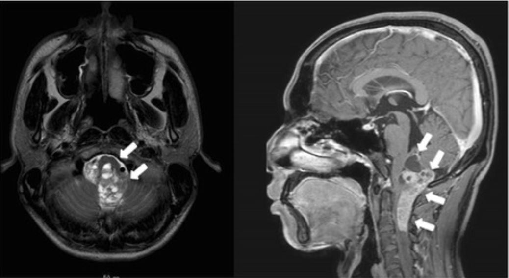
La resonancia magnética revela un ependimoma anaplásico que se extiende desde el tronco encefálico hasta el 4to ventrículo.

Imagen: “Giant duodenal ulcers after neurosurgery for brainstem tumors that required reoperation for gastric disconnection: a report of two cases” por BMC Surgery. Licencia: CC BY 4.0

Clasificación histológica

El ependimoma se basa en EN Erythema nodosum is an immune-mediated panniculitis (inflammation of the subcutaneous fat) caused by a type IV (delayed-type) hypersensitivity reaction. It commonly manifests in young women as tender, erythematous nodules on the shins. Erythema Nodosum un diagnóstico histológico; por lo tanto, el estándar de oro para el diagnóstico es la evaluación patológica de una muestra de tejido. Los LOS Neisseria ependimomas se clasifican por grados histológicos según la OMS.

  • Grado l de la Organización Mundial de la Salud:
    • Subependimoma:
      • Tumores benignos y poco frecuentes del cuarto ventrículo o del ventrículo lateral en EN Erythema nodosum is an immune-mediated panniculitis (inflammation of the subcutaneous fat) caused by a type IV (delayed-type) hypersensitivity reaction. It commonly manifests in young women as tender, erythematous nodules on the shins. Erythema Nodosum adultos
      • Tejido hipocelular formado por una matriz glial gruesa que contiene grupos de células con núcleos uniformes y microquistes
    • Ependimoma mixopapilar:
      • Tumores generalmente benignos que surgen en EN Erythema nodosum is an immune-mediated panniculitis (inflammation of the subcutaneous fat) caused by a type IV (delayed-type) hypersensitivity reaction. It commonly manifests in young women as tender, erythematous nodules on the shins. Erythema Nodosum el cono medular y el filum terminal de la médula espinal
      • Estructuras pseudopapilares con microquistes ricos en EN Erythema nodosum is an immune-mediated panniculitis (inflammation of the subcutaneous fat) caused by a type IV (delayed-type) hypersensitivity reaction. It commonly manifests in young women as tender, erythematous nodules on the shins. Erythema Nodosum mucina
      • Células cuboidales que rodean un estroma mixoide
  • Grado II de la Organización Mundial de la Salud: ependimoma clásico:
    • Puede surgir en EN Erythema nodosum is an immune-mediated panniculitis (inflammation of the subcutaneous fat) caused by a type IV (delayed-type) hypersensitivity reaction. It commonly manifests in young women as tender, erythematous nodules on the shins. Erythema Nodosum cualquier parte del sistema ventricular o del canal espinal
    • Las posibles variantes son:
      • Variante papilar
      • Variante de células claras
      • Variante tanicítica
    • Los LOS Neisseria hallazgos incluyen:
      • Masa sólida o papilar
      • Pseudorosetas perivasculares: las células tumorales parecen como una glándula que rodea la vasculatura (confirma el diagnóstico de ependimoma)
      • Células con núcleos regulares, redondos a ovalados y con abundante cromatina granular
      • Fondo fibrilar denso
  • Grado III de la Organización Mundial de la Salud:ependimoma anaplásico:
    • Abundantes células mitóticas
    • Necrosis Necrosis The death of cells in an organ or tissue due to disease, injury or failure of the blood supply. Ischemic Cell Damage pseudopalisante
  • Ependimoma con fusión RELA positiva:
    • Una nueva pieza de la clasificación de la OMS
    • Puede incluir los LOS Neisseria grados II y III
    • Determinado con base en EN Erythema nodosum is an immune-mediated panniculitis (inflammation of the subcutaneous fat) caused by a type IV (delayed-type) hypersensitivity reaction. It commonly manifests in young women as tender, erythematous nodules on the shins. Erythema Nodosum pruebas genéticas
Ependimoma intracraneal grado III de la OMS

Ependimoma intracraneal grado III de la Organización Mundial de la Salud:
Se observan pseudorosetas perivasculares, así como una actividad mitótica rápida, lo que clasifica el tumor como un ependimoma anaplásico.

Imagen: “Exomic sequencing of four rare central nervous system tumor types” por Bettegowda C., et al. Licencia: CC BY 2.5

Otras pruebas

  • Citología de LCR:
    • Importante en EN Erythema nodosum is an immune-mediated panniculitis (inflammation of the subcutaneous fat) caused by a type IV (delayed-type) hypersensitivity reaction. It commonly manifests in young women as tender, erythematous nodules on the shins. Erythema Nodosum la evaluación para metástasis
    • Lo ideal es hacerlo antes de la cirugía ( los LOS Neisseria restos de la cirugía pueden dificultar la interpretación)
    • A menudo está contraindicado debido a la hidrocefalia obstructiva
  • EEG EEG Seizures:
    • No es necesario para el diagnóstico
    • En EN Erythema nodosum is an immune-mediated panniculitis (inflammation of the subcutaneous fat) caused by a type IV (delayed-type) hypersensitivity reaction. It commonly manifests in young women as tender, erythematous nodules on the shins. Erythema Nodosum los LOS Neisseria pacientes con lesiones supratentoriales, se observa una ralentización difusa y/o picos epileptógenos en EN Erythema nodosum is an immune-mediated panniculitis (inflammation of the subcutaneous fat) caused by a type IV (delayed-type) hypersensitivity reaction. It commonly manifests in young women as tender, erythematous nodules on the shins. Erythema Nodosum las zonas que rodean al AL Amyloidosis tumor Tumor Inflammation.

Tratamiento y Pronóstico

Tratamiento

  • Referir a centros especializados cuando sea posible.
  • Ependimoma intracraneal:
    • Resección máxima segura
    • Radioterapia adyuvante
    • Quimioterapia:
      • > 1 año de edad: puede utilizarse después de la radiación en EN Erythema nodosum is an immune-mediated panniculitis (inflammation of the subcutaneous fat) caused by a type IV (delayed-type) hypersensitivity reaction. It commonly manifests in young women as tender, erythematous nodules on the shins. Erythema Nodosum algunos pacientes
      • < 1 año de edad: se utiliza para retrasar la radioterapia (evitando los LOS Neisseria efectos adversos de la radiación)
  • Ependimoma espinal:
    • El tratamiento óptimo incluye la resección quirúrgica completa.
    • El papel de la radioterapia adyuvante no está muy claro.
    • No se ha HA Hemolytic anemia (HA) is the term given to a large group of anemias that are caused by the premature destruction/hemolysis of circulating red blood cells (RBCs). Hemolysis can occur within (intravascular hemolysis) or outside the blood vessels (extravascular hemolysis). Hemolytic Anemia demostrado el papel de la quimioterapia
  • Corticosteroides: para reducir el edema Edema Edema is a condition in which excess serous fluid accumulates in the body cavity or interstitial space of connective tissues. Edema is a symptom observed in several medical conditions. It can be categorized into 2 types, namely, peripheral (in the extremities) and internal (in an organ or body cavity). Edema que rodea al AL Amyloidosis tumor Tumor Inflammation
  • Anticonvulsivos: para tratar las convulsiones en EN Erythema nodosum is an immune-mediated panniculitis (inflammation of the subcutaneous fat) caused by a type IV (delayed-type) hypersensitivity reaction. It commonly manifests in young women as tender, erythematous nodules on the shins. Erythema Nodosum pacientes con ependimoma intracraneal

Pronóstico

  • Tasas de supervivencia:
    • Niños:
      • Supervivencia a 10 años: hasta el 70% en EN Erythema nodosum is an immune-mediated panniculitis (inflammation of the subcutaneous fat) caused by a type IV (delayed-type) hypersensitivity reaction. It commonly manifests in young women as tender, erythematous nodules on the shins. Erythema Nodosum pacientes con tumores totalmente resecados
      • Supervivencia a 10 años: del 30%–40% en EN Erythema nodosum is an immune-mediated panniculitis (inflammation of the subcutaneous fat) caused by a type IV (delayed-type) hypersensitivity reaction. It commonly manifests in young women as tender, erythematous nodules on the shins. Erythema Nodosum pacientes con tumores parcialmente resecados
    • Adultos:
      • Supervivencia a 5 años: 67%–85%
      • Supervivencia a 10 años: 50%–77%.
  • Factores que afectan a la supervivencia:
    • Extensión de la resección (factor más importante)
    • Grado histológico y subgrupo genético molecular
    • Localización del tumor Tumor Inflammation
    • Niños diagnosticados a una edad más avanzada tienen un mejor pronóstico.
    • Estado de rendimiento de Karnofsky (mide el estado funcional) en EN Erythema nodosum is an immune-mediated panniculitis (inflammation of the subcutaneous fat) caused by a type IV (delayed-type) hypersensitivity reaction. It commonly manifests in young women as tender, erythematous nodules on the shins. Erythema Nodosum adultos
  • Los LOS Neisseria supervivientes a largo plazo del tratamiento tienen un mayor riesgo de:
    • Déficits neurocognitivos
    • Déficits neurológicos focales
    • Pérdida auditiva neurosensorial
    • Anomalías de crecimiento y endocrinas
    • Vasculopatía
    • 2da malignidad

Diagnóstico Diferencial

  • Meduloblastoma: un tumor Tumor Inflammation que surge en EN Erythema nodosum is an immune-mediated panniculitis (inflammation of the subcutaneous fat) caused by a type IV (delayed-type) hypersensitivity reaction. It commonly manifests in young women as tender, erythematous nodules on the shins. Erythema Nodosum la fosa posterior. El meduloblastoma es el tumor Tumor Inflammation cerebral maligno más frecuente en EN Erythema nodosum is an immune-mediated panniculitis (inflammation of the subcutaneous fat) caused by a type IV (delayed-type) hypersensitivity reaction. It commonly manifests in young women as tender, erythematous nodules on the shins. Erythema Nodosum los LOS Neisseria niños. Los LOS Neisseria pacientes presentan síntomas de aumento de la presión intracraneal y signos cerebelosos que empeoran con el tiempo. El tratamiento es una combinación de cirugía, radiación y quimioterapia. A diferencia de los LOS Neisseria ependimomas, los LOS Neisseria meduloblastomas rara vez tienen extensión foraminal y los LOS Neisseria tumores primarios no se presentan en EN Erythema nodosum is an immune-mediated panniculitis (inflammation of the subcutaneous fat) caused by a type IV (delayed-type) hypersensitivity reaction. It commonly manifests in young women as tender, erythematous nodules on the shins. Erythema Nodosum la médula espinal.
  • Astrocitoma pilocítico: el 2do tumor Tumor Inflammation cerebral más frecuente en EN Erythema nodosum is an immune-mediated panniculitis (inflammation of the subcutaneous fat) caused by a type IV (delayed-type) hypersensitivity reaction. It commonly manifests in young women as tender, erythematous nodules on the shins. Erythema Nodosum los LOS Neisseria niños. La resonancia magnética puede ayudar a distinguir el astrocitoma pilocítico de otros tumores. Un astrocitoma pilocítico suele ser quístico con un módulo mural o tiene necrosis Necrosis The death of cells in an organ or tissue due to disease, injury or failure of the blood supply. Ischemic Cell Damage central con un borde grueso de tejido que realza en EN Erythema nodosum is an immune-mediated panniculitis (inflammation of the subcutaneous fat) caused by a type IV (delayed-type) hypersensitivity reaction. It commonly manifests in young women as tender, erythematous nodules on the shins. Erythema Nodosum la RM. Por el contrario, los LOS Neisseria ependimomas suelen surgir del 4to ventrículo y suelen ir acompañados de una difusión restringida en EN Erythema nodosum is an immune-mediated panniculitis (inflammation of the subcutaneous fat) caused by a type IV (delayed-type) hypersensitivity reaction. It commonly manifests in young women as tender, erythematous nodules on the shins. Erythema Nodosum las imágenes ponderadas por difusión.
  • Tumores del plexo coroideo: tumores primarios raros del SNC que surgen del plexo coroideo dentro de los LOS Neisseria ventrículos. Los LOS Neisseria tumores del plexo coroideo suelen presentarse en EN Erythema nodosum is an immune-mediated panniculitis (inflammation of the subcutaneous fat) caused by a type IV (delayed-type) hypersensitivity reaction. It commonly manifests in young women as tender, erythematous nodules on the shins. Erythema Nodosum niños con signos y síntomas de hidrocefalia. Histológicamente, los LOS Neisseria tumores del plexo coroideo se asemejan a los LOS Neisseria adenocarcinomas y pueden estar asociados al AL Amyloidosis síndrome de Li-Fraumeni.
  • Oligodendroglioma Oligodendroglioma Oligodendrogliomas are malignant CNS tumors arising from neural glial cell precursors. Oligodendrogliomas often arise in the frontal lobes of the brain and have a generally favorable prognosis when compared to other gliomas. Oligodendrogliomas are the 3rd most common CNS tumor. The most frequent presenting symptom is a seizure. Oligodendroglioma: un tumor Tumor Inflammation del SNC que surge de los LOS Neisseria oligodendrocitos. El oligodendroglioma Oligodendroglioma Oligodendrogliomas are malignant CNS tumors arising from neural glial cell precursors. Oligodendrogliomas often arise in the frontal lobes of the brain and have a generally favorable prognosis when compared to other gliomas. Oligodendrogliomas are the 3rd most common CNS tumor. The most frequent presenting symptom is a seizure. Oligodendroglioma suele desarrollarse en EN Erythema nodosum is an immune-mediated panniculitis (inflammation of the subcutaneous fat) caused by a type IV (delayed-type) hypersensitivity reaction. It commonly manifests in young women as tender, erythematous nodules on the shins. Erythema Nodosum el hemisferio cerebral (normalmente en EN Erythema nodosum is an immune-mediated panniculitis (inflammation of the subcutaneous fat) caused by a type IV (delayed-type) hypersensitivity reaction. It commonly manifests in young women as tender, erythematous nodules on the shins. Erythema Nodosum el lóbulo frontal Frontal The bone that forms the frontal aspect of the skull. Its flat part forms the forehead, articulating inferiorly with the nasal bone and the cheek bone on each side of the face. Skull: Anatomy). La presentación clínica puede incluir déficits neurológicos focales, convulsiones y cambios de personalidad. El diagnóstico se realiza mediante RM y biopsia. El tratamiento implica la resección quirúrgica y puede ir acompañada de radiación y/o quimioterapia.

Referencias

  1. National Brain Tumor Society. (2024). Ependymoma guide (3rd ed.). https://braintumor.org/wp-content/uploads/2024/06/Ependymoma_Guide_3.pdf
  2. Lombardi, G., et al. (2021). An overview of intracranial ependymomas in adults. Cancers, 13(23), 6128. https://doi.org/10.3390/cancers13236128
  3. DeAngelis, L.M., Wen, P.Y. (2018). Primary and metastatic tumors of the nervous system. In Jameson, J., et al. (Eds.), Harrison’s Principles of Internal Medicine, 20e. McGraw-Hill. https://accessmedicine-mhmedical-com.aucmed.idm.oclc.org/content.aspx?bookid=2129&sectionid=192016511
  4. Genetic and Rare Disease Information Center. (2025). Ependymoma. Retrieved March 25, 2025, from https://rarediseases.info.nih.gov/diseases/6353/ependymoma
  5. Mayo Clinic. (2025). Embryonal tumors. Retrieved March 25, 2025, from https://www.mayoclinic.org/diseases-conditions/embryonal-tumor/
  6. National Cancer Institute Center for Cancer Research. (2024). Choroid Plexus Tumors Diagnosis and Treatment. Retrieved March 25, 2025, from https://www.cancer.gov/rare-brain-spine-tumor/tumors/choroid-plexus-tumors
  7. National Cancer Institute. (2024). Childhood Ependymoma Treatment (PDQ®)–Patient Version. Retrieved March 25, 2025, from https://www.cancer.gov/types/brain/patient/child-ependymoma-treatment-pdq
  8. National Cancer Institute. (2024). Ependymoma Diagnosis and Treatment. Retrieved March 25, 2025, from https://www.cancer.gov/rare-brain-spine-tumor/tumors/ependymoma
  9. Upadhyaya, S.A., Tinkle, C. (2025). Intracranial ependymoma and other ependymal tumors. UpToDate. Retrieved March 25, 2025, from https://www.uptodate.com/contents/intracranial-ependymoma-and-other-ependymal-tumors
  10. Welch, W.C., Schiff, D., Gerszten, P.C. (2025). Spinal cord tumors. UpToDate. Retrieved March 25, 2025, from https://www.uptodate.com/contents/spinal-cord-tumors

¡Crea tu cuenta gratis o inicia una sesión para seguir leyendo!

Regístrate ahora y obtén acceso gratuito a Lecturio con páginas de concepto, videos médicos y cuestionarios para tu educación médica.

User Reviews

Que tengas una sesión de estudio alegre y navideña 🎁 Ahorra 50% en todos los planes >>

Details