El desprendimiento de retina Retina The ten-layered nervous tissue membrane of the eye. It is continuous with the optic nerve and receives images of external objects and transmits visual impulses to the brain. Its outer surface is in contact with the choroid and the inner surface with the vitreous body. The outermost layer is pigmented, whereas the inner nine layers are transparent. Eye: Anatomy es la separación de la retina Retina The ten-layered nervous tissue membrane of the eye. It is continuous with the optic nerve and receives images of external objects and transmits visual impulses to the brain. Its outer surface is in contact with the choroid and the inner surface with the vitreous body. The outermost layer is pigmented, whereas the inner nine layers are transparent. Eye: Anatomy neurosensorial del epitelio pigmentado de la retina Retina The ten-layered nervous tissue membrane of the eye. It is continuous with the optic nerve and receives images of external objects and transmits visual impulses to the brain. Its outer surface is in contact with the choroid and the inner surface with the vitreous body. The outermost layer is pigmented, whereas the inner nine layers are transparent. Eye: Anatomy y la coroides. El desprendimiento de retina Retina The ten-layered nervous tissue membrane of the eye. It is continuous with the optic nerve and receives images of external objects and transmits visual impulses to the brain. Its outer surface is in contact with the choroid and the inner surface with the vitreous body. The outermost layer is pigmented, whereas the inner nine layers are transparent. Eye: Anatomy regmatógeno, el tipo más común, proviene de una ruptura en EN Erythema nodosum is an immune-mediated panniculitis (inflammation of the subcutaneous fat) caused by a type IV (delayed-type) hypersensitivity reaction. It commonly manifests in young women as tender, erythematous nodules on the shins. Erythema Nodosum la retina Retina The ten-layered nervous tissue membrane of the eye. It is continuous with the optic nerve and receives images of external objects and transmits visual impulses to the brain. Its outer surface is in contact with the choroid and the inner surface with the vitreous body. The outermost layer is pigmented, whereas the inner nine layers are transparent. Eye: Anatomy, lo que permite que se acumule líquido en EN Erythema nodosum is an immune-mediated panniculitis (inflammation of the subcutaneous fat) caused by a type IV (delayed-type) hypersensitivity reaction. It commonly manifests in young women as tender, erythematous nodules on the shins. Erythema Nodosum el espacio subretiniano. En EN Erythema nodosum is an immune-mediated panniculitis (inflammation of the subcutaneous fat) caused by a type IV (delayed-type) hypersensitivity reaction. It commonly manifests in young women as tender, erythematous nodules on the shins. Erythema Nodosum el caso de una retina Retina The ten-layered nervous tissue membrane of the eye. It is continuous with the optic nerve and receives images of external objects and transmits visual impulses to the brain. Its outer surface is in contact with the choroid and the inner surface with the vitreous body. The outermost layer is pigmented, whereas the inner nine layers are transparent. Eye: Anatomy intacta, el desprendimiento ocurre cuando el vítreo tira de la retina Retina The ten-layered nervous tissue membrane of the eye. It is continuous with the optic nerve and receives images of external objects and transmits visual impulses to the brain. Its outer surface is in contact with the choroid and the inner surface with the vitreous body. The outermost layer is pigmented, whereas the inner nine layers are transparent. Eye: Anatomy (tracción) o cuando una afección subyacente conduce a una mayor pérdida de líquido (exudativo). Los LOS Neisseria síntomas de fotopsia, partículas flotantes y defectos visuales pueden presentarse en EN Erythema nodosum is an immune-mediated panniculitis (inflammation of the subcutaneous fat) caused by a type IV (delayed-type) hypersensitivity reaction. It commonly manifests in young women as tender, erythematous nodules on the shins. Erythema Nodosum horas o gradualmente durante semanas. El desprendimiento de retina Retina The ten-layered nervous tissue membrane of the eye. It is continuous with the optic nerve and receives images of external objects and transmits visual impulses to the brain. Its outer surface is in contact with the choroid and the inner surface with the vitreous body. The outermost layer is pigmented, whereas the inner nine layers are transparent. Eye: Anatomy con pérdida visual es una emergencia. Una vez que ocurre el desprendimiento macular, el pronóstico visual es malo. El desprendimiento de retina Retina The ten-layered nervous tissue membrane of the eye. It is continuous with the optic nerve and receives images of external objects and transmits visual impulses to the brain. Its outer surface is in contact with the choroid and the inner surface with the vitreous body. The outermost layer is pigmented, whereas the inner nine layers are transparent. Eye: Anatomy regmatógeno sintomático con agudeza central intacta justifica una cirugía urgente. Para los LOS Neisseria desprendimientos de retina Retina The ten-layered nervous tissue membrane of the eye. It is continuous with the optic nerve and receives images of external objects and transmits visual impulses to the brain. Its outer surface is in contact with the choroid and the inner surface with the vitreous body. The outermost layer is pigmented, whereas the inner nine layers are transparent. Eye: Anatomy no regmatógenos, el tratamiento se dirige hacia el proceso primario.
Last updated: Dec 15, 2025
El desprendimiento de retina Retina The ten-layered nervous tissue membrane of the eye. It is continuous with the optic nerve and receives images of external objects and transmits visual impulses to the brain. Its outer surface is in contact with the choroid and the inner surface with the vitreous body. The outermost layer is pigmented, whereas the inner nine layers are transparent. Eye: Anatomy es la separación de la retina Retina The ten-layered nervous tissue membrane of the eye. It is continuous with the optic nerve and receives images of external objects and transmits visual impulses to the brain. Its outer surface is in contact with the choroid and the inner surface with the vitreous body. The outermost layer is pigmented, whereas the inner nine layers are transparent. Eye: Anatomy del epitelio pigmentario retinal subyacente y de la coroides.
Retina Retina The ten-layered nervous tissue membrane of the eye. It is continuous with the optic nerve and receives images of external objects and transmits visual impulses to the brain. Its outer surface is in contact with the choroid and the inner surface with the vitreous body. The outermost layer is pigmented, whereas the inner nine layers are transparent. Eye: Anatomy:
Anatomía del ojo humano
Imagen por Lecturio.
Izquierda: los componentes de las capas más internas, nerviosas y sensoriales del ojo.
Derecha: estructura general del ojo.
La imagen muestra una ruptura retiniana (desgarro) donde el líquido puede ingresar al espacio subretiniano.
Imagen por Lecturio.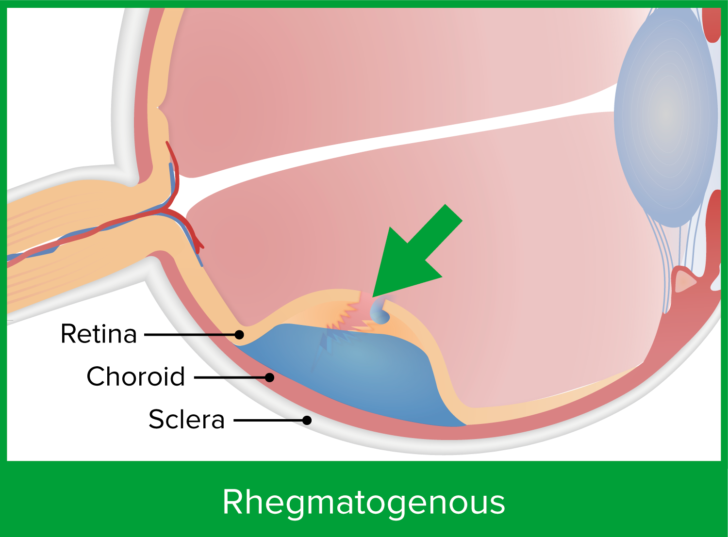
La imagen muestra un desgarro retiniano, lo que permite que entre líquido y se acumule en el espacio subretiniano. Esto predispone al paciente a un tipo regmatógeno de desprendimiento de retina.
Imagen por Lecturio.
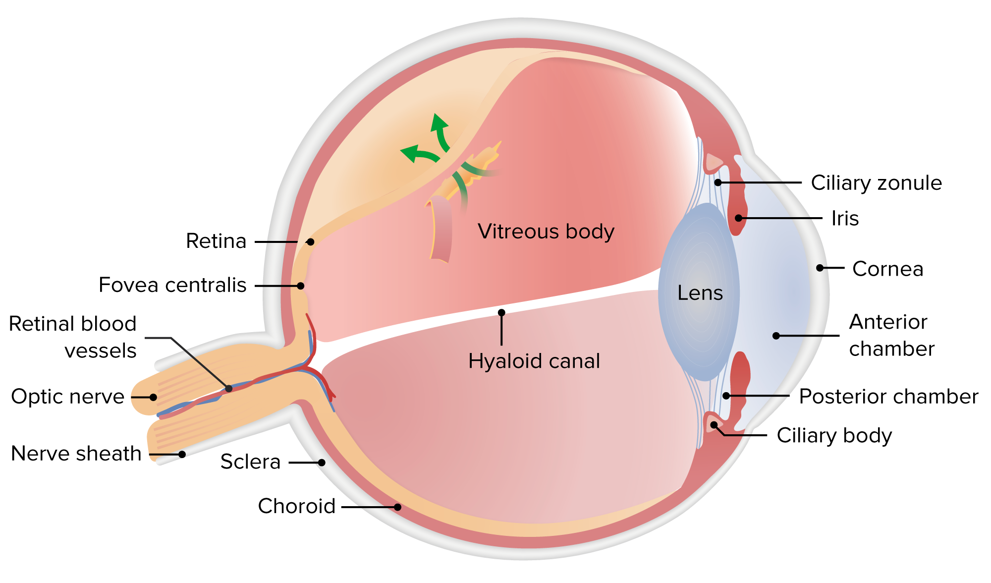
Un vítreo fuertemente adherido o una membrana proliferativa tira de la retina, provocando un desprendimiento por tracción.
Imagen por Lecturio.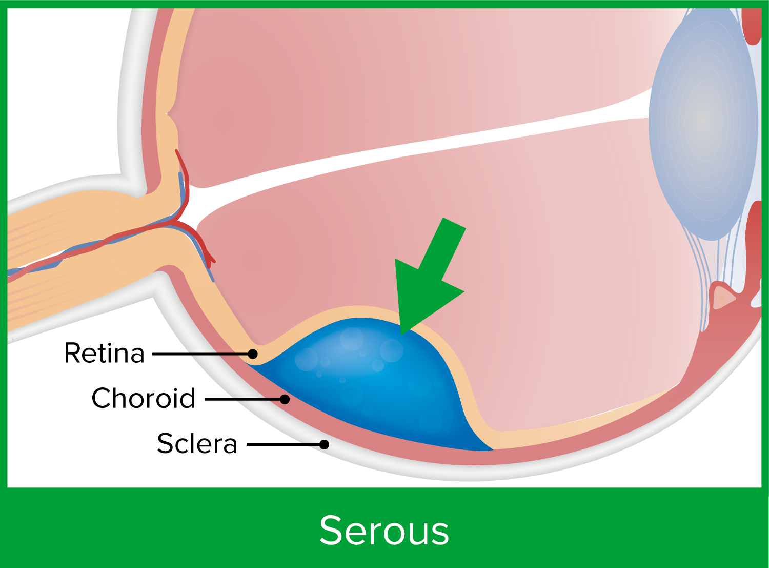
El desprendimiento de retina exudativo o seroso es el resultado de una alteración del flujo de salida de líquido o un aumento de la permeabilidad vascular debido a una afección o enfermedad subyacente.
Imagen por Lecturio.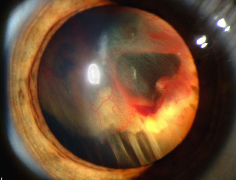
Fotografía con lámpara de hendidura que muestra un desprendimiento de retina con hemorragia vítrea
Imagen : “Slit lamp photograph showing retinal detachment” por National Eye Institute/National Institutes of Health. Licencia: Dominio Público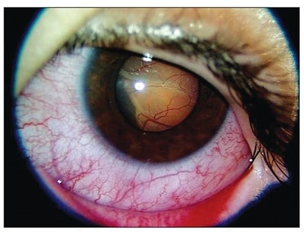
Fotografía con lámpara de hendidura del ojo izquierdo que muestra un desprendimiento de retina exudativo en el área nasal inferior. El desprendimiento de retina es secundario a la leucemia linfoblástica aguda.
Imagen : “Slit-lamp photograph of the left eye at presentation” por Diskapi Children’s Hospital, Department of Pediatric Hematology, Ankara, Turkey. Licencia: CC BY 2.5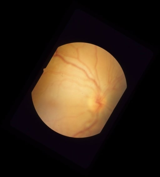
Fotografía en color del fondo de ojo que muestra un desprendimiento de retina regmatógeno mácular debido a un orificio retiniano necrótico de una lesión coroidea plana por debajo de la arcada inferotemporal
Imagen : “Pre-operative color fundus photograph of the right eye” por University of Southern California and Doheny Eye Institute, Los Angeles, California 90033, USA. Licencia: CC BY 2.0
Desprendimiento de retina ampolloso exudativo unilateral en la enfermedad de Behçet. Fundoscopia y angiografía con fluoresceína:
(a) Vaina perivascular extensa con infiltrados retinianos amarillentos dispersos y hemorragias en ambos ojos, con desprendimiento de retina bulloso exudativo que afecta los cuadrantes superior e inferior de la retina temporal en el ojo derecho (flechas blancas)
(b) Angiografía con fluoresceína que muestra tinción tardía de la vasculatura retiniana con fuga difusa de colorante en ambos ojos y acumulación de fluoresceína en el espacio subretiniano del ojo derecho (flechas negras)
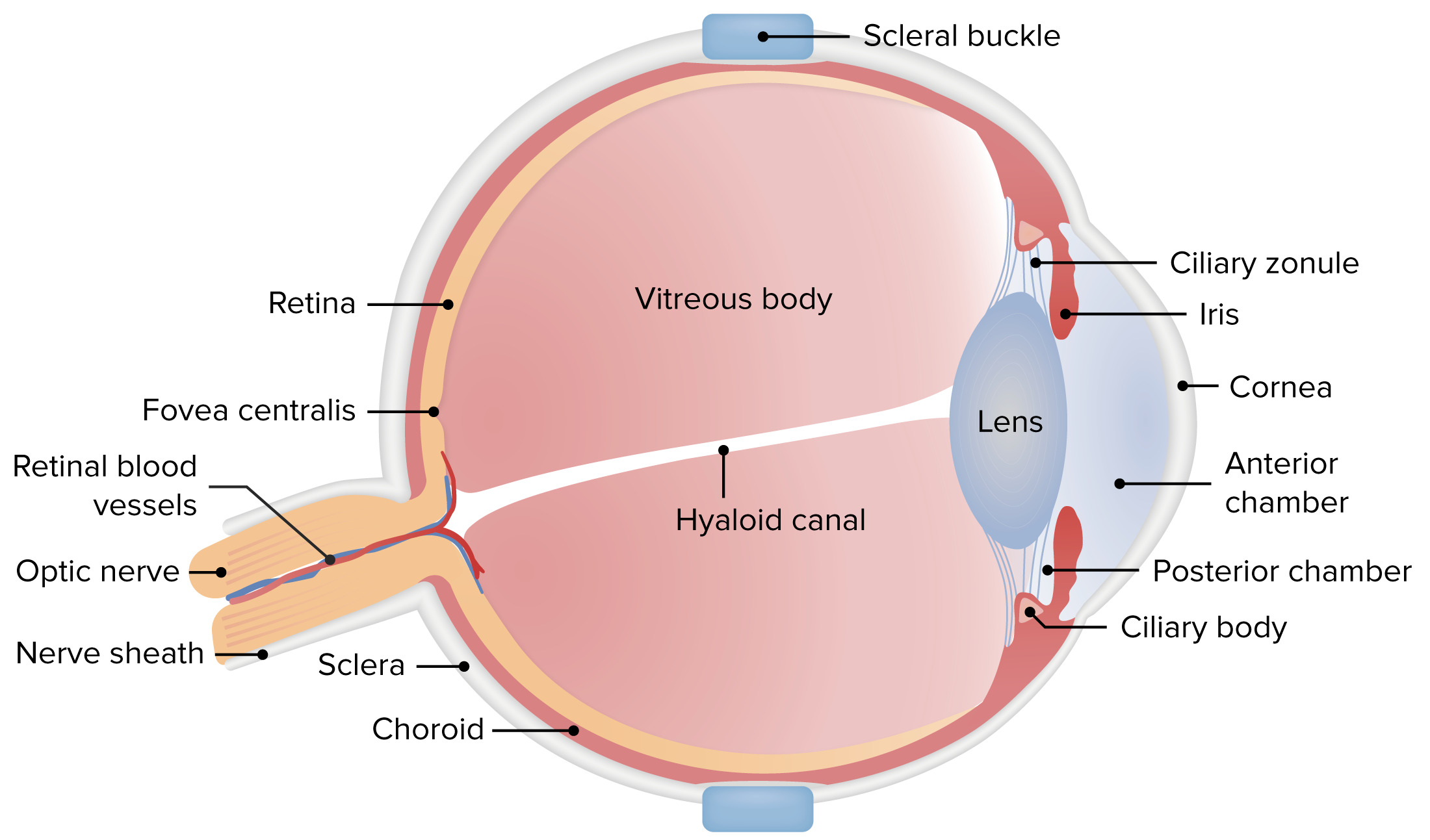
La imagen ilustra el pandeo escleral, la colocación de una silicona flexible suturada a la esclerótica. El pandeo escleral empuja la esclerótica hacia adentro, manteniendo así las estructuras separadas más cerca y permitiendo que la retina se vuelva a unir.
Imagen por Lecturio.