La blastomicosis es una infección causada por la inhalación de las esporas del hongo Blastomyces Blastomyces Blastomycosis is an infection caused by inhalation of the spores of the fungus, Blastomyces. Blastomyces species thrive in moist soil and decaying material and are common in the Ohio and Mississippi River valleys and the Great Lakes regions of the United States and Canada. Although most patients are asymptomatic, some can develop pneumonia. Blastomyces/Blastomycosis. Las especies de Blastomyces Blastomyces Blastomycosis is an infection caused by inhalation of the spores of the fungus, Blastomyces. Blastomyces species thrive in moist soil and decaying material and are common in the Ohio and Mississippi River valleys and the Great Lakes regions of the United States and Canada. Although most patients are asymptomatic, some can develop pneumonia. Blastomyces/Blastomycosis crecen en EN Erythema nodosum is an immune-mediated panniculitis (inflammation of the subcutaneous fat) caused by a type IV (delayed-type) hypersensitivity reaction. It commonly manifests in young women as tender, erythematous nodules on the shins. Erythema Nodosum suelos húmedos y en EN Erythema nodosum is an immune-mediated panniculitis (inflammation of the subcutaneous fat) caused by a type IV (delayed-type) hypersensitivity reaction. It commonly manifests in young women as tender, erythematous nodules on the shins. Erythema Nodosum material en EN Erythema nodosum is an immune-mediated panniculitis (inflammation of the subcutaneous fat) caused by a type IV (delayed-type) hypersensitivity reaction. It commonly manifests in young women as tender, erythematous nodules on the shins. Erythema Nodosum descomposición, además son comunes en EN Erythema nodosum is an immune-mediated panniculitis (inflammation of the subcutaneous fat) caused by a type IV (delayed-type) hypersensitivity reaction. It commonly manifests in young women as tender, erythematous nodules on the shins. Erythema Nodosum los LOS Neisseria valles de los LOS Neisseria ríos Ohio y Mississippi y en EN Erythema nodosum is an immune-mediated panniculitis (inflammation of the subcutaneous fat) caused by a type IV (delayed-type) hypersensitivity reaction. It commonly manifests in young women as tender, erythematous nodules on the shins. Erythema Nodosum las regiones de los LOS Neisseria Grandes Lagos de Estados Unidos y Canadá. Aunque la mayoría de los LOS Neisseria pacientes son asintomáticos, algunos pueden desarrollar neumonía. Pueden producirse enfermedades extrapulmonares, como lesiones cutáneas, osteomielitis, infecciones genitourinarias y meningitis Meningitis Meningitis is inflammation of the meninges, the protective membranes of the brain, and spinal cord. The causes of meningitis are varied, with the most common being bacterial or viral infection. The classic presentation of meningitis is a triad of fever, altered mental status, and nuchal rigidity. Meningitis. El diagnóstico se realiza identificando el organismo en EN Erythema nodosum is an immune-mediated panniculitis (inflammation of the subcutaneous fat) caused by a type IV (delayed-type) hypersensitivity reaction. It commonly manifests in young women as tender, erythematous nodules on the shins. Erythema Nodosum muestras de esputo o de tejido mediante cultivo, PCR PCR Polymerase chain reaction (PCR) is a technique that amplifies DNA fragments exponentially for analysis. The process is highly specific, allowing for the targeting of specific genomic sequences, even with minuscule sample amounts. The PCR cycles multiple times through 3 phases: denaturation of the template DNA, annealing of a specific primer to the individual DNA strands, and synthesis/elongation of new DNA molecules. Polymerase Chain Reaction (PCR) o pruebas de antígenos. Para el tratamiento se utilizan antifúngicos.
Last updated: Dec 15, 2025
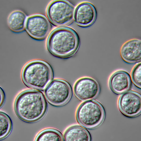
Blastomyces dermatitidis en forma de levadura a 37°C
Imagen: “Blastomyces dermatitidis yeast form” por Medmyco. Licencia: CC0 1.0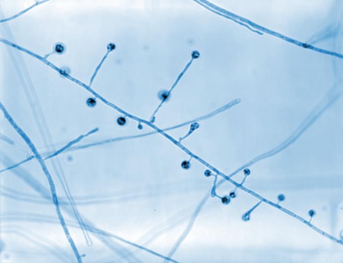
Microfotografía que muestra la morfología ultraestructural exhibida por Blastomyces dermatitidis:
En esta vista, hay un número de conidióforos. Obsérvese cómo cada conidióforo brota directamente de las hifas filamentosas en disposición perpendicular y que cada uno está coronado por un conidio esférico.
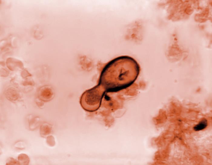
Microfotografía de una célula fúngica de Blastomyces dermatitidis:
La forma de levadura del organismo se somete a un método de reproducción asexual conocido como gemación, en el que el contenido de su pared celular y su contenido interno son extruidos, produciendo así una nueva célula.
La blastomicosis es causada por Blastomyces dermatitidis Blastomyces dermatitidis Blastomyces/Blastomycosis.
Los LOS Neisseria conidios de B. dermatitidis pueden aerosolizarse al AL Amyloidosis perturbar la colonia fúngica y luego son inhalados.
La blastomicosis es más común en EN Erythema nodosum is an immune-mediated panniculitis (inflammation of the subcutaneous fat) caused by a type IV (delayed-type) hypersensitivity reaction. It commonly manifests in young women as tender, erythematous nodules on the shins. Erythema Nodosum individuos inmunocomprometidos, pero también puede ocurrir en EN Erythema nodosum is an immune-mediated panniculitis (inflammation of the subcutaneous fat) caused by a type IV (delayed-type) hypersensitivity reaction. It commonly manifests in young women as tender, erythematous nodules on the shins. Erythema Nodosum individuos inmunocompetentes.
La gravedad de la infección depende del sistema inmunitario de la persona.
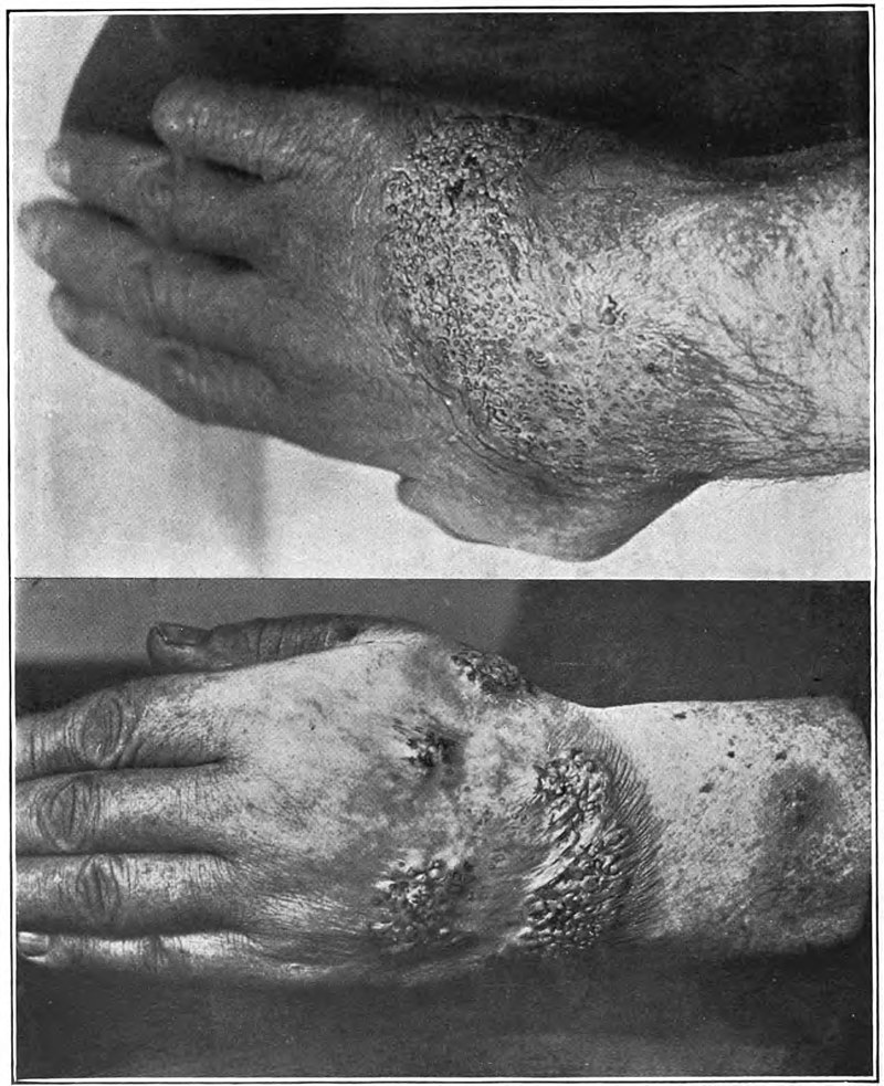
Lesiones verrugosas en las manos de un paciente con blastomicosis
Imagen: “Blastomycosis” por Norman Purvis Walker. Licencia: Dominio Público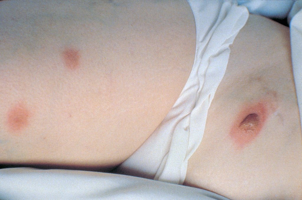
Lesiones cutáneas nodulares por blastomicosis
Imagen: “Nodular skin lesions of blastomycosis” por Centers for Disease Control and Prevention. Licencia: Dominio PúblicoPuede producirse una remisión espontánea, pero debe indicarse a los LOS Neisseria pacientes una terapia antifúngica para reducir las posibilidades de diseminación o recurrencia.