Os hemangioblastomasHemangioblastomasA benign tumor of the nervous system that may occur sporadically or in association with von Hippel-Lindau disease. It accounts for approximately 2% of intracranial tumors, arising most frequently in the cerebellar hemispheres and vermis. Histologically, the tumors are composed of multiple capillary and sinusoidal channels lined with endothelial cells and clusters of lipid-laden pseudoxanthoma cells. Usually solitary, these tumors can be multiple and may also occur in the brain stem, spinal cord, retina, and supratentorial compartment. Cerebellar hemangioblastomas usually present in the third decade with intracranial hypertension, and ataxia.Von Hippel-Lindau Disease são neoplasias vasculares do SNC. Estes são raros e frequentemente associados à doença de von Hippel-Lindau (VHL). A apresentação maisMAISAndrogen Insensitivity Syndrome comum é a cefaleia e, dependendo do tamanho e localização do tumorTumorInflammation, os doentes podem apresentar défices sensitivos e fraqueza motora. A imagem é o principal método de rastreio, sendo necessária uma avaliação histopatológica para diagnóstico definitivo. A cirurgia está, frequentemente, indicada no tratamento de hemangioblastomasHemangioblastomasA benign tumor of the nervous system that may occur sporadically or in association with von Hippel-Lindau disease. It accounts for approximately 2% of intracranial tumors, arising most frequently in the cerebellar hemispheres and vermis. Histologically, the tumors are composed of multiple capillary and sinusoidal channels lined with endothelial cells and clusters of lipid-laden pseudoxanthoma cells. Usually solitary, these tumors can be multiple and may also occur in the brain stem, spinal cord, retina, and supratentorial compartment. Cerebellar hemangioblastomas usually present in the third decade with intracranial hypertension, and ataxia.Von Hippel-Lindau Disease, embora, dependendo do tamanho, número e localização dos tumores, a radioterapia também possa ser necessária. O prognóstico é, geralmente, bom nos hemangioblastomasHemangioblastomasA benign tumor of the nervous system that may occur sporadically or in association with von Hippel-Lindau disease. It accounts for approximately 2% of intracranial tumors, arising most frequently in the cerebellar hemispheres and vermis. Histologically, the tumors are composed of multiple capillary and sinusoidal channels lined with endothelial cells and clusters of lipid-laden pseudoxanthoma cells. Usually solitary, these tumors can be multiple and may also occur in the brain stem, spinal cord, retina, and supratentorial compartment. Cerebellar hemangioblastomas usually present in the third decade with intracranial hypertension, and ataxia.Von Hippel-Lindau Disease solitários, mas os tumores associados à VHL estão, frequentemente, associados a um pior prognóstico e a um maior risco de recorrência.
Os hemangioblastomasHemangioblastomasA benign tumor of the nervous system that may occur sporadically or in association with von Hippel-Lindau disease. It accounts for approximately 2% of intracranial tumors, arising most frequently in the cerebellar hemispheres and vermis. Histologically, the tumors are composed of multiple capillary and sinusoidal channels lined with endothelial cells and clusters of lipid-laden pseudoxanthoma cells. Usually solitary, these tumors can be multiple and may also occur in the brain stem, spinal cord, retina, and supratentorial compartment. Cerebellar hemangioblastomas usually present in the third decade with intracranial hypertension, and ataxia.Von Hippel-Lindau Diseasesão neoplasias do SNC raras, benignas, de crescimento lento, altamente vasculares, que têm alta associação com a doença de von Hippel-Lindau (VHL). A doença de VHL é uma condição autossómica dominante caracterizada por uma variedade de tumores benignos e malignos.
Os tumores são encontrados com maior frequência no cerebelo.
Os hemangioblastomasHemangioblastomasA benign tumor of the nervous system that may occur sporadically or in association with von Hippel-Lindau disease. It accounts for approximately 2% of intracranial tumors, arising most frequently in the cerebellar hemispheres and vermis. Histologically, the tumors are composed of multiple capillary and sinusoidal channels lined with endothelial cells and clusters of lipid-laden pseudoxanthoma cells. Usually solitary, these tumors can be multiple and may also occur in the brain stem, spinal cord, retina, and supratentorial compartment. Cerebellar hemangioblastomas usually present in the third decade with intracranial hypertension, and ataxia.Von Hippel-Lindau Disease são encontrados frequentemente no parênquima, ligados ou junto à pia-mater, uma parte das meningesMeningesThe brain and the spinal cord are enveloped by 3 overlapping layers of connective tissue called the meninges. The layers are, from the most external layer to the most internal layer, the dura mater, arachnoid mater, and pia mater. Between these layers are 3 potential spaces called the epidural, subdural, and subarachnoid spaces. Meninges: Anatomy (composta por 3 camadas fibrosas) que encerram o SNC:
Dura-máter: camada externa, forma uma bainha de colagénio resistente
Aracnoide-máter: camada intermédia, composta por epitélio escamoso simples com uma rede laxa de colagénio e fibras elásticas
Pia-máter: camada interna fina em contacto com o cérebro e com a medula espinhal
Tabela: Classificação dos tumores do sistema nervoso
Categorias
Tumores específicos
Tumores neuroepiteliais no SNC
Astrocitomas, incluindo o glioblastoma multiformeGlioblastoma multiformeGlioblastoma multiforme is a high-grade astrocytoma, an aggressive brain tumor arising from astrocytes, with an unknown cause and a poorly understood link to risk factors. There are two main types: primary, a more aggressive form seen more commonly in older patients, and secondary, developing from lower-grade astrocytomas and seen more commonly in younger patients.Glioblastoma Multiforme
OligodendrogliomaOligodendrogliomaOligodendrogliomas are malignant CNS tumors arising from neural glial cell precursors. Oligodendrogliomas often arise in the frontal lobes of the brain and have a generally favorable prognosis when compared to other gliomas. Oligodendrogliomas are the 3rd most common CNS tumor. The most frequent presenting symptom is a seizure.Oligodendroglioma
Ependimoma e tumores do plexo coroide
Meduloblastomas (tumores embrionários)
Tumores meníngeos
Meningiomas
HemangioblastomasHemangioblastomasA benign tumor of the nervous system that may occur sporadically or in association with von Hippel-Lindau disease. It accounts for approximately 2% of intracranial tumors, arising most frequently in the cerebellar hemispheres and vermis. Histologically, the tumors are composed of multiple capillary and sinusoidal channels lined with endothelial cells and clusters of lipid-laden pseudoxanthoma cells. Usually solitary, these tumors can be multiple and may also occur in the brain stem, spinal cord, retina, and supratentorial compartment. Cerebellar hemangioblastomas usually present in the third decade with intracranial hypertension, and ataxia.Von Hippel-Lindau Disease
MelanomaMelanomaMelanoma is a malignant tumor arising from melanocytes, the melanin-producing cells of the epidermis. These tumors are most common in fair-skinned individuals with a history of excessive sun exposure and sunburns. Melanoma
Tumores periféricos
Schwannomas, incluindo neurinomas acústicos
NeuroblastomaNeuroblastomaNeuroblastoma is a malignancy that arises from the neural crest cell derivatives along the sympathetic chain (neuroblasts) and is most commonly located in the adrenal medulla. The tumor often presents in childhood with a flank mass that crosses the midline.Neuroblastoma
Epidemiologia
Esporádico: 75%
Associado à VHL: 25%
Incidência:
Extremamente raro
2,5% de todas as neoplasias intracranianas
5% dos tumores da medula espinhal
Sexo: A incidência é maior em homens do que em mulheres (2:1).
Idade:
As crianças raramente são afetadas.
A idade de pico de incidência é dos 20 aos 50 anos.
Etiologia
A causa exata do hemangioblastomaHemangioblastomaHemangioblastomas are vascular neoplasms of the CNS. Hemangioblastomas are rare and are often associated with von Hippel-Lindau disease (VHL). The most common presentation is a headache and, depending on the size and location of the tumor, patients may present with sensory deficits and motor weakness.Hemangioblastoma não é conhecida.
Considera-se que a origem etiológica seja uma mutação no geneGeneA category of nucleic acid sequences that function as units of heredity and which code for the basic instructions for the development, reproduction, and maintenance of organisms.Basic Terms of GeneticsVHL devido à elevada associação ao VHL.
Os hemangioblastomasHemangioblastomasA benign tumor of the nervous system that may occur sporadically or in association with von Hippel-Lindau disease. It accounts for approximately 2% of intracranial tumors, arising most frequently in the cerebellar hemispheres and vermis. Histologically, the tumors are composed of multiple capillary and sinusoidal channels lined with endothelial cells and clusters of lipid-laden pseudoxanthoma cells. Usually solitary, these tumors can be multiple and may also occur in the brain stem, spinal cord, retina, and supratentorial compartment. Cerebellar hemangioblastomas usually present in the third decade with intracranial hypertension, and ataxia.Von Hippel-Lindau Disease crescem ligados à pia-máter (camada meníngea maisMAISAndrogen Insensitivity Syndrome interna)
Parênquima do cerebelo
Tronco cerebral
Medula espinhal
Patogénese
Embora os hemangiomas esporádicos não tenham uma patogénese clara, acredita-se que os hemangiomas associados a VHL sejam causados por uma mutação no geneGeneA category of nucleic acid sequences that function as units of heredity and which code for the basic instructions for the development, reproduction, and maintenance of organisms.Basic Terms of GeneticsVHL. Até 50% dos hemangioblastomasHemangioblastomasA benign tumor of the nervous system that may occur sporadically or in association with von Hippel-Lindau disease. It accounts for approximately 2% of intracranial tumors, arising most frequently in the cerebellar hemispheres and vermis. Histologically, the tumors are composed of multiple capillary and sinusoidal channels lined with endothelial cells and clusters of lipid-laden pseudoxanthoma cells. Usually solitary, these tumors can be multiple and may also occur in the brain stem, spinal cord, retina, and supratentorial compartment. Cerebellar hemangioblastomas usually present in the third decade with intracranial hypertension, and ataxia.Von Hippel-Lindau Disease esporádicos apresentam, também, mutações ou deleções no geneGeneA category of nucleic acid sequences that function as units of heredity and which code for the basic instructions for the development, reproduction, and maintenance of organisms.Basic Terms of GeneticsVHL.
GeneGeneA category of nucleic acid sequences that function as units of heredity and which code for the basic instructions for the development, reproduction, and maintenance of organisms.Basic Terms of GeneticsVHL:
GeneGeneA category of nucleic acid sequences that function as units of heredity and which code for the basic instructions for the development, reproduction, and maintenance of organisms.Basic Terms of Genetics supressor tumoral encontrado no cromossoma 3p
Responsável pela inibição do fator-2α induzido por hipóxia (HIF-2α, pela sigla em inglês), por degradação proteassómica mediada por ubiquitina
HIF-2α:
Parte de um grande complexo proteico do fator de transcrição denominado HIFHIFHypoxia-inducible factor 1, alpha subunit is a basic helix-loop-helix transcription factor that is regulated by oxygen availability and is targeted for degradation by VHL tumor suppressor protein.Von Hippel-Lindau Disease, que está envolvido na regulação da capacidade do corpo em se adaptar às mudanças nos níveis de oxigénio
O HIFHIFHypoxia-inducible factor 1, alpha subunit is a basic helix-loop-helix transcription factor that is regulated by oxygen availability and is targeted for degradation by VHL tumor suppressor protein.Von Hippel-Lindau Disease pode induzir a expressão de maisMAISAndrogen Insensitivity Syndrome de 70 genes-alvo, incluindo:
Fator de crescimento endotelial vascular (VEGF, pela sigla em inglês)
Fator de crescimento derivado de plaquetas (PDGF, pela sigla em inglês)
Eritropoietina
Fator de crescimento transformador α (TGF-α, pela sigla em inglês)
Quando está disponível a concentração de oxigénio adequada, a proteína VHL ajuda a suprimir o HIFHIFHypoxia-inducible factor 1, alpha subunit is a basic helix-loop-helix transcription factor that is regulated by oxygen availability and is targeted for degradation by VHL tumor suppressor protein.Von Hippel-Lindau Disease.
Um geneGeneA category of nucleic acid sequences that function as units of heredity and which code for the basic instructions for the development, reproduction, and maintenance of organisms.Basic Terms of GeneticsVHL disfuncional (causado por mutação ou deleção) pode levar à acumulação de HIF-α devido à incapacidade de degradar o HIF-2α.
A estabilização do HIF-2α permite que ele induza a expressão dos seus genesGenesA category of nucleic acid sequences that function as units of heredity and which code for the basic instructions for the development, reproduction, and maintenance of organisms.DNA Types and Structure alvo → ↑ fatores angiogénicos → crescimento tumoral
Desenvolvimento de sintomas
O desenvolvimento de sintomas é causado por:
Compressão direta do tumorTumorInflammation nas estruturas neuronais
AtaxiaAtaxiaImpairment of the ability to perform smoothly coordinated voluntary movements. This condition may affect the limbs, trunk, eyes, pharynx, larynx, and other structures. Ataxia may result from impaired sensory or motor function. Sensory ataxia may result from posterior column injury or peripheral nerve diseases. Motor ataxia may be associated with cerebellar diseases; cerebral cortex diseases; thalamic diseases; basal ganglia diseases; injury to the red nucleus; and other conditions.Ataxia-telangiectasia cerebelar e descoordenação
Aumento da pressão intracraniana (PIC) devido a hidrocefalia obstrutiva
Disfunção do nervo oculomotor
Fraqueza motora
Défices sensitivos
HemangioblastomasHemangioblastomasA benign tumor of the nervous system that may occur sporadically or in association with von Hippel-Lindau disease. It accounts for approximately 2% of intracranial tumors, arising most frequently in the cerebellar hemispheres and vermis. Histologically, the tumors are composed of multiple capillary and sinusoidal channels lined with endothelial cells and clusters of lipid-laden pseudoxanthoma cells. Usually solitary, these tumors can be multiple and may also occur in the brain stem, spinal cord, retina, and supratentorial compartment. Cerebellar hemangioblastomas usually present in the third decade with intracranial hypertension, and ataxia.Von Hippel-Lindau Disease esporádicos versus associados a VHL
Tabela: Apresentações clínicas típicas de hemangioblastomasHemangioblastomasA benign tumor of the nervous system that may occur sporadically or in association with von Hippel-Lindau disease. It accounts for approximately 2% of intracranial tumors, arising most frequently in the cerebellar hemispheres and vermis. Histologically, the tumors are composed of multiple capillary and sinusoidal channels lined with endothelial cells and clusters of lipid-laden pseudoxanthoma cells. Usually solitary, these tumors can be multiple and may also occur in the brain stem, spinal cord, retina, and supratentorial compartment. Cerebellar hemangioblastomas usually present in the third decade with intracranial hypertension, and ataxia.Von Hippel-Lindau Disease esporádicos versus hemangioblastomasHemangioblastomasA benign tumor of the nervous system that may occur sporadically or in association with von Hippel-Lindau disease. It accounts for approximately 2% of intracranial tumors, arising most frequently in the cerebellar hemispheres and vermis. Histologically, the tumors are composed of multiple capillary and sinusoidal channels lined with endothelial cells and clusters of lipid-laden pseudoxanthoma cells. Usually solitary, these tumors can be multiple and may also occur in the brain stem, spinal cord, retina, and supratentorial compartment. Cerebellar hemangioblastomas usually present in the third decade with intracranial hypertension, and ataxia.Von Hippel-Lindau Disease associados a VHL
HemangioblastomaHemangioblastomaHemangioblastomas are vascular neoplasms of the CNS. Hemangioblastomas are rare and are often associated with von Hippel-Lindau disease (VHL). The most common presentation is a headache and, depending on the size and location of the tumor, patients may present with sensory deficits and motor weakness.Hemangioblastoma esporádico
HemangioblastomaHemangioblastomaHemangioblastomas are vascular neoplasms of the CNS. Hemangioblastomas are rare and are often associated with von Hippel-Lindau disease (VHL). The most common presentation is a headache and, depending on the size and location of the tumor, patients may present with sensory deficits and motor weakness.Hemangioblastoma associado a VHL
Frequentemente apresenta-se como um tumorTumorInflammation isolado no cerebelo
50% dos tumores estão localizados na medula espinhal, 40% no cerebelo e 10% no tronco cerebral.
Complicações
As complicações geralmente ocorrem devido ao aumento do tamanho do tumorTumorInflammation (> 1,5 cm), por causar compressão ou hemorragia espontânea:
Sintomas de aumento da PIC/hidrocefalia obstrutiva rápida
Alteração do estado de consciência
Policitemia devido à produção ectópica de eritropoietina (síndrome paraneoplásica)
Tetraplegia
Diagnóstico
Embora a principal ferramenta diagnóstica seja a imagem, o exame diagnóstico de eleição requer uma análise histopatológica de uma amostra obtida por biópsia.
Imagiologia
Todo o eixo neuronal deve ser visualizado para descartar múltiplas lesões, que são comuns em casos de VHL.
RMN com contraste:
A RMN com gadolínio é o método de imagem maisMAISAndrogen Insensitivity Syndrome sensível para diagnosticar o hemangioblastomaHemangioblastomaHemangioblastomas are vascular neoplasms of the CNS. Hemangioblastomas are rare and are often associated with von Hippel-Lindau disease (VHL). The most common presentation is a headache and, depending on the size and location of the tumor, patients may present with sensory deficits and motor weakness.Hemangioblastoma.
Características típicas:
Nódulo, com captação de contraste, associado a um quisto (60% dos tumores)
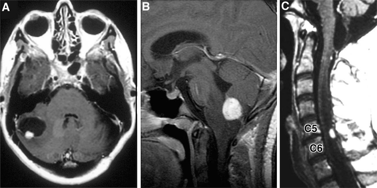
Imagens radiográficas de hemangioblastomas: A: RMN axial com contraste, ponderada em T1, a demonstrar um hemangioblastoma cerebelar com um nódulo mural captante e um quisto peritumoral B: RMN sagital com contraste, ponderada em T1, a revelar um hemangioblastoma medular captante, com edema vasogénico circundante C: RMN sagital com contraste, ponderada em T1, a demonstrar um hemangioblastoma posterior/dorsal captante, com siringe associada
Imagem: “Hemangioblastoma” por Department of Neurological Surgery, Ohio State University Wexner Medical Center , Columbus, OH , USA. Licença: CC BY 4.0
Exame oftalmológico
Os hemangioblastomasHemangioblastomasA benign tumor of the nervous system that may occur sporadically or in association with von Hippel-Lindau disease. It accounts for approximately 2% of intracranial tumors, arising most frequently in the cerebellar hemispheres and vermis. Histologically, the tumors are composed of multiple capillary and sinusoidal channels lined with endothelial cells and clusters of lipid-laden pseudoxanthoma cells. Usually solitary, these tumors can be multiple and may also occur in the brain stem, spinal cord, retina, and supratentorial compartment. Cerebellar hemangioblastomas usually present in the third decade with intracranial hypertension, and ataxia.Von Hippel-Lindau Disease têm uma alta associação com VHL, que está associada aos hemangioblastomasHemangioblastomasA benign tumor of the nervous system that may occur sporadically or in association with von Hippel-Lindau disease. It accounts for approximately 2% of intracranial tumors, arising most frequently in the cerebellar hemispheres and vermis. Histologically, the tumors are composed of multiple capillary and sinusoidal channels lined with endothelial cells and clusters of lipid-laden pseudoxanthoma cells. Usually solitary, these tumors can be multiple and may also occur in the brain stem, spinal cord, retina, and supratentorial compartment. Cerebellar hemangioblastomas usually present in the third decade with intracranial hypertension, and ataxia.Von Hippel-Lindau Disease da retinaRetinaThe ten-layered nervous tissue membrane of the eye. It is continuous with the optic nerve and receives images of external objects and transmits visual impulses to the brain. Its outer surface is in contact with the choroid and the inner surface with the vitreous body. The outermost layer is pigmented, whereas the inner nine layers are transparent.Eye: Anatomy.
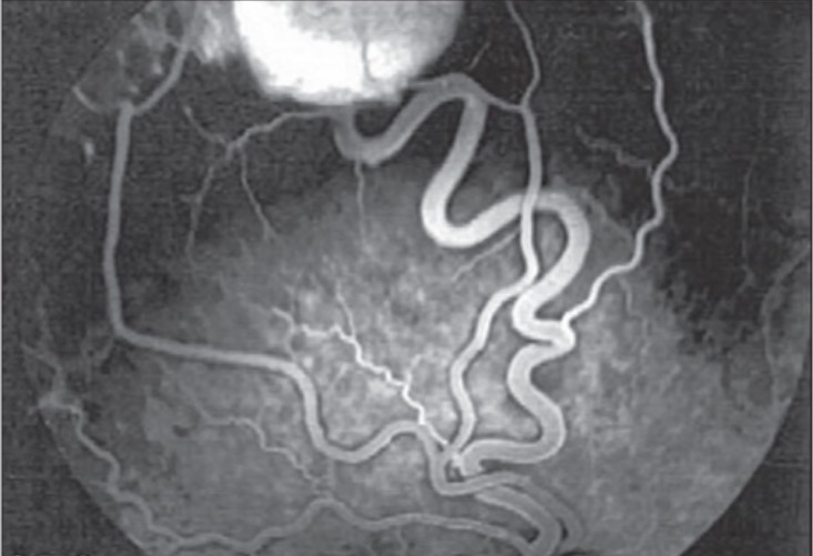
A angiografia retiniana pode ser realizada para descartar a presença de um hemangioblastomaHemangioblastomaHemangioblastomas are vascular neoplasms of the CNS. Hemangioblastomas are rare and are often associated with von Hippel-Lindau disease (VHL). The most common presentation is a headache and, depending on the size and location of the tumor, patients may present with sensory deficits and motor weakness.Hemangioblastoma retiniano.
Angiografia de fluoresceína do olho direito a demonstrar um hemangioblastoma temporal superior da retina com a artéria e veia de drenagem
Imagem: “Fluorescein angiogram of right eye” por Vitreoretinal Division, Department of Ophthalmology, Al-Hussein Hospital, King Hussein Medical Center, Amman, Jordan. Licença: CC BY 2.0
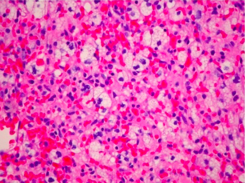
Histopatologia
Exame de eleição para diagnóstico
Exame macroscópico: nódulos vermelhos bem circunscritos e altamente vasculares
Exame microscópico:
Extensas redes vasculares com capilares estruturalmente normais
Células estromais neoplásicas:
Núcleos hipercromáticos e pleomórficos
Baixa taxa mitótica
Sem atipias
Fibras de Rosenthal
Podem surgir 2 componentes celulares distintos no mesmo tumorTumorInflammation:
Tipo 1: pequenas células endoteliais perivasculares com núcleos hipercromáticos e citoplasma esparso
Tipo 2: citoplasma vacuolado e rico em lípidos
Fotomicrografia de H&E de um hemangioblastoma do nervo óptico (localização incomum) a revelar um tumor marcadamente vascular com células estromais lipidizadas (×200)
Imagem: “Optic nerve hemangioblastoma” por Department of Surgery, Division of Neurosurgery, University of Alabama at Birmingham, Birmingham, AL 35294, USA. Licença: CC BY 3.0
Tratamento e Prognóstico
A resseção cirúrgica é a principal abordagem no tratamento de hemangioblastomasHemangioblastomasA benign tumor of the nervous system that may occur sporadically or in association with von Hippel-Lindau disease. It accounts for approximately 2% of intracranial tumors, arising most frequently in the cerebellar hemispheres and vermis. Histologically, the tumors are composed of multiple capillary and sinusoidal channels lined with endothelial cells and clusters of lipid-laden pseudoxanthoma cells. Usually solitary, these tumors can be multiple and may also occur in the brain stem, spinal cord, retina, and supratentorial compartment. Cerebellar hemangioblastomas usually present in the third decade with intracranial hypertension, and ataxia.Von Hippel-Lindau Disease. Terapias adjuvantes são frequentemente necessárias nos hemangioblastomasHemangioblastomasA benign tumor of the nervous system that may occur sporadically or in association with von Hippel-Lindau disease. It accounts for approximately 2% of intracranial tumors, arising most frequently in the cerebellar hemispheres and vermis. Histologically, the tumors are composed of multiple capillary and sinusoidal channels lined with endothelial cells and clusters of lipid-laden pseudoxanthoma cells. Usually solitary, these tumors can be multiple and may also occur in the brain stem, spinal cord, retina, and supratentorial compartment. Cerebellar hemangioblastomas usually present in the third decade with intracranial hypertension, and ataxia.Von Hippel-Lindau Disease associados a VHL, e as opções incluem radioterapia, embolização endovascular e terapia antiangiogénica.
Tratamento cirúrgico
A cirurgia é a principal abordagem definitiva para tratar os hemangioblastomasHemangioblastomasA benign tumor of the nervous system that may occur sporadically or in association with von Hippel-Lindau disease. It accounts for approximately 2% of intracranial tumors, arising most frequently in the cerebellar hemispheres and vermis. Histologically, the tumors are composed of multiple capillary and sinusoidal channels lined with endothelial cells and clusters of lipid-laden pseudoxanthoma cells. Usually solitary, these tumors can be multiple and may also occur in the brain stem, spinal cord, retina, and supratentorial compartment. Cerebellar hemangioblastomas usually present in the third decade with intracranial hypertension, and ataxia.Von Hippel-Lindau Disease, pois são tumores benignos e não invasivos.
Os tumores geralmente são bem demarcados das estruturas circundantes, mas são altamente vasculares e localizados em áreas neurologicamente sensíveis.
A angiografia pré-operatória é útil para identificar as artérias de alimentação.
Necessidade de terapia adjuvante:
Geralmente não é necessária em tumores únicos esporádicos
Normalmente necessária em tumores associados a VHL e/ou múltiplos
Outras modalidades terapêuticas
Radioterapia:
A radioterapia pós-operatória reduz a recorrência de hemangioblastomasHemangioblastomasA benign tumor of the nervous system that may occur sporadically or in association with von Hippel-Lindau disease. It accounts for approximately 2% of intracranial tumors, arising most frequently in the cerebellar hemispheres and vermis. Histologically, the tumors are composed of multiple capillary and sinusoidal channels lined with endothelial cells and clusters of lipid-laden pseudoxanthoma cells. Usually solitary, these tumors can be multiple and may also occur in the brain stem, spinal cord, retina, and supratentorial compartment. Cerebellar hemangioblastomas usually present in the third decade with intracranial hypertension, and ataxia.Von Hippel-Lindau Disease.
Indicações:
Lesões inacessíveis cirurgicamente
Lesões múltiplas
Embolização endovascular:
Objetivo: diminuir a vascularização do tumorTumorInflammation e reduzir as complicações intraoperatórias da hemorragia
Tumores grandes podem ser consideravelmente reduzidos para facilitar a resseção.
Mecanismo de ação: um anticorpo monoclonal liga-se e inibe os efeitos do VEGF → ↓ angiogénese
Usos: doentes com tumores VHL, que não respondem ou não são recetivos à cirurgia ou radiação
Inibidor HIF-2ɑ (belzutifan):
Para indivíduos com doença de VHL
Usado para atrasar a cirurgia naqueles cujos tumores apresentam um crescimento rápido
Também benéfico em indivíduos com tumores refratários ou recorrentes
Prognóstico
O prognóstico é bom, na maioria dos hemangioblastomasHemangioblastomasA benign tumor of the nervous system that may occur sporadically or in association with von Hippel-Lindau disease. It accounts for approximately 2% of intracranial tumors, arising most frequently in the cerebellar hemispheres and vermis. Histologically, the tumors are composed of multiple capillary and sinusoidal channels lined with endothelial cells and clusters of lipid-laden pseudoxanthoma cells. Usually solitary, these tumors can be multiple and may also occur in the brain stem, spinal cord, retina, and supratentorial compartment. Cerebellar hemangioblastomas usually present in the third decade with intracranial hypertension, and ataxia.Von Hippel-Lindau Disease tratados:
A resseção cirúrgica limpa (com margens cirúrgicas negativas), principalmente em tumores solitários esporádicos, garante um bom prognóstico.
A deteção e intervenção precoces são favoráveis.
HemangioblastomasHemangioblastomasA benign tumor of the nervous system that may occur sporadically or in association with von Hippel-Lindau disease. It accounts for approximately 2% of intracranial tumors, arising most frequently in the cerebellar hemispheres and vermis. Histologically, the tumors are composed of multiple capillary and sinusoidal channels lined with endothelial cells and clusters of lipid-laden pseudoxanthoma cells. Usually solitary, these tumors can be multiple and may also occur in the brain stem, spinal cord, retina, and supratentorial compartment. Cerebellar hemangioblastomas usually present in the third decade with intracranial hypertension, and ataxia.Von Hippel-Lindau Disease associados à VHL:
Maior probabilidade de recorrência do que tumores esporádicos
Pior prognóstico
Maior associação com défices neurológicos
Diagnóstico Diferencial
MeningiomaMeningiomaMeningiomas are slow-growing tumors that arise from the meninges of the brain and spinal cord. The vast majority are benign. These tumors commonly occur in individuals with a history of high doses of skull radiation, head trauma, and neurofibromatosis 2. Meningioma: tumores que surgem das meningesMeningesThe brain and the spinal cord are enveloped by 3 overlapping layers of connective tissue called the meninges. The layers are, from the most external layer to the most internal layer, the dura mater, arachnoid mater, and pia mater. Between these layers are 3 potential spaces called the epidural, subdural, and subarachnoid spaces. Meninges: Anatomy do cérebro e da medula espinhal. Os meningiomas são frequentemente assintomáticos, mas podem apresentar-se com cefaleia, convulsões e distúrbios visuais. O diagnóstico é realizado por RMN e biópsia. Os casos assintomáticos são normalmente mantidos em observação, enquanto que os doentes sintomáticos são tratados cirurgicamente ou com radiação. Ao contrário dos hemangioblastomasHemangioblastomasA benign tumor of the nervous system that may occur sporadically or in association with von Hippel-Lindau disease. It accounts for approximately 2% of intracranial tumors, arising most frequently in the cerebellar hemispheres and vermis. Histologically, the tumors are composed of multiple capillary and sinusoidal channels lined with endothelial cells and clusters of lipid-laden pseudoxanthoma cells. Usually solitary, these tumors can be multiple and may also occur in the brain stem, spinal cord, retina, and supratentorial compartment. Cerebellar hemangioblastomas usually present in the third decade with intracranial hypertension, and ataxia.Von Hippel-Lindau Disease, os meningiomas estão sempre próximos às meningesMeningesThe brain and the spinal cord are enveloped by 3 overlapping layers of connective tissue called the meninges. The layers are, from the most external layer to the most internal layer, the dura mater, arachnoid mater, and pia mater. Between these layers are 3 potential spaces called the epidural, subdural, and subarachnoid spaces. Meninges: Anatomy e geralmente apresentam achados na imagem de fixação dural (e.g., sinal da cauda dural).
Glioblastoma multiformeGlioblastoma multiformeGlioblastoma multiforme is a high-grade astrocytoma, an aggressive brain tumor arising from astrocytes, with an unknown cause and a poorly understood link to risk factors. There are two main types: primary, a more aggressive form seen more commonly in older patients, and secondary, developing from lower-grade astrocytomas and seen more commonly in younger patients.Glioblastoma Multiforme: astrocitoma de grau IV segundo a classificação da OMS, rapidamente progressivo que surge de astrócitos (células gliais no cérebro) e apresenta-se clinicamente como dor de cabeça, náuseas, sonolência, visão turva, alterações de personalidade e convulsões. A imagem, a apresentação clínica e a biópsia são os pilares diagnósticos. O tratamento inclui radioterapia, quimioterapia e excisão cirúrgica. O prognóstico é mau, mesmo com o tratamento. Ao contrário do hemangioblastomaHemangioblastomaHemangioblastomas are vascular neoplasms of the CNS. Hemangioblastomas are rare and are often associated with von Hippel-Lindau disease (VHL). The most common presentation is a headache and, depending on the size and location of the tumor, patients may present with sensory deficits and motor weakness.Hemangioblastoma, o glioblastoma multiformeGlioblastoma multiformeGlioblastoma multiforme is a high-grade astrocytoma, an aggressive brain tumor arising from astrocytes, with an unknown cause and a poorly understood link to risk factors. There are two main types: primary, a more aggressive form seen more commonly in older patients, and secondary, developing from lower-grade astrocytomas and seen more commonly in younger patients.Glioblastoma Multiforme não está associado à VHL.
OligodendrogliomaOligodendrogliomaOligodendrogliomas are malignant CNS tumors arising from neural glial cell precursors. Oligodendrogliomas often arise in the frontal lobes of the brain and have a generally favorable prognosis when compared to other gliomas. Oligodendrogliomas are the 3rd most common CNS tumor. The most frequent presenting symptom is a seizure.Oligodendroglioma:tumorTumorInflammation do SNC que se origina a partir de oligodendrócitos. O oligodendrogliomaOligodendrogliomaOligodendrogliomas are malignant CNS tumors arising from neural glial cell precursors. Oligodendrogliomas often arise in the frontal lobes of the brain and have a generally favorable prognosis when compared to other gliomas. Oligodendrogliomas are the 3rd most common CNS tumor. The most frequent presenting symptom is a seizure.Oligodendroglioma pode apresentar-se com défices neurológicos focais, convulsões e alterações de personalidade, dependendo da localização exata. O diagnóstico é realizado por RMN e biópsia. Os oligodendrogliomas desenvolvem-se frequentemente nos hemisférios cerebrais, normalmente no lobo frontalFrontalThe bone that forms the frontal aspect of the skull. Its flat part forms the forehead, articulating inferiorly with the nasal bone and the cheek bone on each side of the face.Skull: Anatomy, e raramente são encontrados na região infratentorial ou na medula espinhal. O tratamento envolve resseção cirúrgica, possivelmente acompanhada de radioterapia e/ou quimioterapia.
Aneurisma cerebral: fraqueza na parede de um vaso sanguíneo que abastece o cérebro, que dilata e corre o risco de romper. Um aneurisma não roto geralmente é assintomático e a sua rutura causa cefaleia súbita e intensa. Os aneurismas cerebrais são diagnosticados com TC, angiografia ou ultrassonografia. O tratamento depende da localização e é tipicamente tratado com endoprótese endovascular.
Dengler, V. L., Galbraith, M., Espinosa, J. M. (2014). Transcriptional regulation by hypoxia inducible factors. Critical reviews in biochemistry and molecular biology, 49(1):1–15. Retrieved June 1, 2021, from https://www.ncbi.nlm.nih.gov/pmc/articles/PMC4342852/
Crie sua conta gratuita ou faça login para continuar lendo!
A Lecturio Medical complementa o teu estudo através de métodos de ensino baseados em evidência, vídeos de palestras, perguntas e muito mais – tudo combinado num só lugar e fácil de usar.
User Reviews
Let's celebrate you becoming a doctor! 🏥 Save 30% now!
Lecturio Premium dá-lhe acesso total a todos os conteúdos e características
Obtenha Premium para ver todos os vídeos
Verifique agora o seu e-mail para obter um teste gratuito.
Crie uma conta gratuita para testar os seus conhecimentos
Lecturio Premium dá-lhe acesso total a todos os conteúdos e características - incluindo o Qbank de Lecturio com perguntas actualizadas ao estilo do board-.