La trombosis venosa cerebral (TVC) es un coágulo sanguíneo en EN Erythema nodosum is an immune-mediated panniculitis (inflammation of the subcutaneous fat) caused by a type IV (delayed-type) hypersensitivity reaction. It commonly manifests in young women as tender, erythematous nodules on the shins. Erythema Nodosum las venas cerebrales o los LOS Neisseria senos durales que afecta predominantemente a adultos jóvenes. Entre los LOS Neisseria factores de riesgo figuran estados hipercoagulables, infecciones, traumatismos encefalocraneanos, afecciones inflamatorias y medicamentos. La TVC puede tener presentaciones clínicas variadas. Los LOS Neisseria síntomas incluyen cefalea, déficits neurológicos focales, convulsiones y signos generales de aumento de la PIC. Los LOS Neisseria pacientes con esta sospecha diagnóstica deben someterse a una neuroimagen urgente con venografía cerebral por resonancia magnética. La anticoagulación es el pilar del tratamiento. La TVC puede ser mortal durante la fase aguda; sin embargo, el pronóstico general a largo plazo es favorable en EN Erythema nodosum is an immune-mediated panniculitis (inflammation of the subcutaneous fat) caused by a type IV (delayed-type) hypersensitivity reaction. It commonly manifests in young women as tender, erythematous nodules on the shins. Erythema Nodosum los LOS Neisseria pacientes que sobreviven.
Last updated: Dec 15, 2025
La trombosis venosa cerebral (TVC) es un coágulo de sangre en EN Erythema nodosum is an immune-mediated panniculitis (inflammation of the subcutaneous fat) caused by a type IV (delayed-type) hypersensitivity reaction. It commonly manifests in young women as tender, erythematous nodules on the shins. Erythema Nodosum las venas cerebrales o los LOS Neisseria senos durales.

Esta vista sagital a través del cráneo ilustra el sistema de drenaje venoso. Las flechas muestran el flujo de sangre desde las venas y los senos cerebrales hasta la confluencia de los senos y su retorno final a la vena yugular a través del seno sigmoideo.
Imagen: “Blood drains from the brain through a series of sinuses that connect to the jugular veins” por OpenStax College, editado por Lecturio. Licencia: CC BY 4.0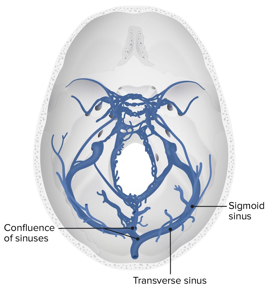
Vista transversal del sistema venoso profundo cerebral, concretamente de los senos sigmoideos y transversos.
Imagen de Lecturio.Las presentaciones clínicas pueden variar significativamente. Algunos casos pueden presentarse con signos aislados de PIC, mientras que otros presentan síntomas isquémicos. El inicio de los LOS Neisseria síntomas también puede diferir (agudo, subagudo, crónico).
Los LOS Neisseria estudios de imagen urgentes están indicados para quienes presenten signos preocupantes de TVC. Las pruebas de laboratorio no desempeñan ningún papel en EN Erythema nodosum is an immune-mediated panniculitis (inflammation of the subcutaneous fat) caused by a type IV (delayed-type) hypersensitivity reaction. It commonly manifests in young women as tender, erythematous nodules on the shins. Erythema Nodosum el diagnóstico de la TVC, pero pueden ayudar a identificar la causa subyacente.
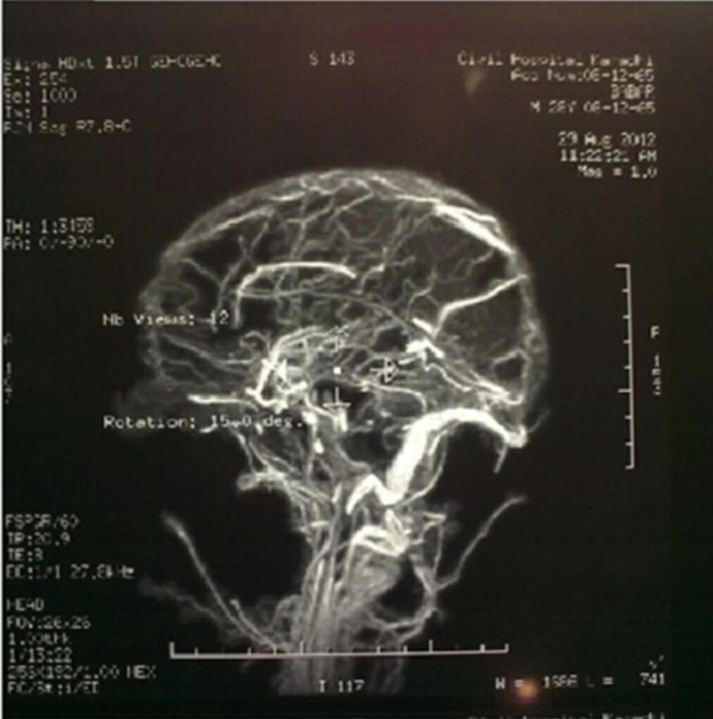
Esta venografía por RM del cerebro revela una trombosis venosa cerebral extensa.
Imagen: “Magnetic Resonance Venography” por el Departamento de Medicina, Dow University of Medical Sciences, Karachi, Pakistán. Licencia: CC BY 4.0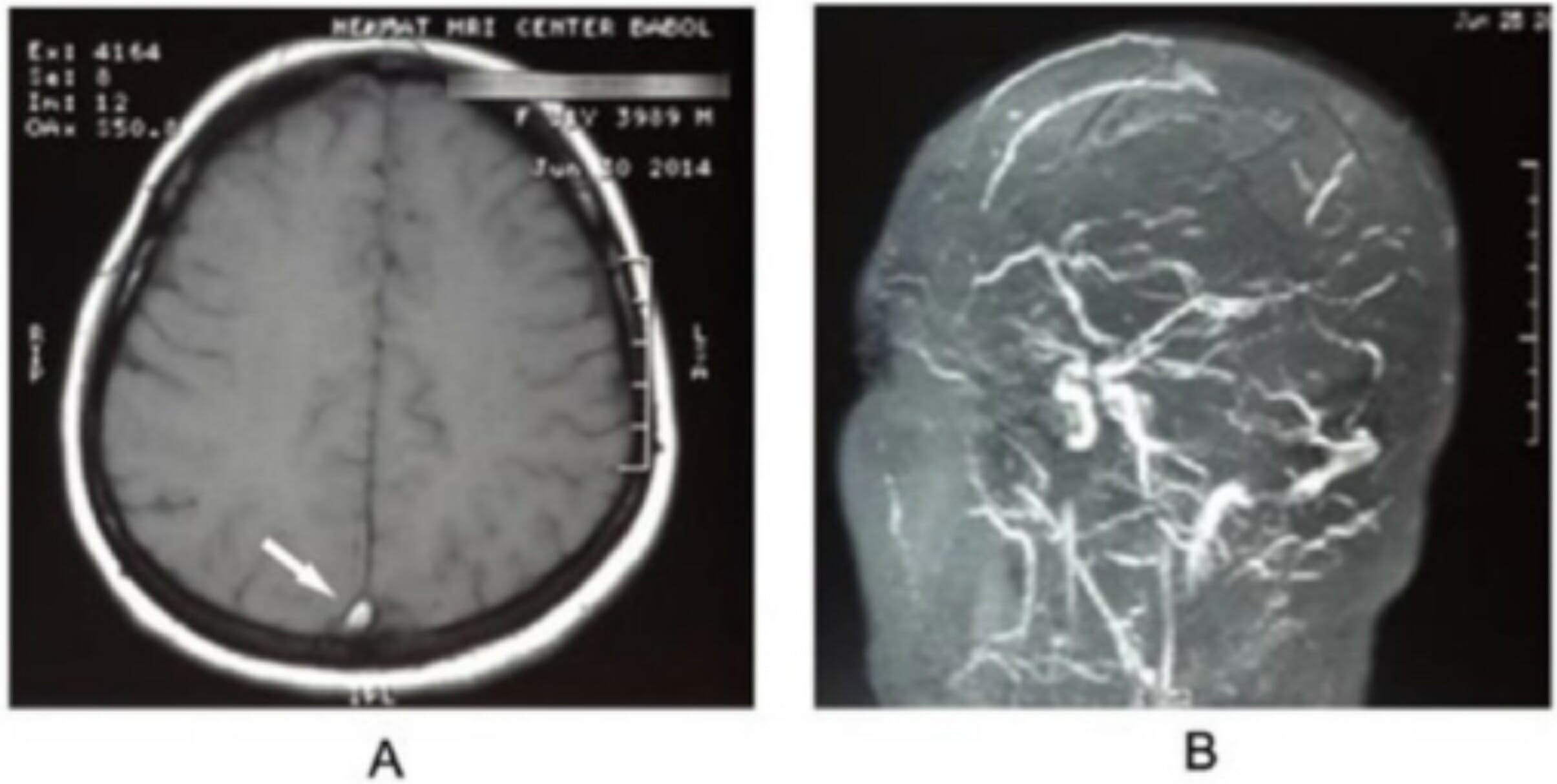
Trombosis del seno transverso:
La RM del cerebro muestra un signo delta vacío (panel A, flecha), y la venografía por RM (panel B) confirma el diagnóstico de trombosis venosa cerebral.
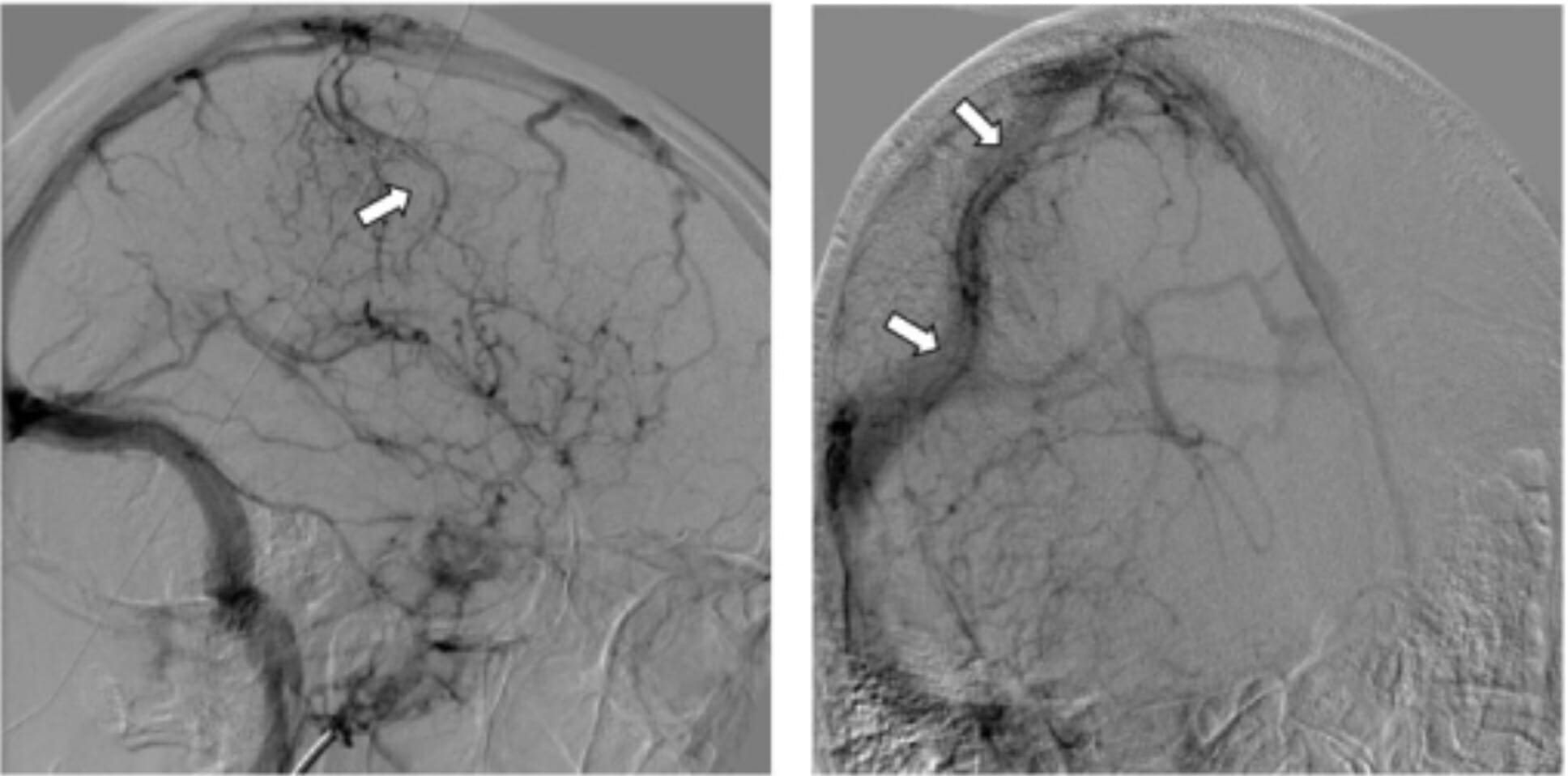
Angiografía cerebral:
La imagen en fase venosa de la arteria carótida interna derecha muestra un defecto de llenado tubular dentro de la vena cortical superficial de Trolard (flecha) y congestión de las vénulas circundantes (flechas), sugestivo de trombosis.