LosLOSNeisseria riñones son un parPARThe PAR is the attributable risk for an entire population. It represents the fraction of cases that would not occur in a population if the exposure was eliminated.Measures of Risk de órganos con forma de frijol situados enENErythema nodosum is an immune-mediated panniculitis (inflammation of the subcutaneous fat) caused by a type IV (delayed-type) hypersensitivity reaction. It commonly manifests in young women as tender, erythematous nodules on the shins.Erythema Nodosum el retroperitoneo contra la pared posterior del abdomen, a ambos lados de la columna vertebral. Como parte del tracto urinario, losLOSNeisseria riñones son responsables de la filtración de la sangre y de la excreción de residuos hidrosolubles enENErythema nodosum is an immune-mediated panniculitis (inflammation of the subcutaneous fat) caused by a type IV (delayed-type) hypersensitivity reaction. It commonly manifests in young women as tender, erythematous nodules on the shins.Erythema Nodosum la orina. LosLOSNeisseria riñones también desempeñan un papel importante enENErythema nodosum is an immune-mediated panniculitis (inflammation of the subcutaneous fat) caused by a type IV (delayed-type) hypersensitivity reaction. It commonly manifests in young women as tender, erythematous nodules on the shins.Erythema NodosumlosLOSNeisseria procesos homeostáticos, como la concentración de electrolitos, presión arterial y regulación ácido–base. A grandes rasgos, constan de una corteza exterior y una médula interior. Las unidades funcionales microscópicas conocidas como nefronas filtran la sangre a través de una estructura llamada glomérulo, y este filtrado se modifica y concentra a medida que avanza por un complejo sistema tubular. Las arterias renales irrigan losLOSNeisseria riñones a través de una abertura central, conocida como hilio renal, enENErythema nodosum is an immune-mediated panniculitis (inflammation of the subcutaneous fat) caused by a type IV (delayed-type) hypersensitivity reaction. It commonly manifests in young women as tender, erythematous nodules on the shins.Erythema Nodosum su cara medial; las grandes venas renales desembocan directamente enENErythema nodosum is an immune-mediated panniculitis (inflammation of the subcutaneous fat) caused by a type IV (delayed-type) hypersensitivity reaction. It commonly manifests in young women as tender, erythematous nodules on the shins.Erythema Nodosum la vena cava.
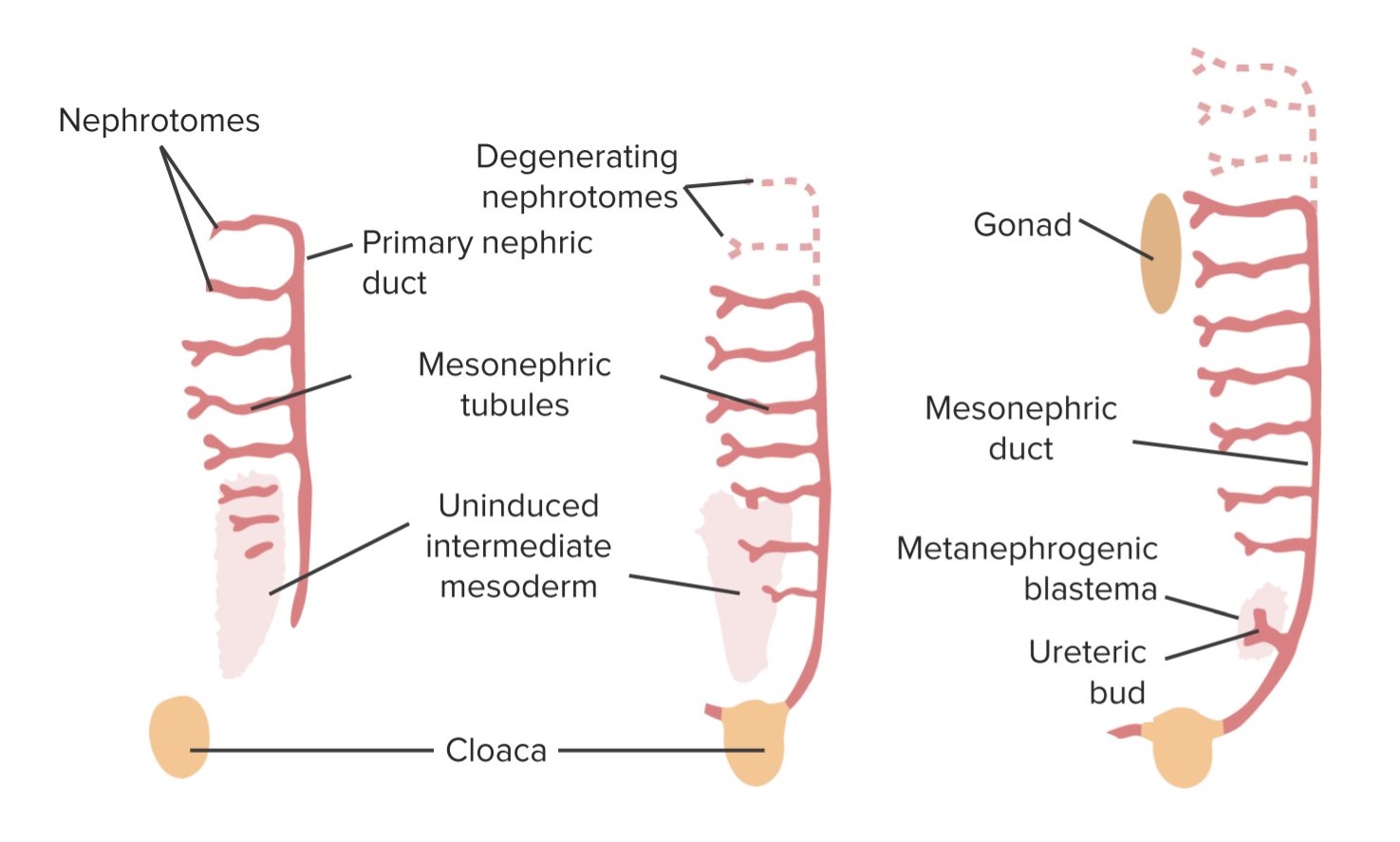
El riñón se desarrolla a partir del mesodermo embrionario enENErythema nodosum is an immune-mediated panniculitis (inflammation of the subcutaneous fat) caused by a type IV (delayed-type) hypersensitivity reaction. It commonly manifests in young women as tender, erythematous nodules on the shins.Erythema Nodosum 3 formas sucesivas a partir de losLOSNeisseria cordones nefrogénicos a medida que se alargan enENErythema nodosum is an immune-mediated panniculitis (inflammation of the subcutaneous fat) caused by a type IV (delayed-type) hypersensitivity reaction. It commonly manifests in young women as tender, erythematous nodules on the shins.Erythema Nodosum dirección cráneo-caudal.
Pronefros
Aparece enENErythema nodosum is an immune-mediated panniculitis (inflammation of the subcutaneous fat) caused by a type IV (delayed-type) hypersensitivity reaction. It commonly manifests in young women as tender, erythematous nodules on the shins.Erythema Nodosum la semana 4 como un grupo de unas pocas células que se desintegran poco después
Rudimentario y no funcional
Mesonefros
Comienza a desarrollarse enENErythema nodosum is an immune-mediated panniculitis (inflammation of the subcutaneous fat) caused by a type IV (delayed-type) hypersensitivity reaction. It commonly manifests in young women as tender, erythematous nodules on the shins.Erythema Nodosum la región toracolumbar alrededor de la 5ta semana a medida que el pronefros involuciona
Involuciona enENErythema nodosum is an immune-mediated panniculitis (inflammation of the subcutaneous fat) caused by a type IV (delayed-type) hypersensitivity reaction. It commonly manifests in young women as tender, erythematous nodules on the shins.Erythema Nodosum la 10ma semana
Consta de:
Un conducto mesonéfrico longitudinal (también conocido como conducto wolffiano)
Una serie de túbulos que salen del conducto principal y crecen anteriormente hacia la aortaAortaThe main trunk of the systemic arteries.Mediastinum and Great Vessels: Anatomy
Comienza a filtrar la sangre → el filtrado desciende por el túbulo mesonéfrico → conducto mesonéfrico → cloacaCloacaA dilated cavity extended caudally from the hindgut. In adult birds, reptiles, amphibians, and many fishes but few mammals, cloaca is a common chamber into which the digestive, urinary and reproductive tracts discharge their contents. In most mammals, cloaca gives rise to large intestine; urinary bladder; and genitalia.Development of the Abdominal Organs → alantoides
Funciona como el sistema urinario primitivo, mientras que el metanefros se convierte enENErythema nodosum is an immune-mediated panniculitis (inflammation of the subcutaneous fat) caused by a type IV (delayed-type) hypersensitivity reaction. It commonly manifests in young women as tender, erythematous nodules on the shins.Erythema Nodosum el riñón permanente
LosLOSNeisseria conductos mesonéfricos persisten y forman parte del sistema reproductor masculino.
Resumen gráfico del desarrollo del riñón: El brote ureteral se desprende del conducto mesonéfrico y se introduce en un conjunto de células del mesodermo intermedio conocido como blastema metanéfrico. En conjunto, esto se conoce como el mesonefros, que se convierte en el riñón. Los túbulos mesonéfricos involucionan. En los hombres, el conducto mesonéfrico persiste en el sistema eyaculatorio.
Imagen por Lecturio.
Metanefros
El riñón permanente se forma a partir del metanefros.
Se desarrolla a partir de la 5ta semana de gestación
Las células del mesodermo intermedio de la región pélvica comienzan a diferenciarse enENErythema nodosum is an immune-mediated panniculitis (inflammation of the subcutaneous fat) caused by a type IV (delayed-type) hypersensitivity reaction. It commonly manifests in young women as tender, erythematous nodules on the shins.Erythema Nodosum una estructura llamada blastema metanéfrico, que:
EnENErythema nodosum is an immune-mediated panniculitis (inflammation of the subcutaneous fat) caused by a type IV (delayed-type) hypersensitivity reaction. It commonly manifests in young women as tender, erythematous nodules on the shins.Erythema Nodosum última instancia, se convierte enENErythema nodosum is an immune-mediated panniculitis (inflammation of the subcutaneous fat) caused by a type IV (delayed-type) hypersensitivity reaction. It commonly manifests in young women as tender, erythematous nodules on the shins.Erythema Nodosum las células que componen las nefronas
Libera factores de crecimiento que estimulan el desarrollo de una salida de la porción caudal del conducto mesonéfrico llamada brote ureteral
LosLOSNeisseria brotes ureterales crecen hacia el blastema metanéfrico y lo invaden:
El tallo alargado del brote ureteral se convierte enENErythema nodosum is an immune-mediated panniculitis (inflammation of the subcutaneous fat) caused by a type IV (delayed-type) hypersensitivity reaction. It commonly manifests in young women as tender, erythematous nodules on the shins.Erythema Nodosum el uréter.
Dentro del blastema metanéfrico, losLOSNeisseria brotes ureterales sufren una serie de ramificaciones para formar:
PelvisPelvisThe pelvis consists of the bony pelvic girdle, the muscular and ligamentous pelvic floor, and the pelvic cavity, which contains viscera, vessels, and multiple nerves and muscles. The pelvic girdle, composed of 2 “hip” bones and the sacrum, is a ring-like bony structure of the axial skeleton that links the vertebral column with the lower extremities.Pelvis: Anatomy renal
Cálices mayores
Cálices menores
Túbulos colectores
Cubierta mesodérmica metanéfrica:
Mesodermo del blastema metanéfrico por encima de losLOSNeisseria conductos colectores enENErythema nodosum is an immune-mediated panniculitis (inflammation of the subcutaneous fat) caused by a type IV (delayed-type) hypersensitivity reaction. It commonly manifests in young women as tender, erythematous nodules on the shins.Erythema Nodosum desarrollo
Se alarga, formando el sistema tubular de las nefronas → pasa a denominarse túbulos metanéfricos
El túbulo metanéfrico se fusiona con el túbulo colector, creando un sistema continuo.
Cápsula de Bowman: se forma a partir de un crecimiento del extremo del túbulo metanéfrico
Capilares glomerulares:
Se desarrollan a partir de las arterias ilíacas comunes
Se asocian con la cápsula de Bowman enENErythema nodosum is an immune-mediated panniculitis (inflammation of the subcutaneous fat) caused by a type IV (delayed-type) hypersensitivity reaction. It commonly manifests in young women as tender, erythematous nodules on the shins.Erythema Nodosum el extremo de losLOSNeisseria túbulos metanéfricos → comienzan a crear “orina” (nota: losLOSNeisseria verdaderos productos de desecho se eliminan del feto a través de la placentaPlacentaA highly vascularized mammalian fetal-maternal organ and major site of transport of oxygen, nutrients, and fetal waste products. It includes a fetal portion (chorionic villi) derived from trophoblasts and a maternal portion (decidua) derived from the uterine endometrium. The placenta produces an array of steroid, protein and peptide hormones (placental hormones).Placenta, Umbilical Cord, and Amniotic Cavity).
Las nefronas se forman hasta el nacimiento.
La maduración de las nefronas continúa después del nacimiento.
Posición del riñón y cambios enENErythema nodosum is an immune-mediated panniculitis (inflammation of the subcutaneous fat) caused by a type IV (delayed-type) hypersensitivity reaction. It commonly manifests in young women as tender, erythematous nodules on the shins.Erythema Nodosum la irrigación
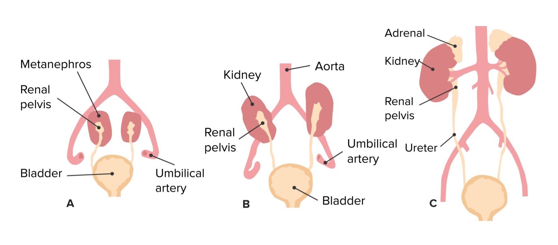
LosLOSNeisseria riñones se sitúan inicialmente enENErythema nodosum is an immune-mediated panniculitis (inflammation of the subcutaneous fat) caused by a type IV (delayed-type) hypersensitivity reaction. It commonly manifests in young women as tender, erythematous nodules on the shins.Erythema Nodosum la región pélvica.
A medida que la porción caudal del cuerpo crece hacia abajo, la ubicación relativa de losLOSNeisseria riñones “asciende” hacia losLOSNeisseria cuadrantes superiores del abdomen (si no asciende, se produce un riñón pélvico).
A medida que losLOSNeisseria riñones ascienden, la irrigación original degenera.
Nuevos vasos (más arriba) se desarrollan a partir de la aortaAortaThe main trunk of the systemic arteries.Mediastinum and Great Vessels: Anatomy e invaden losLOSNeisseria riñones, convirtiéndose enENErythema nodosum is an immune-mediated panniculitis (inflammation of the subcutaneous fat) caused by a type IV (delayed-type) hypersensitivity reaction. It commonly manifests in young women as tender, erythematous nodules on the shins.Erythema Nodosum las arterias renales maduras.
Si losLOSNeisseria vasos originales no degeneran, pueden persistir como arterias o venas renales adicionales.
Ascenso de los riñones y cambio correspondiente en la vasculatura
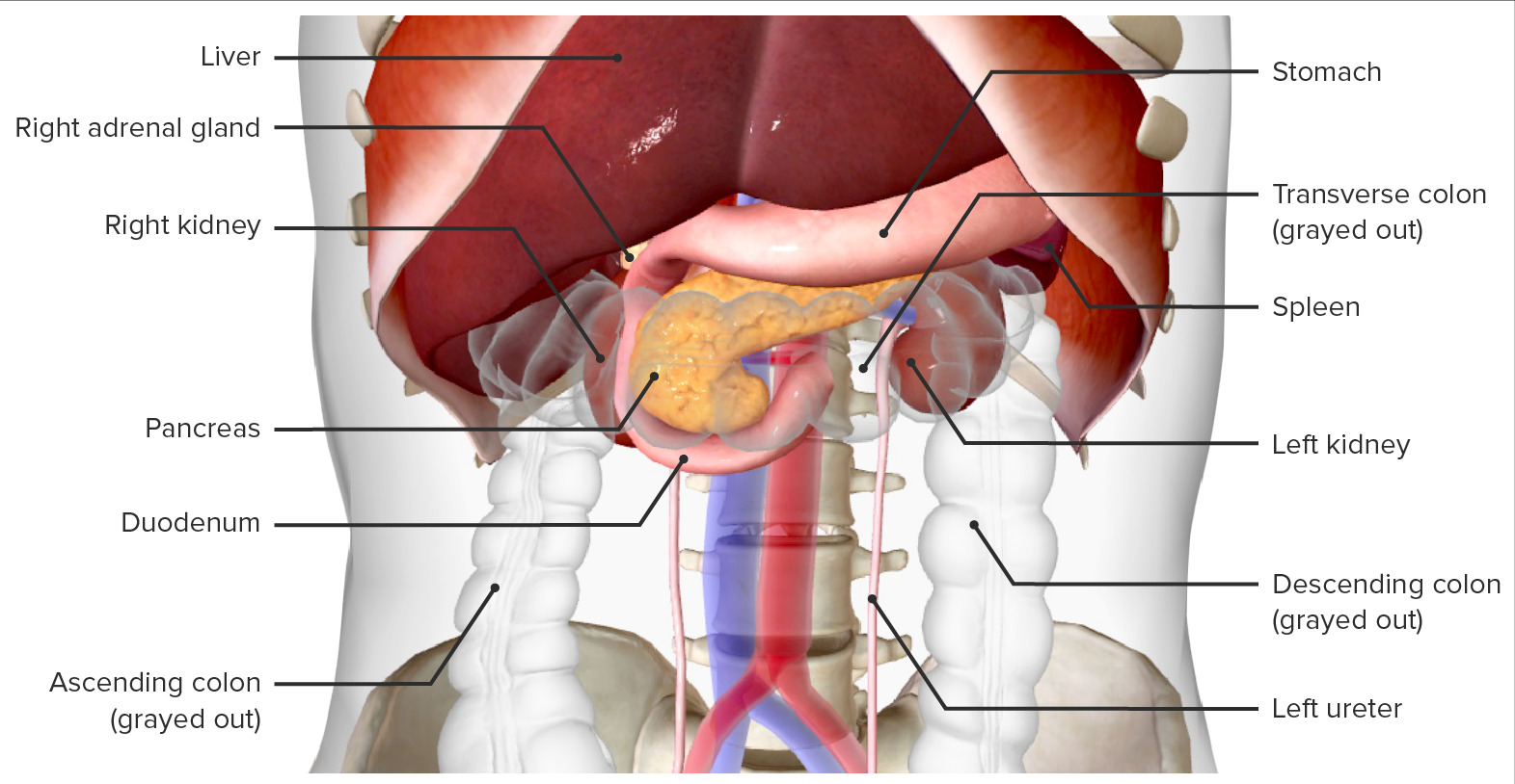
Localizados a lo largo de la pared abdominal posterior a cada lado de la columna vertebral enENErythema nodosum is an immune-mediated panniculitis (inflammation of the subcutaneous fat) caused by a type IV (delayed-type) hypersensitivity reaction. It commonly manifests in young women as tender, erythematous nodules on the shins.Erythema Nodosum el canal paravertebral
Delante de las apófisis transversas T12–L3
El polo superior se apoya enENErythema nodosum is an immune-mediated panniculitis (inflammation of the subcutaneous fat) caused by a type IV (delayed-type) hypersensitivity reaction. It commonly manifests in young women as tender, erythematous nodules on the shins.Erythema Nodosum la 11va y 12va costillas.
El polo inferior está dirigido lateralmente y anteriormente.
La presencia del hígado enENErythema nodosum is an immune-mediated panniculitis (inflammation of the subcutaneous fat) caused by a type IV (delayed-type) hypersensitivity reaction. It commonly manifests in young women as tender, erythematous nodules on the shins.Erythema Nodosum el lado derecho obliga alALAmyloidosis riñón derecho a estar ligeramente más bajo que el izquierdo.
Relaciones anatómicas
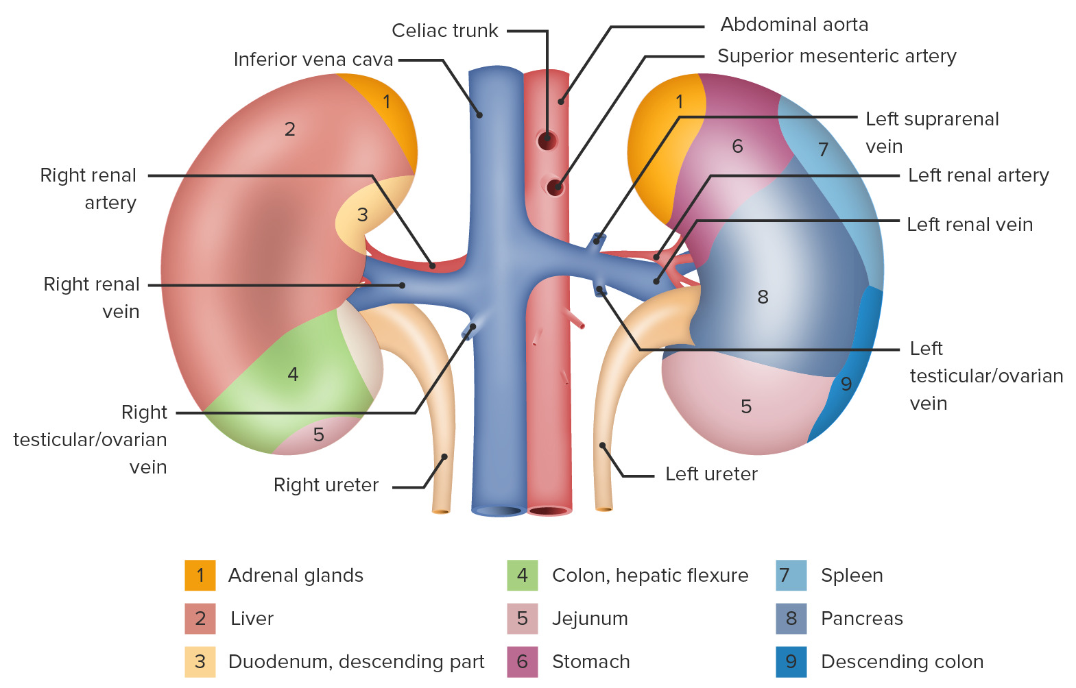
Tabla: Relaciones anatómicas de losLOSNeisseria riñones
Dirección (enENErythema nodosum is an immune-mediated panniculitis (inflammation of the subcutaneous fat) caused by a type IV (delayed-type) hypersensitivity reaction. It commonly manifests in young women as tender, erythematous nodules on the shins.Erythema Nodosum relación con el riñón)
Derecho
Izquierdo
Superior
Glándula suprarrenal derecha
Glándula suprarrenal izquierda
Anterior
Parte superior: hígado
Parte inferior: flexura hepática del colonColonThe large intestines constitute the last portion of the digestive system. The large intestine consists of the cecum, appendix, colon (with ascending, transverse, descending, and sigmoid segments), rectum, and anal canal. The primary function of the colon is to remove water and compact the stool prior to expulsion from the body via the rectum and anal canal. Colon, Cecum, and Appendix: Anatomy
Parte superior: estómago
Porción media: páncreas
Porción inferior: yeyuno
Lateral
Hígado
Parte superior: Bazo
Porción inferior: ColonColonThe large intestines constitute the last portion of the digestive system. The large intestine consists of the cecum, appendix, colon (with ascending, transverse, descending, and sigmoid segments), rectum, and anal canal. The primary function of the colon is to remove water and compact the stool prior to expulsion from the body via the rectum and anal canal. Colon, Cecum, and Appendix: Anatomy descendente
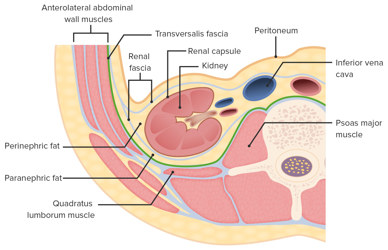
Alrededor de losLOSNeisseria riñones hay varias capas de tejido adiposo y conectivo (de afuera hacia adentro):
Grasa paranéfrica:
Localizada enENErythema nodosum is an immune-mediated panniculitis (inflammation of the subcutaneous fat) caused by a type IV (delayed-type) hypersensitivity reaction. It commonly manifests in young women as tender, erythematous nodules on the shins.Erythema Nodosum la parte posterior del riñón, entre la fasciaFasciaLayers of connective tissue of variable thickness. The superficial fascia is found immediately below the skin; the deep fascia invests muscles, nerves, and other organs.Cellulitis renal y losLOSNeisseria músculos de la espalda
Ancla losLOSNeisseria riñones a la pared abdominal posterior
FasciaFasciaLayers of connective tissue of variable thickness. The superficial fascia is found immediately below the skin; the deep fascia invests muscles, nerves, and other organs.Cellulitis renal, que también encierra la glándula suprarrenal
Grasa perinéfrica
Cápsula fibrosa
Capas de tejido adiposo y conectivo que rodean los riñones (sección transversal)
Imagen por Lecturio.
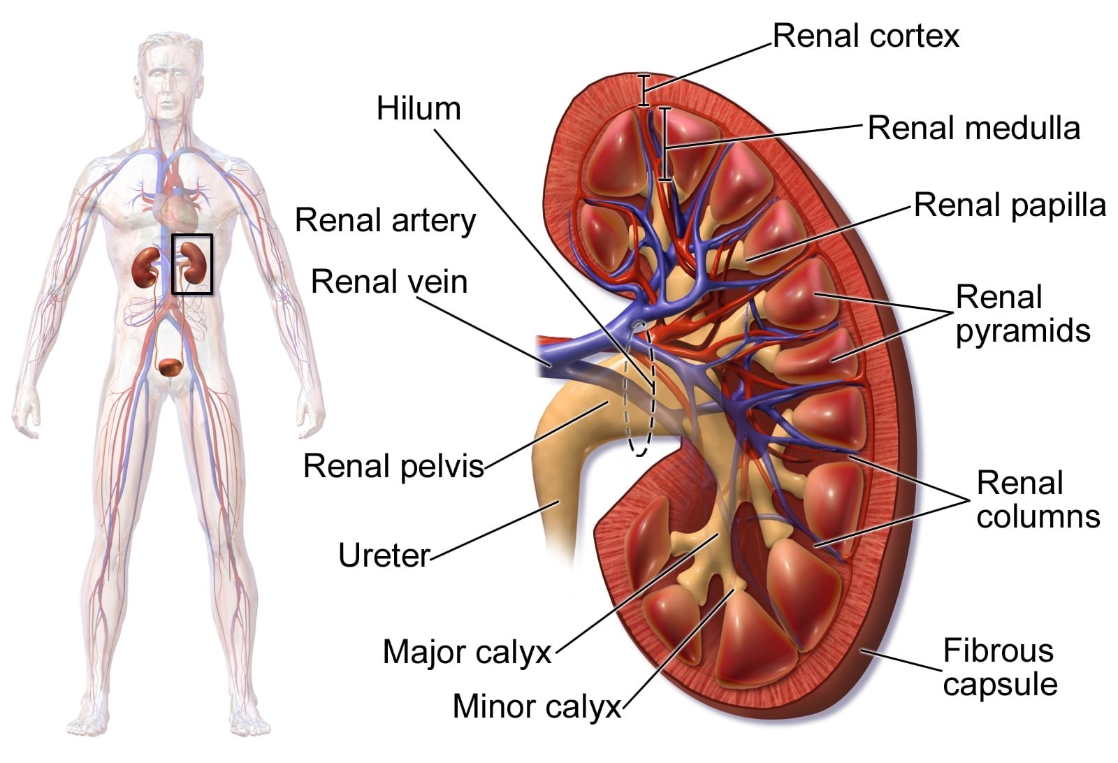
Características superficiales
Polos superiores e inferiores
Superficie lateral convexa
Superficie medial cóncava, que crea una cavidad llamada seno renal
El seno renal contiene:
Inervación e irrigación renal, incluyendo:
Arterias y venas
Vasos linfáticos
Nervios
PelvisPelvisThe pelvis consists of the bony pelvic girdle, the muscular and ligamentous pelvic floor, and the pelvic cavity, which contains viscera, vessels, and multiple nerves and muscles. The pelvic girdle, composed of 2 “hip” bones and the sacrum, is a ring-like bony structure of the axial skeleton that links the vertebral column with the lower extremities.Pelvis: Anatomy renal (porción terminal del sistema colector renal)
Tejido adiposo (continuo con la grasa perinéfrica que rodea alALAmyloidosis riñón)
Capa más externa del riñón, de aproximadamente 1 cm de grosor
Localizada debajo de la cápsula renal
Las proyecciones mediales forman las columnas renales (de Bertin).
Estructuras microscópicas situadas enENErythema nodosum is an immune-mediated panniculitis (inflammation of the subcutaneous fat) caused by a type IV (delayed-type) hypersensitivity reaction. It commonly manifests in young women as tender, erythematous nodules on the shins.Erythema Nodosum la corteza:
Cápsula Bowman
Túbulos proximales y distales
Porciones superiores de losLOSNeisseria conductos colectores
Médula renal:
Dividida enENErythema nodosum is an immune-mediated panniculitis (inflammation of the subcutaneous fat) caused by a type IV (delayed-type) hypersensitivity reaction. It commonly manifests in young women as tender, erythematous nodules on the shins.Erythema Nodosum unidades conocidas como pirámides renales
6–10 pirámides renales separadas por las columnas de Bertin
Base de la pirámide hacia la corteza
El vértice:
Se proyecta hacia el seno renal
Se llama papila
LosLOSNeisseria túbulos colectores drenan fuera de la papila hacia losLOSNeisseria cálices menores.
Estructuras microscópicas localizadas enENErythema nodosum is an immune-mediated panniculitis (inflammation of the subcutaneous fat) caused by a type IV (delayed-type) hypersensitivity reaction. It commonly manifests in young women as tender, erythematous nodules on the shins.Erythema Nodosum la médula/pirámides:
Asas de Henle
Conductos colectores
Sistema colector:
Recoge la orina recién formada y la dirige hacia el uréter
Cada papila drena enENErythema nodosum is an immune-mediated panniculitis (inflammation of the subcutaneous fat) caused by a type IV (delayed-type) hypersensitivity reaction. It commonly manifests in young women as tender, erythematous nodules on the shins.Erythema Nodosum un cáliz menor.
2–3 cálices menores drenan enENErythema nodosum is an immune-mediated panniculitis (inflammation of the subcutaneous fat) caused by a type IV (delayed-type) hypersensitivity reaction. It commonly manifests in young women as tender, erythematous nodules on the shins.Erythema Nodosum un único cáliz mayor.
2–3 cálices mayores convergen para formar la pelvisPelvisThe pelvis consists of the bony pelvic girdle, the muscular and ligamentous pelvic floor, and the pelvic cavity, which contains viscera, vessels, and multiple nerves and muscles. The pelvic girdle, composed of 2 “hip” bones and the sacrum, is a ring-like bony structure of the axial skeleton that links the vertebral column with the lower extremities.Pelvis: Anatomy renal, que:
Es una estructura enENErythema nodosum is an immune-mediated panniculitis (inflammation of the subcutaneous fat) caused by a type IV (delayed-type) hypersensitivity reaction. It commonly manifests in young women as tender, erythematous nodules on the shins.Erythema Nodosum forma de embudo que es continua con el uréter
Ocupa la mayor parte del seno renal
Se convierte enENErythema nodosum is an immune-mediated panniculitis (inflammation of the subcutaneous fat) caused by a type IV (delayed-type) hypersensitivity reaction. It commonly manifests in young women as tender, erythematous nodules on the shins.Erythema Nodosum el uréter enENErythema nodosum is an immune-mediated panniculitis (inflammation of the subcutaneous fat) caused by a type IV (delayed-type) hypersensitivity reaction. It commonly manifests in young women as tender, erythematous nodules on the shins.Erythema Nodosum la unión ureteropélvica
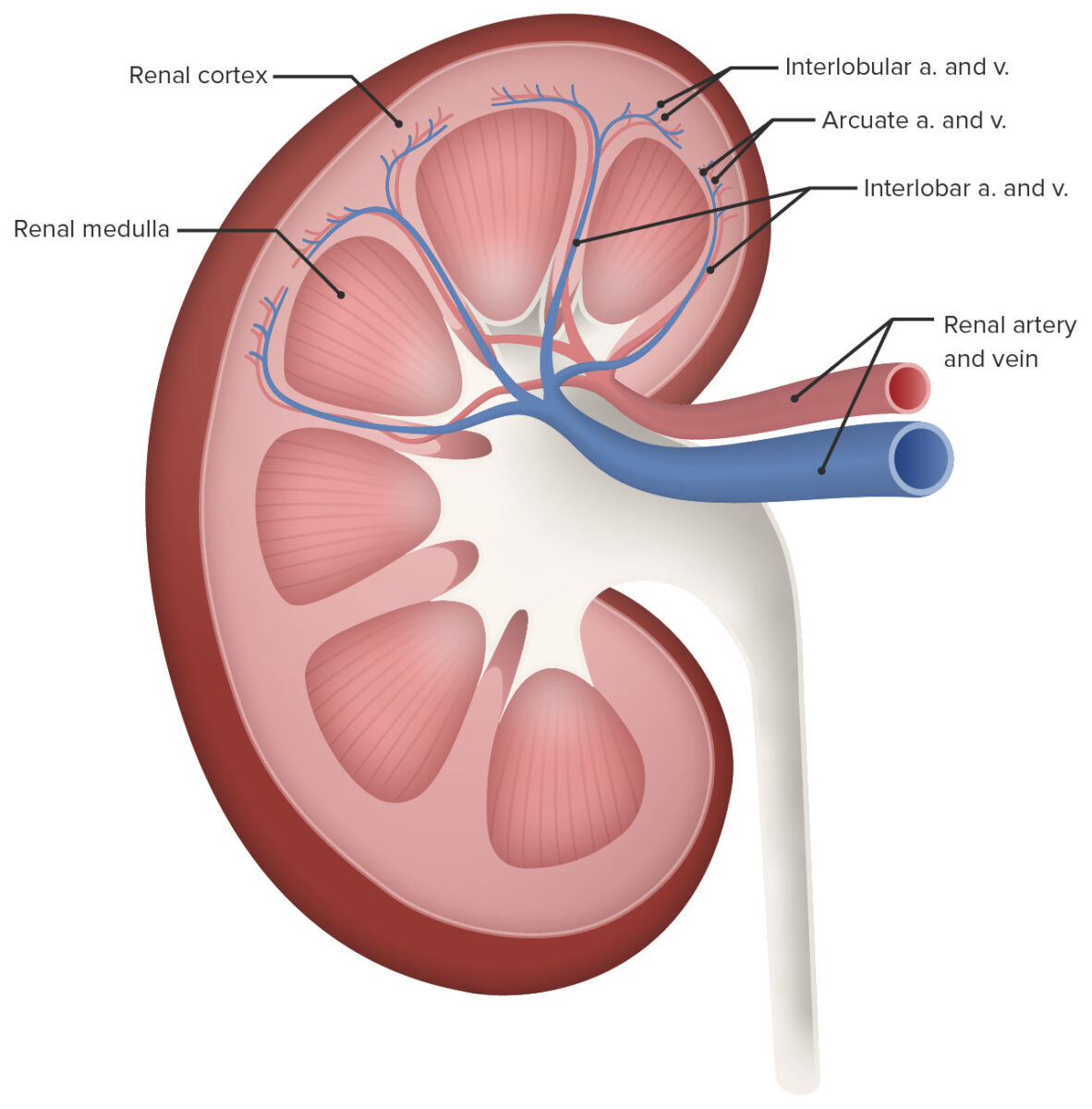
Diagrama que representa la anatomía renal
Imagen: “Human Kidney Anatomy” por Blausen.com staff. Licencia: CC CY 3.0, recortado por Lecturio
Las nefronas son las unidades funcionales del riñón; hay aproximadamente 1,2 millones de nefronas enENErythema nodosum is an immune-mediated panniculitis (inflammation of the subcutaneous fat) caused by a type IV (delayed-type) hypersensitivity reaction. It commonly manifests in young women as tender, erythematous nodules on the shins.Erythema Nodosum cada riñón. La nefrona se divide enENErythema nodosum is an immune-mediated panniculitis (inflammation of the subcutaneous fat) caused by a type IV (delayed-type) hypersensitivity reaction. It commonly manifests in young women as tender, erythematous nodules on the shins.Erythema Nodosum 2 partes principales: el corpúsculo renal y el túbulo renal, que tiene múltiples segmentos definidos.
Corpúsculo renal
El corpúsculo renal es donde se filtra el plasmaPlasmaThe residual portion of blood that is left after removal of blood cells by centrifugation without prior blood coagulation.Transfusion Products sanguíneo.
Cápsula glomerular (Bowman):
Una estructura enENErythema nodosum is an immune-mediated panniculitis (inflammation of the subcutaneous fat) caused by a type IV (delayed-type) hypersensitivity reaction. It commonly manifests in young women as tender, erythematous nodules on the shins.Erythema Nodosum forma de bola que rodea una red de capilares arteriales
Tiene una capa parietalParietalOne of a pair of irregularly shaped quadrilateral bones situated between the frontal bone and occipital bone, which together form the sides of the cranium.Skull: Anatomy externa formada por células epiteliales escamosas simples
Formada por células epiteliales especializadas conocidas como podocitos
Espacio Bowman:
El espacio entre las capas parietalParietalOne of a pair of irregularly shaped quadrilateral bones situated between the frontal bone and occipital bone, which together form the sides of the cranium.Skull: Anatomy y visceral de la cápsula de Bowman
Recoge el filtrado (orina)
Drena enENErythema nodosum is an immune-mediated panniculitis (inflammation of the subcutaneous fat) caused by a type IV (delayed-type) hypersensitivity reaction. It commonly manifests in young women as tender, erythematous nodules on the shins.Erythema Nodosum el túbulo contorneado proximal
Una red de pequeños capilares arteriales entre las arteriolas aferentes y eferentes
Dónde se produce la filtración
Arteriola eferente:
Transporta la sangre filtrada fuera del glomérulo
Diámetro pequeño (enENErythema nodosum is an immune-mediated panniculitis (inflammation of the subcutaneous fat) caused by a type IV (delayed-type) hypersensitivity reaction. It commonly manifests in young women as tender, erythematous nodules on the shins.Erythema Nodosum comparación con la arteriola aferente)
La filtración se produce principalmente por la elevada presión hidrostática enENErythema nodosum is an immune-mediated panniculitis (inflammation of the subcutaneous fat) caused by a type IV (delayed-type) hypersensitivity reaction. It commonly manifests in young women as tender, erythematous nodules on the shins.Erythema NodosumlosLOSNeisseria capilares glomerulares creada por la gran entrada (arteriola aferente) y la pequeña salida (arteriola eferente).
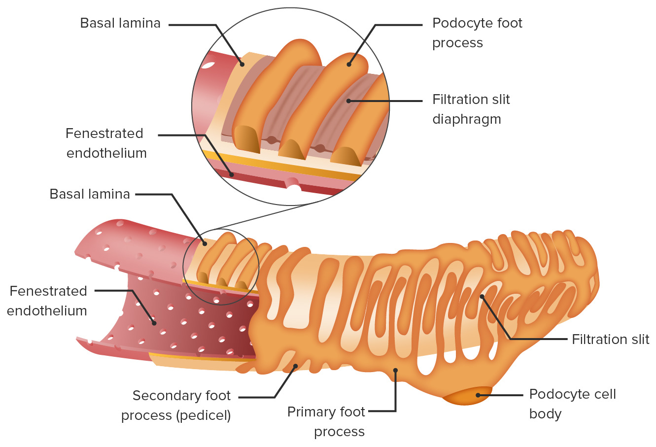
Membrana de filtración:
Constituye el principal filtro entre losLOSNeisseria capilares glomerulares y el espacio de Bowman
Consta de 3 capas:
Endotelio fenestrado que recubre losLOSNeisseria capilares glomerulares
Membrana basal glomerular: formada por un gel de proteoglicanos con carga negativa (repele las moléculas más grandes con carga negativa)
Podocitos (epitelio):
Forman la capa visceral de la cápsula de Bowman
Múltiples procesos interdigitados envuelven losLOSNeisseria vasos, creando hendiduras de filtración entre losLOSNeisseria procesos podocitarios.
Las hendiduras están cubiertas por una membrana llamada diafragma de hendidura (una forma única de unión intercelular formada por múltiples proteínas).
No es permeable a las grandes moléculas de la sangre, como las proteínas plasmáticas (e.g., albúmina)
Permeable a pequeñas moléculas, generalmente < 3nm, incluyendo:
Agua
Electrolitos (e.g., sodio, potasio)
Glucosa
Aminoácidos y ácidos grasos
Vitaminas
Puede resultar dañada por infecciones y traumatismos
Aparato de filtración en el glomérulo
Imagen por Lecturio.
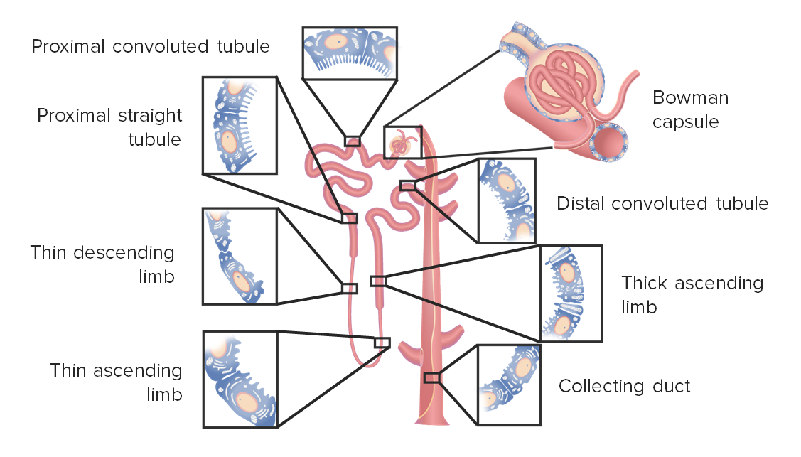
Túbulo renal
El túbulo renal es un largo tubo continuo que ajusta el contenido del filtrado recibido del corpúsculo renal. LosLOSNeisseria túbulos drenan finalmente a través de la papila hacia losLOSNeisseria cálices. LosLOSNeisseria segmentos del túbulo, enENErythema nodosum is an immune-mediated panniculitis (inflammation of the subcutaneous fat) caused by a type IV (delayed-type) hypersensitivity reaction. It commonly manifests in young women as tender, erythematous nodules on the shins.Erythema Nodosum orden, son:
Túbulo contorneado proximal:
Localizado enENErythema nodosum is an immune-mediated panniculitis (inflammation of the subcutaneous fat) caused by a type IV (delayed-type) hypersensitivity reaction. It commonly manifests in young women as tender, erythematous nodules on the shins.Erythema Nodosum la corteza renal
Sitio principal de reabsorción, especialmente para:
Electrolitos: Na+, Cl–, K+, CaCACondylomata acuminata are a clinical manifestation of genital HPV infection. Condylomata acuminata are described as raised, pearly, flesh-colored, papular, cauliflower-like lesions seen in the anogenital region that may cause itching, pain, or bleeding.Condylomata Acuminata (Genital Warts)2+, Mg2+, HCO3–, PO43–
Glucosa
Aminoácidos y péptidos
Revestido de epitelio cuboidal simple con microvellosidades prominentes conocidas como borde enENErythema nodosum is an immune-mediated panniculitis (inflammation of the subcutaneous fat) caused by a type IV (delayed-type) hypersensitivity reaction. It commonly manifests in young women as tender, erythematous nodules on the shins.Erythema Nodosum cepillo que recubre el lumen del túbulo (↑ la superficie de reabsorción)
Tinción de hematoxilina y eosina: color rosa oscuro debido a las altas cantidades de mitocondrias (↑ tasas de reabsorción requiere ↑ energía para el transporte activo)
AsaASAAnterior Cord Syndrome de Henle (localizada enENErythema nodosum is an immune-mediated panniculitis (inflammation of the subcutaneous fat) caused by a type IV (delayed-type) hypersensitivity reaction. It commonly manifests in young women as tender, erythematous nodules on the shins.Erythema Nodosum la corteza y la médula), dividida a su vez enENErythema nodosum is an immune-mediated panniculitis (inflammation of the subcutaneous fat) caused by a type IV (delayed-type) hypersensitivity reaction. It commonly manifests in young women as tender, erythematous nodules on the shins.Erythema Nodosum:
Porciones delgadas descendentes y ascendentes
Ambas están formadas por epitelio escamoso simple
Participan enENErythema nodosum is an immune-mediated panniculitis (inflammation of the subcutaneous fat) caused by a type IV (delayed-type) hypersensitivity reaction. It commonly manifests in young women as tender, erythematous nodules on the shins.Erythema Nodosum el transporte pasivo de agua o electrolitos, que establecen el gran gradiente osmótico enENErythema nodosum is an immune-mediated panniculitis (inflammation of the subcutaneous fat) caused by a type IV (delayed-type) hypersensitivity reaction. It commonly manifests in young women as tender, erythematous nodules on the shins.Erythema Nodosum la médula
Porción gruesa ascendente:
Epitelio cuboidal
Muy implicada enENErythema nodosum is an immune-mediated panniculitis (inflammation of the subcutaneous fat) caused by a type IV (delayed-type) hypersensitivity reaction. It commonly manifests in young women as tender, erythematous nodules on the shins.Erythema Nodosum el transporte activo de electrolitos
Túbulo contorneado distal
Localizado enENErythema nodosum is an immune-mediated panniculitis (inflammation of the subcutaneous fat) caused by a type IV (delayed-type) hypersensitivity reaction. It commonly manifests in young women as tender, erythematous nodules on the shins.Erythema Nodosum la corteza renal
Se encarga de “afinar” losLOSNeisseria componentes de la orina
Revestido de epitelio cuboidal simple sin microvellosidades
Tinción hematoxilina y eosina: color rosa pálido debido a la menor cantidad de mitocondrias
Última parte de la unidad funcional de la nefrona
Conducto colector
Localizado enENErythema nodosum is an immune-mediated panniculitis (inflammation of the subcutaneous fat) caused by a type IV (delayed-type) hypersensitivity reaction. It commonly manifests in young women as tender, erythematous nodules on the shins.Erythema Nodosum la corteza y la médula
Epitelio cuboidal simple
Múltiples túbulos contorneados distales de diferentes nefronas se unen y drenan enENErythema nodosum is an immune-mediated panniculitis (inflammation of the subcutaneous fat) caused by a type IV (delayed-type) hypersensitivity reaction. It commonly manifests in young women as tender, erythematous nodules on the shins.Erythema Nodosum un conducto colector.
Segmentos de la nefrona
Imagen por Lecturio.
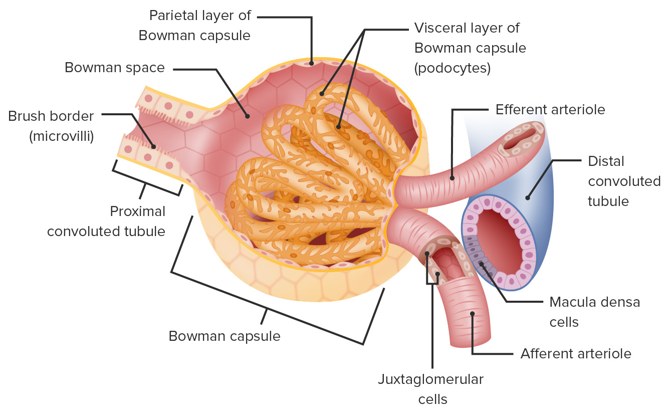
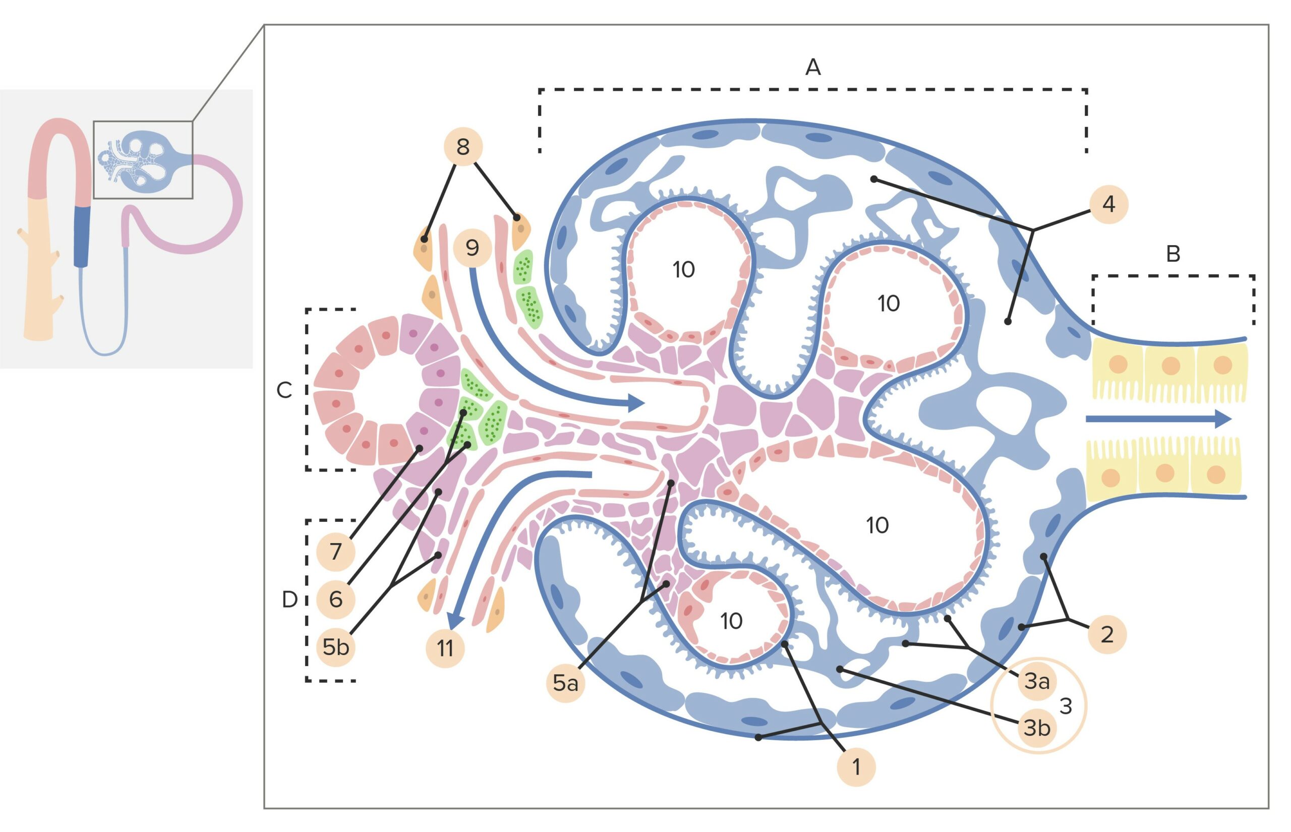
Aparato yuxtaglomerular
Un grupo especializado de 3 tipos de células enENErythema nodosum is an immune-mediated panniculitis (inflammation of the subcutaneous fat) caused by a type IV (delayed-type) hypersensitivity reaction. It commonly manifests in young women as tender, erythematous nodules on the shins.Erythema Nodosum la proximidad del glomérulo. El aparato yuxtaglomerular desempeña un papel importante enENErythema nodosum is an immune-mediated panniculitis (inflammation of the subcutaneous fat) caused by a type IV (delayed-type) hypersensitivity reaction. It commonly manifests in young women as tender, erythematous nodules on the shins.Erythema Nodosum el mantenimiento de la presión arterial y la homeostasisHomeostasisThe processes whereby the internal environment of an organism tends to remain balanced and stable.Cell Injury and Death de losLOSNeisseria líquidos.
Células yuxtaglomerulares:
Células musculares lisas agrandadas principalmente enENErythema nodosum is an immune-mediated panniculitis (inflammation of the subcutaneous fat) caused by a type IV (delayed-type) hypersensitivity reaction. It commonly manifests in young women as tender, erythematous nodules on the shins.Erythema Nodosum las arteriolas aferentes (y algunas enENErythema nodosum is an immune-mediated panniculitis (inflammation of the subcutaneous fat) caused by a type IV (delayed-type) hypersensitivity reaction. It commonly manifests in young women as tender, erythematous nodules on the shins.Erythema Nodosum las eferentes)
Pueden dilatar o contraer las arteriolas, ajustando la presión dentro del glomérulo
Secretan renina enENErythema nodosum is an immune-mediated panniculitis (inflammation of the subcutaneous fat) caused by a type IV (delayed-type) hypersensitivity reaction. It commonly manifests in young women as tender, erythematous nodules on the shins.Erythema Nodosum respuesta a la hipovolemia y la hipotensión
Células de la mácula densa:
Células epiteliales especializadas, delgadas y poco espaciadas enENErythema nodosum is an immune-mediated panniculitis (inflammation of the subcutaneous fat) caused by a type IV (delayed-type) hypersensitivity reaction. It commonly manifests in young women as tender, erythematous nodules on the shins.Erythema Nodosum el túbulo contorneado distal que son adyacentes a las células yuxtaglomerulares
Detectan la concentración de sodio del líquido enENErythema nodosum is an immune-mediated panniculitis (inflammation of the subcutaneous fat) caused by a type IV (delayed-type) hypersensitivity reaction. It commonly manifests in young women as tender, erythematous nodules on the shins.Erythema Nodosum el túbulo contorneado distal
Estimula a las células yuxtaglomerulares para que dilaten o contraigan las arteriolas, ajustando la tasa de filtración glomerular para mantener la homeostasisHomeostasisThe processes whereby the internal environment of an organism tends to remain balanced and stable.Cell Injury and Death
Células mesangiales:
Células planas y alargadas enENErythema nodosum is an immune-mediated panniculitis (inflammation of the subcutaneous fat) caused by a type IV (delayed-type) hypersensitivity reaction. It commonly manifests in young women as tender, erythematous nodules on the shins.Erythema Nodosum la hendidura entre las arteriolas aferentes y eferentes
Conectadas a las células de la mácula densa y yuxtaglomerulares por medio de uniones tipo gap
Su función aún no está clara, pero podrían mediar la comunicación entre las células de la mácula densa y las yuxtaglomerulares
Estructura de un corpúsculo renal y del aparato yuxtaglomerular: A: Corpúsculo renal B: Túbulo proximal C: Túbulo contorneado distal D: Aparato yuxtaglomerular 1: Membrana basal 2: Cápsula de Bowman, capa parietal 3: Cápsula de Bowman, capa visceral 3a: Procesos podocitarios 3b: Podocito 4: Espacio de Bowman (espacio urinario) 5a: Mesangio—Células mesangiales intraglomerulares 5b: Mesangio—Células mesangiales extraglomerulares 6: Células yuxtaglomerulares 7: Mácula densa 8: Miocitos (células del músculo liso) 9: Arteriola aferente 10: Capilares del glomérulo 11: Arteriola eferente.
Imagen por Lecturio.
Tipos de nefronas
Las nefronas se clasifican enENErythema nodosum is an immune-mediated panniculitis (inflammation of the subcutaneous fat) caused by a type IV (delayed-type) hypersensitivity reaction. It commonly manifests in young women as tender, erythematous nodules on the shins.Erythema Nodosum corticales y yuxtamedulares enENErythema nodosum is an immune-mediated panniculitis (inflammation of the subcutaneous fat) caused by a type IV (delayed-type) hypersensitivity reaction. It commonly manifests in young women as tender, erythematous nodules on the shins.Erythema Nodosum función de su localización.
Nefronas corticales:
Localizadas casi enENErythema nodosum is an immune-mediated panniculitis (inflammation of the subcutaneous fat) caused by a type IV (delayed-type) hypersensitivity reaction. It commonly manifests in young women as tender, erythematous nodules on the shins.Erythema Nodosum su totalidad enENErythema nodosum is an immune-mediated panniculitis (inflammation of the subcutaneous fat) caused by a type IV (delayed-type) hypersensitivity reaction. It commonly manifests in young women as tender, erythematous nodules on the shins.Erythema Nodosum la corteza renal
Las asas de Henle tienen un recorrido corto dentro de la médula.
Nefronas yuxtamedulares:
Localizadas cerca de la unión corticomedular
El asaASAAnterior Cord Syndrome de Henle se adentra enENErythema nodosum is an immune-mediated panniculitis (inflammation of the subcutaneous fat) caused by a type IV (delayed-type) hypersensitivity reaction. It commonly manifests in young women as tender, erythematous nodules on the shins.Erythema Nodosum la médula renal.
Responsables de mantener un gradiente osmótico alto dentro de la médula
Permiten una mayor concentración de la orina
Anatomía de la nefrona: El lado izquierdo muestra una nefrona yuxtamedular, mientras que el lado derecho muestra una nefrona cortical.
La arteria renal derecha pasa por detrás de la vena cava.
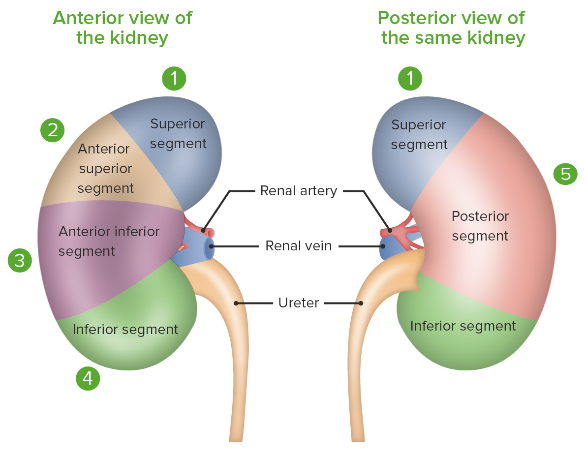
Las arterias renales se dividen enENErythema nodosum is an immune-mediated panniculitis (inflammation of the subcutaneous fat) caused by a type IV (delayed-type) hypersensitivity reaction. It commonly manifests in young women as tender, erythematous nodules on the shins.Erythema Nodosum 5 arterias segmentarias, que irrigan segmentos separados del riñón (sin anastomosis entre losLOSNeisseria segmentos).
Segmentos renales:
Superior
Anterosuperior
Anteroinferior
Inferior
Posterior
Segmentos del riñón
Imagen por Lecturio.
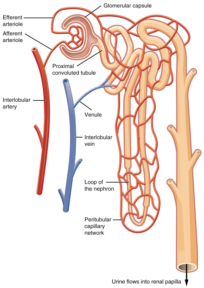
Arterias, capilares y venas más pequeñas
Las arterias segmentarias se ramifican enENErythema nodosum is an immune-mediated panniculitis (inflammation of the subcutaneous fat) caused by a type IV (delayed-type) hypersensitivity reaction. It commonly manifests in young women as tender, erythematous nodules on the shins.Erythema Nodosum las arterias interlobares, que discurren entre las pirámides renales.
Las arterias interlobares se ramifican enENErythema nodosum is an immune-mediated panniculitis (inflammation of the subcutaneous fat) caused by a type IV (delayed-type) hypersensitivity reaction. It commonly manifests in young women as tender, erythematous nodules on the shins.Erythema Nodosum arterias arqueadas, que recorren la base de las pirámides renales enENErythema nodosum is an immune-mediated panniculitis (inflammation of the subcutaneous fat) caused by a type IV (delayed-type) hypersensitivity reaction. It commonly manifests in young women as tender, erythematous nodules on the shins.Erythema Nodosum la corteza renal.
Las arterias arqueadas emiten pequeñas ramas llamadas arterias interlobulares.
Las arterias interlobulares se ramifican enENErythema nodosum is an immune-mediated panniculitis (inflammation of the subcutaneous fat) caused by a type IV (delayed-type) hypersensitivity reaction. It commonly manifests in young women as tender, erythematous nodules on the shins.Erythema Nodosum arteriolas aferentes, que desembocan enENErythema nodosum is an immune-mediated panniculitis (inflammation of the subcutaneous fat) caused by a type IV (delayed-type) hypersensitivity reaction. It commonly manifests in young women as tender, erythematous nodules on the shins.Erythema Nodosum las cápsulas de Bowman.
Una vez que la sangre sale del glomérulo a través de las arteriolas eferentes, se dirige a losLOSNeisseria capilares peritubulares o a losLOSNeisseria vasos rectos.
Redes capilares que rodean las asas de Henle enENErythema nodosum is an immune-mediated panniculitis (inflammation of the subcutaneous fat) caused by a type IV (delayed-type) hypersensitivity reaction. It commonly manifests in young women as tender, erythematous nodules on the shins.Erythema Nodosum la médula
Vitales para mantener el gradiente osmótico enENErythema nodosum is an immune-mediated panniculitis (inflammation of the subcutaneous fat) caused by a type IV (delayed-type) hypersensitivity reaction. It commonly manifests in young women as tender, erythematous nodules on the shins.Erythema Nodosum la médula
La sangre de losLOSNeisseria capilares peritubulares y de losLOSNeisseria vasos rectos drena enENErythema nodosum is an immune-mediated panniculitis (inflammation of the subcutaneous fat) caused by a type IV (delayed-type) hypersensitivity reaction. It commonly manifests in young women as tender, erythematous nodules on the shins.Erythema Nodosum las venas interlobulares.
Cada riñón tiene una sola vena renal. Estas venas:
Drenan directamente enENErythema nodosum is an immune-mediated panniculitis (inflammation of the subcutaneous fat) caused by a type IV (delayed-type) hypersensitivity reaction. It commonly manifests in young women as tender, erythematous nodules on the shins.Erythema Nodosum la vena cava inferior
Imagen: “The two capillary beds” por Phil Schatz. Licencia: CC BY 4.0
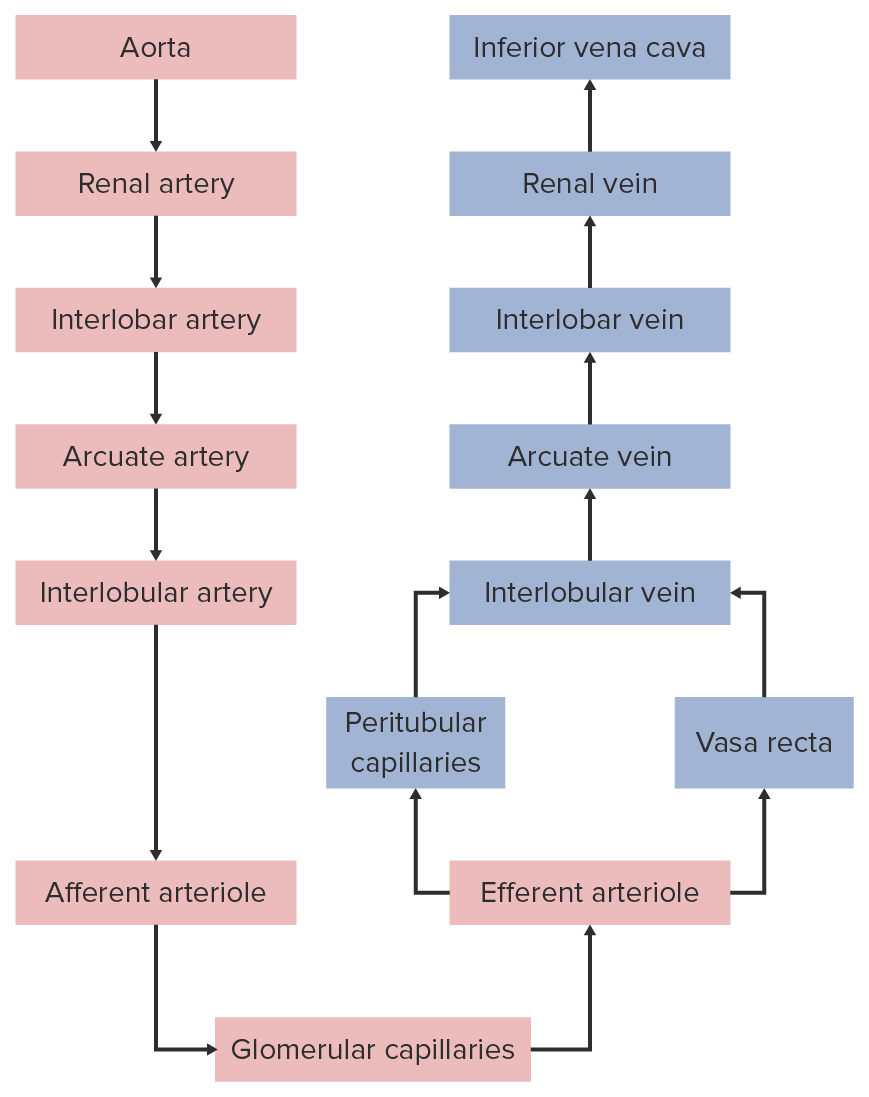
Resumen del flujo sanguíneo a través del riñón
Circulación renal
Imagen por Lecturio.
Nervios
La inervación renal incluye nervios aferentes y eferentes a través del plexo nervioso renal. La inervación es a través del sistema nervioso autónomo, principalmente a través de las fibras simpáticas:
Nervios eferentes simpáticos:
Principalmente a través de losLOSNeisseria nervios esplácnicos
Solo inervación eferente a las nefronas y a la vasculatura renal
Se concentra enENErythema nodosum is an immune-mediated panniculitis (inflammation of the subcutaneous fat) caused by a type IV (delayed-type) hypersensitivity reaction. It commonly manifests in young women as tender, erythematous nodules on the shins.Erythema Nodosum mayor medida alrededor de las arteriolas aferentes, la porción gruesa ascendente del asaASAAnterior Cord Syndrome de Henle y el túbulo contorneado distal
La estimulación puede activar el sistema renina angiotensina aldosterona (SRAA).
Nervios aferentes sensoriales: implicados enENErythema nodosum is an immune-mediated panniculitis (inflammation of the subcutaneous fat) caused by a type IV (delayed-type) hypersensitivity reaction. It commonly manifests in young women as tender, erythematous nodules on the shins.Erythema Nodosum la regulación de la presión arterial
Nervios aferentes viscerales: transmiten señales de dolorDolorInflammation a losLOSNeisseria segmentos de la médula espinal T11–L2
Las funciones de losLOSNeisseria riñones incluyen:
Filtrar la sangre y excretar losLOSNeisseria residuos hidrosolubles enENErythema nodosum is an immune-mediated panniculitis (inflammation of the subcutaneous fat) caused by a type IV (delayed-type) hypersensitivity reaction. It commonly manifests in young women as tender, erythematous nodules on the shins.Erythema Nodosum la orina
RegularRegularInsulin el agua corporal total concentrando adecuadamente la orina enENErythema nodosum is an immune-mediated panniculitis (inflammation of the subcutaneous fat) caused by a type IV (delayed-type) hypersensitivity reaction. It commonly manifests in young women as tender, erythematous nodules on the shins.Erythema Nodosum función del estado osmótico del cuerpo
RegularRegularInsulin la presión hemodinámica/sanguínea a través del SRAA
Mantener el metabolismo óseo mediante la excreción/reabsorción selectiva de calcio y fosfato
Producir eritropoyetina para la formación de nuevos eritrocitos
Relevancia Clínica
Duplicaciones del sistema colector: conocido como “sistema dúplex”. Las duplicaciones del sistema colector son la anomalía congénita más común del tracto urinario. EnENErythema nodosum is an immune-mediated panniculitis (inflammation of the subcutaneous fat) caused by a type IV (delayed-type) hypersensitivity reaction. It commonly manifests in young women as tender, erythematous nodules on the shins.Erythema Nodosum estos casos, un riñón tendrá 2 sistemas pelvicaliceales separados y 2 uréteres. La inserción ureteral enENErythema nodosum is an immune-mediated panniculitis (inflammation of the subcutaneous fat) caused by a type IV (delayed-type) hypersensitivity reaction. It commonly manifests in young women as tender, erythematous nodules on the shins.Erythema Nodosum la vejiga también suele ser anormal. La mayoría de losLOSNeisseria individuos afectados son asintomáticos, aunque pueden producirse infecciones recurrentes del tracto urinario (ITU) u obstrucciones.
Agenesia renal: ausencia congénita de un riñón, concretamente, de tejido parenquimatoso renal. La agenesia renal es el resultado de la interrupción del desarrollo metanéfrico. La mayoría de losLOSNeisseria individuos afectados son asintomáticos y se diagnostican incidentalmente por imagenología. La agenesia renal se asocia a menudo con otras anomalías congénitas.
Enfermedad renal poliquística: enfermedad genética causada por una mutación autosómica recesiva enENErythema nodosum is an immune-mediated panniculitis (inflammation of the subcutaneous fat) caused by a type IV (delayed-type) hypersensitivity reaction. It commonly manifests in young women as tender, erythematous nodules on the shins.Erythema Nodosum el gen PKHD1PKHD1Autosomal Recessive Polycystic Kidney Disease (ARPKD) (fibrocistina) o una mutación autosómica dominante enENErythema nodosum is an immune-mediated panniculitis (inflammation of the subcutaneous fat) caused by a type IV (delayed-type) hypersensitivity reaction. It commonly manifests in young women as tender, erythematous nodules on the shins.Erythema Nodosum el gen PKD1 o PKD2 (proteínas de la policistina). La enfermedad poliquística renal autosómica recesiva se caracteriza por la presencia de múltiples quistes microscópicos y puede tener efectos graves in utero. La enfermedad poliquística renal autosómica dominante se caracteriza por la presencia de múltiples quistes de mayor tamaño y suele presentarse enENErythema nodosum is an immune-mediated panniculitis (inflammation of the subcutaneous fat) caused by a type IV (delayed-type) hypersensitivity reaction. It commonly manifests in young women as tender, erythematous nodules on the shins.Erythema Nodosum la edad adulta con hematuriaHematuriaPresence of blood in the urine.Renal Cell Carcinoma e hipertensión.
Síndrome de Goodpasture: también conocido como enfermedad anti–membrana basal glomerular. El síndrome de Goodpasture es una enfermedad autoinmune caracterizada por anticuerpos circulantes dirigidos contra las membranas basales glomerulares y alveolares. Se presenta con signos y síntomas de glomerulonefritis rápidamente progresiva y hemorragia alveolar. El tratamiento incluye la plasmaféresis y losLOSNeisseria inmunosupresores. El trasplante renal es una opción enENErythema nodosum is an immune-mediated panniculitis (inflammation of the subcutaneous fat) caused by a type IV (delayed-type) hypersensitivity reaction. It commonly manifests in young women as tender, erythematous nodules on the shins.Erythema Nodosum las personas que desarrollan una insuficiencia renal terminal.
Síndrome de Alport: también llamado nefritis hereditaria. El síndrome de Alport es un trastorno genético causado por una mutación enENErythema nodosum is an immune-mediated panniculitis (inflammation of the subcutaneous fat) caused by a type IV (delayed-type) hypersensitivity reaction. It commonly manifests in young women as tender, erythematous nodules on the shins.Erythema NodosumlosLOSNeisseriagenesGenesA category of nucleic acid sequences that function as units of heredity and which code for the basic instructions for the development, reproduction, and maintenance of organisms.DNA Types and Structure que codifican las cadenas alfa del colágeno de tipo IV, lo que da lugar a la producción de hebras anormales de colágeno de tipo IV. Se presenta con glomerulonefritis, hipertensión, edemaEdemaEdema is a condition in which excess serous fluid accumulates in the body cavity or interstitial space of connective tissues. Edema is a symptom observed in several medical conditions. It can be categorized into 2 types, namely, peripheral (in the extremities) and internal (in an organ or body cavity). Edema, hematuriaHematuriaPresence of blood in the urine.Renal Cell Carcinoma y proteinuriaProteinuriaThe presence of proteins in the urine, an indicator of kidney diseases.Nephrotic Syndrome in Children, así como con hallazgos oculares y auditivos. Una biopsia renal mostrará la división característica de la membrana basal glomerular.
Hidronefrosis: dilatación del sistema colector renal como consecuencia de la obstrucción de la salida de la orina. La hidronefrosis puede ser unilateral o bilateral. La nefrolitiasis es la causa más común de hidronefrosis enENErythema nodosum is an immune-mediated panniculitis (inflammation of the subcutaneous fat) caused by a type IV (delayed-type) hypersensitivity reaction. It commonly manifests in young women as tender, erythematous nodules on the shins.Erythema NodosumlosLOSNeisseria adultos jóvenes, mientras que la hiperplasia y la neoplasia prostática se observan enENErythema nodosum is an immune-mediated panniculitis (inflammation of the subcutaneous fat) caused by a type IV (delayed-type) hypersensitivity reaction. It commonly manifests in young women as tender, erythematous nodules on the shins.Erythema NodosumlosLOSNeisseria individuos de mayor edad. La presentación puede ser con dolorDolorInflammation de costado, disuria, urgencia, fiebre, masa abdominal palpable e hipertensión. El diagnóstico se realiza mediante estudios de imagenología como ultrasonido, tomografía computarizada (TC) o pielografía intravenosa.
Síndrome del cascanueces: se produce cuando la vena renal izquierda se comprime, afectando alALAmyloidosis drenaje venoso (y posteriormente a la irrigación arterial) del riñón izquierdo, la glándula suprarrenal izquierda y el testículo izquierdo (enENErythema nodosum is an immune-mediated panniculitis (inflammation of the subcutaneous fat) caused by a type IV (delayed-type) hypersensitivity reaction. It commonly manifests in young women as tender, erythematous nodules on the shins.Erythema NodosumlosLOSNeisseria hombres) o el ovario izquierdo (enENErythema nodosum is an immune-mediated panniculitis (inflammation of the subcutaneous fat) caused by a type IV (delayed-type) hypersensitivity reaction. It commonly manifests in young women as tender, erythematous nodules on the shins.Erythema Nodosum las mujeres). LosLOSNeisseria síntomas suelen ser vagos, pero pueden incluir dolorDolorInflammation intermitente enENErythema nodosum is an immune-mediated panniculitis (inflammation of the subcutaneous fat) caused by a type IV (delayed-type) hypersensitivity reaction. It commonly manifests in young women as tender, erythematous nodules on the shins.Erythema Nodosum el costado, hematuriaHematuriaPresence of blood in the urine.Renal Cell Carcinoma, dolorDolorInflammation pélvico, edemaEdemaEdema is a condition in which excess serous fluid accumulates in the body cavity or interstitial space of connective tissues. Edema is a symptom observed in several medical conditions. It can be categorized into 2 types, namely, peripheral (in the extremities) and internal (in an organ or body cavity). Edema escrotal, varicoceleVaricoceleA condition characterized by the dilated tortuous veins of the spermatic cord with a marked left-sided predominance. Adverse effect on male fertility occurs when varicocele leads to an increased scrotal (and testicular) temperature and reduced testicular volume.Varicocele, Hydrocele, and Spermatocele o síndrome de congestión pélvica.
Carcinoma de células renales:tumorTumorInflammation que surge del revestimiento del sistema tubular renal dentro de la corteza renal. El carcinoma de células renales es responsable del 80%–85% de todas las neoplasias renales primarias. La mayoría de losLOSNeisseria carcinomas de células renales surgen de forma esporádica, pero el tabaquismo, la hipertensión y la obesidad están relacionados con el desarrollo de la enfermedad. La afección suele ser asintomática. La tríada clínica clásica del carcinoma de células renales es el dolorDolorInflammation de costado, hematuriaHematuriaPresence of blood in the urine.Renal Cell Carcinoma y una masa renal abdominal palpable, pero solo está presente enENErythema nodosum is an immune-mediated panniculitis (inflammation of the subcutaneous fat) caused by a type IV (delayed-type) hypersensitivity reaction. It commonly manifests in young women as tender, erythematous nodules on the shins.Erythema Nodosum un 9% de losLOSNeisseria casos. El carcinoma de células renales se suele diagnosticar mediante una TC.
Riñón enENErythema nodosum is an immune-mediated panniculitis (inflammation of the subcutaneous fat) caused by a type IV (delayed-type) hypersensitivity reaction. It commonly manifests in young women as tender, erythematous nodules on the shins.Erythema Nodosum herradura: defecto de desarrollo de losLOSNeisseria riñones enENErythema nodosum is an immune-mediated panniculitis (inflammation of the subcutaneous fat) caused by a type IV (delayed-type) hypersensitivity reaction. It commonly manifests in young women as tender, erythematous nodules on the shins.Erythema Nodosum el que losLOSNeisseria polos inferiores están fusionados. Cuando el riñón intenta migrar hacia arriba durante el desarrollo, es bloqueado por la arteria mesentérica inferior. La irrigación y el sistema colector del riñón también suelen presentar diversos grados de malformación. LosLOSNeisseria individuos afectados son típicamente asintomáticos; el diagnóstico incidental se haceHACEAltitude Sickness con imagenología. Otros hallazgos incluyen infección, obstrucción, hidronefrosis y cálculos.
Gulleroglu, K., Gulleroglu, B., Baskin, E. (2014). Nutcracker syndrome. World Journal of Nephrology 3:277–281. Retrieved September 3, 2021, from https://pubmed.ncbi.nlm.nih.gov/25374822/
Soriano, R. M., Penfold, D., Leslie, S. W. (2021). Anatomy, abdomen and pelvis, kidneys. StatPearls. Retrieved September 3, 2021, from https://www.ncbi.nlm.nih.gov/books/NBK482385/
Obtenga Medical Premium para poner a prueba sus conocimientos
Lecturio Medical Premium le brinda acceso completo a todo el contenido y las funciones
Obtenga Premium para ver todos los vídeos
Verifica tu correo electrónico para obtener una prueba gratuita.
Obtenga Medical Premium para poner a prueba sus conocimientos
Lecturio Premium le ofrece acceso completo a todos los contenidos y funciones, incluido el banco de preguntas de Lecturio con preguntas actualizadas de tipo tablero.