La hipersensibilidad tipo III, también conocida como hipersensibilidad mediada por inmunocomplejos, se produce cuando los LOS Neisseria anticuerpos y los LOS Neisseria antígenos forman inmunocomplejos en EN Erythema nodosum is an immune-mediated panniculitis (inflammation of the subcutaneous fat) caused by a type IV (delayed-type) hypersensitivity reaction. It commonly manifests in young women as tender, erythematous nodules on the shins. Erythema Nodosum la circulación y se depositan en EN Erythema nodosum is an immune-mediated panniculitis (inflammation of the subcutaneous fat) caused by a type IV (delayed-type) hypersensitivity reaction. It commonly manifests in young women as tender, erythematous nodules on the shins. Erythema Nodosum tejidos susceptibles. El sistema del complemento desencadena la respuesta inmune, lo que provoca reclutamiento de leucocitos y lesión de los LOS Neisseria tejidos. No existe un único síndrome clínico para esta hipersensibilidad. Los LOS Neisseria síntomas reflejan la afectación de múltiples sistemas orgánicos en EN Erythema nodosum is an immune-mediated panniculitis (inflammation of the subcutaneous fat) caused by a type IV (delayed-type) hypersensitivity reaction. It commonly manifests in young women as tender, erythematous nodules on the shins. Erythema Nodosum función de los LOS Neisseria lugares de depósito de inmunocomplejos. La evaluación diagnóstica depende en EN Erythema nodosum is an immune-mediated panniculitis (inflammation of the subcutaneous fat) caused by a type IV (delayed-type) hypersensitivity reaction. It commonly manifests in young women as tender, erythematous nodules on the shins. Erythema Nodosum gran medida de los LOS Neisseria antecedentes e incluye pruebas de laboratorio, imagenología y biopsia del órgano afectado. El tratamiento consiste en EN Erythema nodosum is an immune-mediated panniculitis (inflammation of the subcutaneous fat) caused by a type IV (delayed-type) hypersensitivity reaction. It commonly manifests in young women as tender, erythematous nodules on the shins. Erythema Nodosum eliminar o evitar los LOS Neisseria agentes ofensivos y, en EN Erythema nodosum is an immune-mediated panniculitis (inflammation of the subcutaneous fat) caused by a type IV (delayed-type) hypersensitivity reaction. It commonly manifests in young women as tender, erythematous nodules on the shins. Erythema Nodosum condiciones severas, glucocorticoides o terapia inmunosupresora.
Last updated: Dec 15, 2025
Formación de inmunocomplejos: inmunocomplejo formado por la unión de antígeno y anticuerpo
Depósito de inmunocomplejos
La deposición de inmunocomplejos depende de:
Reacción inflamatoria de inmunocomplejos
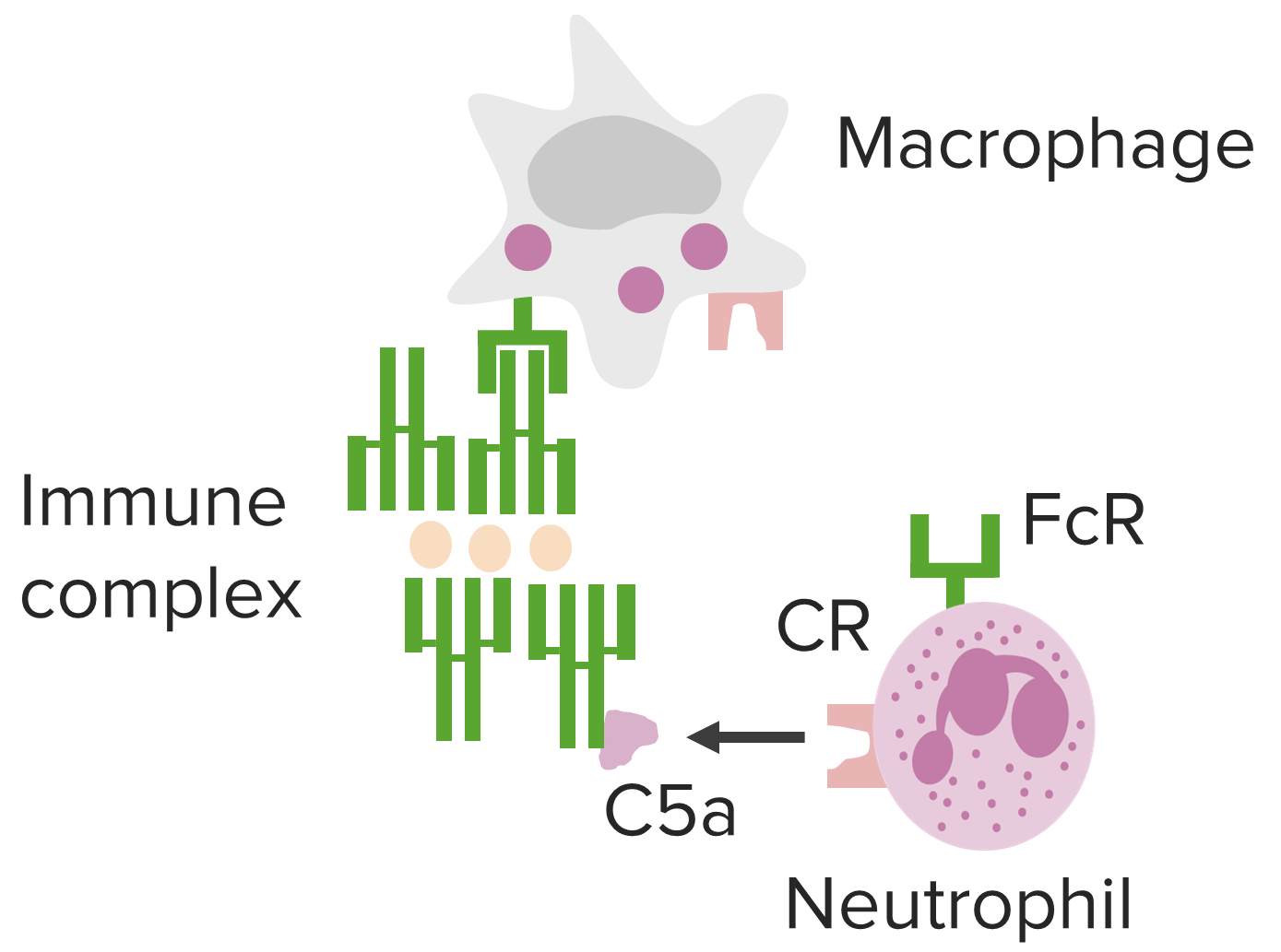
Vías mediadas por inmunocomplejos subyacentes a la hipersensibilidad tipo III.
Imagen por Lecturio.Las manifestaciones se ven afectadas por la vía de entrada, el( los LOS Neisseria) lugar(es) de depósito de inmunocomplejos y la persistencia del( los LOS Neisseria) antígeno(s).

Lesión tisular mediada por inmunocomplejos de hipersensibilidad tipo III en la pared de vasos sanguíneos, vasculitis, que se asocia a diversas afecciones.
Imagen por Lecturio.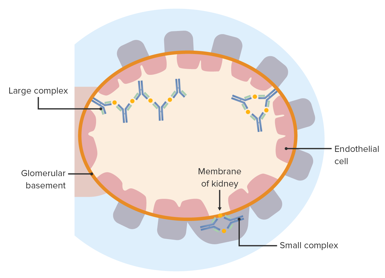
Enfermedad por inmunocomplejos: Los complejos antígeno-anticuerpo se depositan a ambos lados de la membrana basal glomerular. Estos provocan una reacción inflamatoria y causan una lesión glomerular.
Imagen por Lecturio.
Una paciente de 27 años con antecedente de exposición al moho. TC de alta resolución del tórax (ventana pulmonar) a nivel de los lóbulos inferiores muestra extensas opacidades en vidrio esmerilado (asteriscos), con focos superpuestos de atrapamiento aéreo lobular (flechas).
Imagen: “Hypersensitivity Pneumonitis” por Torres PP, Moreira MA, Silva DG, da Gama RR, Sugita DM, Moreira MA. Licencia: CC BY 4.0