Domina Conceptos Médicos

Estudia para la escuela de medicina y tus examenes con Lecturio.

Inmunoterapia Oncológica

La inmunoterapia oncológica es una terapia médica que avanza rápidamente y que aprovecha el sistema inmunitario para contener o eliminar las células cancerosas. Actualmente, las inmunoterapias se han incorporado a los LOS Neisseria regímenes de tratamiento para diferentes tipos de cáncer. Existen varios enfoques terapéuticos, incluido el uso de citoquinas, vacunas, virus Virus Viruses are infectious, obligate intracellular parasites composed of a nucleic acid core surrounded by a protein capsid. Viruses can be either naked (non-enveloped) or enveloped. The classification of viruses is complex and based on many factors, including type and structure of the nucleoid and capsid, the presence of an envelope, the replication cycle, and the host range. Virology oncolíticos, manipulación de células T o inmunoterapia celular adoptiva, o anticuerpos contra moléculas de puntos de control inmunitarios. Estas terapias brindan nuevas opciones para los LOS Neisseria cánceres avanzados, incluidos el melanoma Melanoma Melanoma is a malignant tumor arising from melanocytes, the melanin-producing cells of the epidermis. These tumors are most common in fair-skinned individuals with a history of excessive sun exposure and sunburns. Melanoma, el carcinoma de células renales, el adenocarcinoma de próstata, el cáncer de pulmón, el carcinoma urotelial, el linfoma de Hodgkin y la leucemia linfocítica aguda de células B refractaria. Con el sistema inmunológico involucrado, estos agentes conllevan efectos secundarios y toxicidades graves y potencialmente mortales.

Last updated: Dec 15, 2025

Editorial responsibility: Stanley Oiseth, Lindsay Jones, Evelin Maza

Inmunología Tumoral

Sistema inmune

El sistema inmune brinda defensa (inmunidad) contra patógenos invasores que van desde virus Virus Viruses are infectious, obligate intracellular parasites composed of a nucleic acid core surrounded by a protein capsid. Viruses can be either naked (non-enveloped) or enveloped. The classification of viruses is complex and based on many factors, including type and structure of the nucleoid and capsid, the presence of an envelope, the replication cycle, and the host range. Virology hasta parásitos, y los LOS Neisseria componentes están interconectados por la circulación sanguínea y linfática.

Las 2 líneas de defensa (que se superponen):

  • La inmunidad innata (que no es específica) implica:
    • Células dendríticas
    • Células asesinas naturales
    • Macrófagos
    • Neutrófilos
    • Eosinófilos
    • Basófilos
    • Mastocitos
  • Inmunidad adaptativa (basada en EN Erythema nodosum is an immune-mediated panniculitis (inflammation of the subcutaneous fat) caused by a type IV (delayed-type) hypersensitivity reaction. It commonly manifests in young women as tender, erythematous nodules on the shins. Erythema Nodosum el reconocimiento de antígenos específicos):
    • Inmunidad mediada por células: respuesta adaptativa en EN Erythema nodosum is an immune-mediated panniculitis (inflammation of the subcutaneous fat) caused by a type IV (delayed-type) hypersensitivity reaction. It commonly manifests in young women as tender, erythematous nodules on the shins. Erythema Nodosum las células/tejidos que involucran a las células T (que incluyen células T colaboradoras CD4+ y células T citotóxicas CD8+)
    • Inmunidad humoral: respuesta adaptativa en EN Erythema nodosum is an immune-mediated panniculitis (inflammation of the subcutaneous fat) caused by a type IV (delayed-type) hypersensitivity reaction. It commonly manifests in young women as tender, erythematous nodules on the shins. Erythema Nodosum los LOS Neisseria fluidos (“ humor Humor Defense Mechanisms”) que involucra células B e inmunoglobulinas

Reconocimiento de tumores

Cada día ocurren más de 20 000 eventos que dañan el ADN, los LOS Neisseria cuales se reparan a través de vías específicas, por lo que no causan un efecto duradero.

  • Las células no reparadas tienen el potencial de tener cambios malignos.
  • Pueden ocurrir más de 11 000 mutaciones genómicas y es posible que se expresen muchos antígenos tumorales nuevos.
    • Antígenos asociados a tumores: proteínas que se pueden expresar en EN Erythema nodosum is an immune-mediated panniculitis (inflammation of the subcutaneous fat) caused by a type IV (delayed-type) hypersensitivity reaction. It commonly manifests in young women as tender, erythematous nodules on the shins. Erythema Nodosum o sobre células tumorales o normales, a las que el sistema inmunitario es tolerante. Estos antígenos pueden incluir:
      • Productos de protooncogenes mutados y genes Genes A category of nucleic acid sequences that function as units of heredity and which code for the basic instructions for the development, reproduction, and maintenance of organisms. DNA Types and Structure supresores de tumores (e.g., mutaciones de p53)
      • Oncogenes Oncogenes Genes whose gain-of-function alterations lead to neoplastic cell transformation. They include, for example, genes for activators or stimulators of cell proliferation such as growth factors, growth factor receptors, protein kinases, signal transducers, nuclear phosphoproteins, and transcription factors. A prefix of ‘v-‘ before oncogene symbols indicates oncogenes captured and transmitted by retroviruses; the prefix ‘c-‘ before the gene symbol of an oncogene indicates it is the cellular homolog (proto-oncogenes) of a v-oncogene. Carcinogenesis sobreexpresados (e.g., HER2 HER2 A cell surface protein-tyrosine kinase receptor that is overexpressed in a variety of adenocarcinomas. It has extensive homology to and heterodimerizes with the EGF receptor, the ERBB-3 receptor, and the ERBB-4 receptor. Activation of the erbB-2 receptor occurs through heterodimer formation with a ligand-bound erbB receptor family member. Targeted and Other Nontraditional Antineoplastic Therapy/ Neu Neu A cell surface protein-tyrosine kinase receptor that is overexpressed in a variety of adenocarcinomas. It has extensive homology to and heterodimerizes with the EGF receptor, the ERBB-3 receptor, and the ERBB-4 receptor. Activation of the erbB-2 receptor occurs through heterodimer formation with a ligand-bound erbB receptor family member. Targeted and Other Nontraditional Antineoplastic Therapy)
      • Proteínas no oncogénicas sobreexpresadas o expresadas de manera aberrante (e.g., gp100 en EN Erythema nodosum is an immune-mediated panniculitis (inflammation of the subcutaneous fat) caused by a type IV (delayed-type) hypersensitivity reaction. It commonly manifests in young women as tender, erythematous nodules on the shins. Erythema Nodosum melanoma Melanoma Melanoma is a malignant tumor arising from melanocytes, the melanin-producing cells of the epidermis. These tumors are most common in fair-skinned individuals with a history of excessive sun exposure and sunburns. Melanoma)
      • Antígenos tumorales producidos por virus Virus Viruses are infectious, obligate intracellular parasites composed of a nucleic acid core surrounded by a protein capsid. Viruses can be either naked (non-enveloped) or enveloped. The classification of viruses is complex and based on many factors, including type and structure of the nucleoid and capsid, the presence of an envelope, the replication cycle, and the host range. Virology oncogénicos
      • Antígenos oncofetales (sustancias producidas por tumores y por tejidos fetales)
      • Glicolípidos y glicoproteínas alterados
      • Antígenos de diferenciación específicos de tipo celular (e.g., antígeno prostático específico)
    • Antígenos específicos de tumor Tumor Inflammation: restringidos a células cancerosas (no se encuentran en EN Erythema nodosum is an immune-mediated panniculitis (inflammation of the subcutaneous fat) caused by a type IV (delayed-type) hypersensitivity reaction. It commonly manifests in young women as tender, erythematous nodules on the shins. Erythema Nodosum células sanas)
  • Los LOS Neisseria linfocitos T son capaces de distinguir los LOS Neisseria antígenos propios de los LOS Neisseria no propios (vigilancia inmunológica).
    • Esta respuesta requiere señales coestimuladoras adicionales:
      • (1) El receptor Receptor Receptors are proteins located either on the surface of or within a cell that can bind to signaling molecules known as ligands (e.g., hormones) and cause some type of response within the cell. Receptors de células T reconoce su antígeno afín, tal TAL Renal Sodium and Water Regulation como lo presenta la célula presentadora de antígeno (e.g., célula dendrítica).
      • (2) Coestimulación: mejor caracterizada por la proteína B7 (CD80/86) en EN Erythema nodosum is an immune-mediated panniculitis (inflammation of the subcutaneous fat) caused by a type IV (delayed-type) hypersensitivity reaction. It commonly manifests in young women as tender, erythematous nodules on the shins. Erythema Nodosum la célula presentadora de antígeno que interactúa con el CD28 CD28 Costimulatory t-lymphocyte receptors that have specificity for CD80 antigen and CD86 antigen. Activation of this receptor results in increased t-cell proliferation, cytokine production and promotion of t-cell survival. T cells: Types and Functions de la célula T
    • Esta combinación de receptor Receptor Receptors are proteins located either on the surface of or within a cell that can bind to signaling molecules known as ligands (e.g., hormones) and cause some type of response within the cell. Receptors CD28 CD28 Costimulatory t-lymphocyte receptors that have specificity for CD80 antigen and CD86 antigen. Activation of this receptor results in increased t-cell proliferation, cytokine production and promotion of t-cell survival. T cells: Types and Functions y ligando B7, o “sinapsis inmunológica”, estimula las células T.
    • Son posibles otras combinaciones de receptor-ligando entre las células T activadas y otras células.
  • Regulación de la respuesta:
    • La estimulación de la respuesta es a través de las acciones de moléculas agonistas (e.g., OX40).
    • Las células T reguladoras (Tregs) amortiguan la respuesta de las células T.
    • La inhibición de la señal coestimuladora (y, por lo tanto, la reducción de la actividad de las células T) se realiza a través de moléculas de punto de control inmunitario, como:
      • Muerte celular programada-1 ( PD-1 PD-1 An inhibitory t-lymphocyte receptor that has specificity for CD274 antigen and programmed cell death 1 ligand 2 protein. Signaling by the receptor limits T cell proliferation and interferon gamma synthesis. The receptor also may play an essential role in the regulatory pathway that induces peripheral tolerance. T cells: Types and Functions)
      • Proteína 4 asociada a linfocitos T citotóxicos (CTLA-4)
    • La eliminación de esta inhibición (inmunoterapia de bloqueo de puntos de control) mejora la respuesta inmunitaria contra el tumor Tumor Inflammation.
El modelo de 2 señales de la dependencia de las células T de la coestimulación

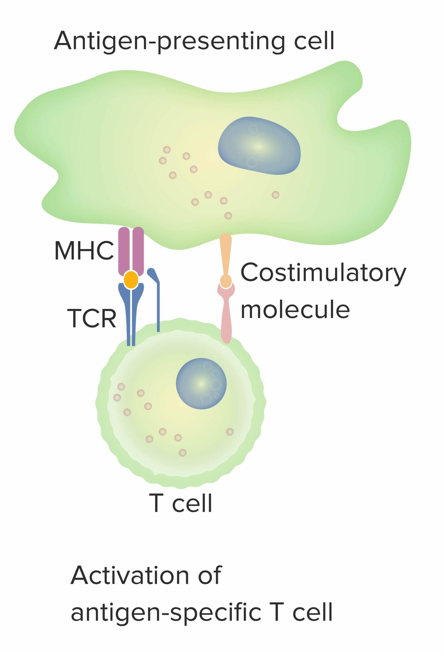
El modelo de 2 señales de la dependencia de las células T a la coestimulación:
Cuando tanto la señal 1 (receptor de células T que se une al antígeno afín presentado por la molécula del CMH en la célula presentadora de antígeno) como la señal 2 (interacción de la molécula coestimuladora entre la célula presentadora de antígeno y la célula T) están presentes, la célula T madura está totalmente activada.
El punto naranja indica la unión adecuada entre el antígeno y el receptor de células T.

Imagen por Lecturio.

Inmunoedición del cáncer

La inmunoedición es un proceso que consiste en EN Erythema nodosum is an immune-mediated panniculitis (inflammation of the subcutaneous fat) caused by a type IV (delayed-type) hypersensitivity reaction. It commonly manifests in young women as tender, erythematous nodules on the shins. Erythema Nodosum la supresión inmunológica del tumor Tumor Inflammation, pero puede conducir a la progresión del tumor Tumor Inflammation. La inmunoedición ocurre en EN Erythema nodosum is an immune-mediated panniculitis (inflammation of the subcutaneous fat) caused by a type IV (delayed-type) hypersensitivity reaction. It commonly manifests in young women as tender, erythematous nodules on the shins. Erythema Nodosum 3 fases principales:

  • Eliminación:
    • El daño inicial y la destrucción de las células tumorales por la inmunidad innata y adaptativa
    • La finalización de la fase significa que no hay crecimiento del cáncer.
  • Equilibrio:
    • Ocurre cuando las células tumorales sobreviven al intento de eliminación inicial
    • Estas células no pueden progresar, siendo mantenidas en un estado de latencia por el sistema inmune adaptativo.
    • En esta fase, se edita la inmunogenicidad del tumor, donde las células T continúan atacando selectivamente a las células tumorales altamente inmunogénicas.
    • Este ataque deja a otras células con menos inmunogenicidad para desarrollar potencialmente resistencia a la respuesta inmune.
  • Escape:
    • Con mecanismos inmunitarios constantes que mantienen en equilibrio a las células tumorales inestables, pueden surgir variantes de células tumorales.
    • Estas células cancerosas pueden expresar menos antígenos en sus superficies o perder su expresión de del CMH de clase I.
    • Las variantes también pueden protegerse del ataque de las células T mediante la expresión de moléculas de punto de control en EN Erythema nodosum is an immune-mediated panniculitis (inflammation of the subcutaneous fat) caused by a type IV (delayed-type) hypersensitivity reaction. It commonly manifests in young women as tender, erythematous nodules on the shins. Erythema Nodosum sus superficies, como las células normales.
    • La creación de un estado inmunosupresor en EN Erythema nodosum is an immune-mediated panniculitis (inflammation of the subcutaneous fat) caused by a type IV (delayed-type) hypersensitivity reaction. It commonly manifests in young women as tender, erythematous nodules on the shins. Erythema Nodosum el microambiente es otra forma de crecer sin interferencia inmunológica.
Carcinogénesis por evasión inmune

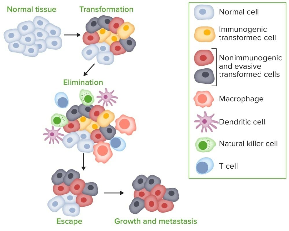
Carcinogénesis por evasión inmune:
Cuando las células tumorales se transforman a partir de células normales, los sistemas inmunitarios innato y adaptativo detectan y eliminan las células transformadas incluso antes de que la enfermedad sea clínicamente aparente (eliminación). El proceso entra en equilibrio, ya que es posible que las variantes de células tumorales no se eliminen por completo. Sin embargo, el sistema inmunitario intenta controlar el crecimiento de las células tumorales ejerciendo una presión selectiva sobre las células tumorales altamente inmunogénicas. Se edita la inmunogenicidad de las células tumorales, dejando que las células con inmunogenicidad reducida crezcan y evadan la inmunovigilancia, lo que lleva a la progresión de las células a la fase de escape, donde las células menos inmunogénicas crecen progresivamente y se vuelven clínicamente cancerosas.

Imagen por Lecturio.

Inmunoterapia contra el cáncer

La inmunoterapia contra el cáncer estimula el sistema inmunitario para que responda a una neoplasia maligna, activando diferentes aspectos del sistema inmunitario para atacar las células cancerosas.

  • El sistema inmunológico es manipulado a través de:
    • Fortalecimiento de las respuestas inmunitarias del huésped contra los LOS Neisseria tumores.
    • Suministro de componentes modificados del sistema inmunitario
    • Contrarrestar las señales producidas por las células cancerosas que suprimen las respuestas inmunitarias
  • Objetivos de la inmunoterapia:
    • Utilizar la especificidad y la memoria a largo plazo de la respuesta inmunitaria adaptativa.
    • Lograr una regresión tumoral duradera.
    • Curar la enfermedad.
    • Mejorar la supervivencia global.
  • Opciones terapéuticas:
    • Algunos agentes actúan sobre diferentes componentes del sistema inmunitario:
      • Citoquinas
      • Células T y otros tipos de células
      • Moléculas de punto de control
    • Otros agentes utilizan la respuesta que producen virus Virus Viruses are infectious, obligate intracellular parasites composed of a nucleic acid core surrounded by a protein capsid. Viruses can be either naked (non-enveloped) or enveloped. The classification of viruses is complex and based on many factors, including type and structure of the nucleoid and capsid, the presence of an envelope, the replication cycle, and the host range. Virology o bacterias sobre el sistema inmunitario.

Terapéutica

Citoquinas y vacunas

  • Interleucina-2 (IL-2)
    • Tiene efectos pleiotrópicos tanto en EN Erythema nodosum is an immune-mediated panniculitis (inflammation of the subcutaneous fat) caused by a type IV (delayed-type) hypersensitivity reaction. It commonly manifests in young women as tender, erythematous nodules on the shins. Erythema Nodosum las células T citotóxicas como en EN Erythema nodosum is an immune-mediated panniculitis (inflammation of the subcutaneous fat) caused by a type IV (delayed-type) hypersensitivity reaction. It commonly manifests in young women as tender, erythematous nodules on the shins. Erythema Nodosum las Tregs.
    • Altas dosis:
      • Promueven la actividad de las células T efectoras CD8+ y las células asesinas naturales
      • Promueven la diferenciación de las células CD4+ en EN Erythema nodosum is an immune-mediated panniculitis (inflammation of the subcutaneous fat) caused by a type IV (delayed-type) hypersensitivity reaction. It commonly manifests in young women as tender, erythematous nodules on the shins. Erythema Nodosum las subclases T colaboradora tipo 1 ( Th1 Th1 A subset of helper-inducer T-lymphocytes which synthesize and secrete interleukin-2; interferon-gamma; and interleukin-12. Due to their ability to kill antigen-presenting cells and their lymphokine-mediated effector activity, th1 cells are associated with vigorous delayed-type hypersensitivity reactions. T cells: Types and Functions) y colaboradora tipo 2
    • Dosis bajas:
      • ↑ Expansión de Tregs
      • Inhiben la formación de células Th17 Th17 A subset of helper-effector T-lymphocytes which synthesize and secrete interleukins il-17; il-17f; and il-22. These cytokines are involved in host defenses and tissue inflammation in autoimmune diseases. T cells: Types and Functions implicadas en EN Erythema nodosum is an immune-mediated panniculitis (inflammation of the subcutaneous fat) caused by a type IV (delayed-type) hypersensitivity reaction. It commonly manifests in young women as tender, erythematous nodules on the shins. Erythema Nodosum la autoinmunidad
    • Se utiliza para el melanoma Melanoma Melanoma is a malignant tumor arising from melanocytes, the melanin-producing cells of the epidermis. These tumors are most common in fair-skinned individuals with a history of excessive sun exposure and sunburns. Melanoma y el carcinoma de células renales
  • Interferón alfa-2b ( IFN IFN Interferon (IFN) is a cytokine with antiviral properties (it interferes with viral infections) and various roles in immunoregulation. The different types are type I IFN (IFN-ɑ and IFN-β), type II IFN (IFN-ɣ), and type III IFN (IFN-ƛ). Interferons⍺-2b):
    • Mejora los LOS Neisseria efectos mediados por Th1 Th1 A subset of helper-inducer T-lymphocytes which synthesize and secrete interleukin-2; interferon-gamma; and interleukin-12. Due to their ability to kill antigen-presenting cells and their lymphokine-mediated effector activity, th1 cells are associated with vigorous delayed-type hypersensitivity reactions. T cells: Types and Functions
    • Efectos citolíticos y antiproliferativos
    • Utilizado como tratamiento adyuvante para el melanoma Melanoma Melanoma is a malignant tumor arising from melanocytes, the melanin-producing cells of the epidermis. These tumors are most common in fair-skinned individuals with a history of excessive sun exposure and sunburns. Melanoma de alto riesgo
  • Lenalidomida y pomalidomida:
    • Actividad inmunomoduladora (↓ factor de necrosis Necrosis The death of cells in an organ or tissue due to disease, injury or failure of the blood supply. Ischemic Cell Damage tumoral-⍺, ↑ células asesinas naturales e IL-2) y antiangiogénica
    • Inducen la detención del ciclo celular y muerte → inhiben el crecimiento tumoral
  • Vacunas (terapéuticas):
    • Sipuleucel-T Sipuleucel-T An approved vaccine-based therapy for advanced (castration-resistant hormone-refractory) prostate cancer. First, there is an autologous dendritic cell preparation targeting prostatic acid phosphatase (PAP). Blood mononuclear cells are obtained via leukapheresis. Antigen-presenting cells are isolated and then activated in vitro by PAP fused to granulocyte macrophage colony-stimulating factor (GM-CSF). The reinfusion of the product produces T-cell activity against tumors expressing PAP. Cancer Immunotherapy ( provenge Provenge An approved vaccine-based therapy for advanced (castration-resistant hormone-refractory) prostate cancer. First, there is an autologous dendritic cell preparation targeting prostatic acid phosphatase (PAP). Blood mononuclear cells are obtained via leukapheresis. Antigen-presenting cells are isolated and then activated in vitro by PAP fused to granulocyte macrophage colony-stimulating factor (GM-CSF). The reinfusion of the product produces T-cell activity against tumors expressing PAP. Cancer Immunotherapy):
      • Actualmente, la terapia basada en EN Erythema nodosum is an immune-mediated panniculitis (inflammation of the subcutaneous fat) caused by a type IV (delayed-type) hypersensitivity reaction. It commonly manifests in young women as tender, erythematous nodules on the shins. Erythema Nodosum vacunas aprobada para el cáncer de próstata avanzado (resistente a la castración y refractario a las hormonas)
      • Preparación de células dendríticas autólogas dirigida a la fosfatasa ácida prostática
      • Las células mononucleares sanguíneas se obtienen mediante leucoféresis.
      • Las células presentadoras de antígeno se aíslan y luego se activan in vitro mediante fosfatasa ácida prostática fusionada con el factor estimulante de colonias de granulocitos y macrófagos.
      • La reinfusión del producto produce actividad de células T contra tumores que expresan fosfatasa ácida prostática.
    • Bacilo de Calmette-Guérin ( BCG BCG An active immunizing agent and a viable avirulent attenuated strain of Mycobacterium bovis, which confers immunity to mycobacterial infections. It is used also in immunotherapy of neoplasms due to its stimulation of antibodies and non-specific immunity. Cancer Immunotherapy)
      • Derivado de una cepa atenuada de Mycobacterium bovis Mycobacterium bovis The bovine variety of the tubercle Bacillus. It is called also Mycobacterium tuberculosis var. Bovis. Mycobacterium
      • Aprobado para el tratamiento intravesical del cáncer de vejiga en EN Erythema nodosum is an immune-mediated panniculitis (inflammation of the subcutaneous fat) caused by a type IV (delayed-type) hypersensitivity reaction. It commonly manifests in young women as tender, erythematous nodules on the shins. Erythema Nodosum estadio temprano
      • Activa una reacción inmune contra el tumor Tumor Inflammation.

Virus Virus Viruses are infectious, obligate intracellular parasites composed of a nucleic acid core surrounded by a protein capsid. Viruses can be either naked (non-enveloped) or enveloped. The classification of viruses is complex and based on many factors, including type and structure of the nucleoid and capsid, the presence of an envelope, the replication cycle, and the host range. Virology oncolíticos

  • Clase emergente de terapias contra el cáncer que utiliza virus Virus Viruses are infectious, obligate intracellular parasites composed of a nucleic acid core surrounded by a protein capsid. Viruses can be either naked (non-enveloped) or enveloped. The classification of viruses is complex and based on many factors, including type and structure of the nucleoid and capsid, the presence of an envelope, the replication cycle, and the host range. Virology para infectar y destruir células tumorales
  • Los LOS Neisseria virus Virus Viruses are infectious, obligate intracellular parasites composed of a nucleic acid core surrounded by a protein capsid. Viruses can be either naked (non-enveloped) or enveloped. The classification of viruses is complex and based on many factors, including type and structure of the nucleoid and capsid, the presence of an envelope, the replication cycle, and the host range. Virology están modificados genéticamente para lisar preferentemente las células cancerosas y evitar las células normales.
  • Talimogén laherparepvec
    • La 1ra inmunoterapia oncolítica aprobada
    • Virus Virus Viruses are infectious, obligate intracellular parasites composed of a nucleic acid core surrounded by a protein capsid. Viruses can be either naked (non-enveloped) or enveloped. The classification of viruses is complex and based on many factors, including type and structure of the nucleoid and capsid, the presence of an envelope, the replication cycle, and the host range. Virology del herpes simple modificado genéticamente, modificado para expresar factor estimulante de colonias de granulocitos y macrófagos
    • Utilizado en EN Erythema nodosum is an immune-mediated panniculitis (inflammation of the subcutaneous fat) caused by a type IV (delayed-type) hypersensitivity reaction. It commonly manifests in young women as tender, erythematous nodules on the shins. Erythema Nodosum melanoma Melanoma Melanoma is a malignant tumor arising from melanocytes, the melanin-producing cells of the epidermis. These tumors are most common in fair-skinned individuals with a history of excessive sun exposure and sunburns. Melanoma no resecable
    • Se puede combinar con moléculas de punto de control

Inmunoterapia adoptiva celular

Estas terapias se desarrollan con base en EN Erythema nodosum is an immune-mediated panniculitis (inflammation of the subcutaneous fat) caused by a type IV (delayed-type) hypersensitivity reaction. It commonly manifests in young women as tender, erythematous nodules on the shins. Erythema Nodosum la manipulación de las células del individuo a través de la replicación in vitro de células T específicas del tumor Tumor Inflammation, que se reinfunden mediante trasplante autólogo.

Técnicas de manipulación de células T (y otros tipos de células inmunitarias):

  • Células T del receptor Receptor Receptors are proteins located either on the surface of or within a cell that can bind to signaling molecules known as ligands (e.g., hormones) and cause some type of response within the cell. Receptors de antígeno quimérico:
    • Las células T están equipadas con el receptor Receptor Receptors are proteins located either on the surface of or within a cell that can bind to signaling molecules known as ligands (e.g., hormones) and cause some type of response within the cell. Receptors de antígeno quimérico, un receptor Receptor Receptors are proteins located either on the surface of or within a cell that can bind to signaling molecules known as ligands (e.g., hormones) and cause some type of response within the cell. Receptors sintético que se une a las células cancerosas.
    • Estas células T se dirigen a las proteínas de las células B leucémicas.
    • Cuando se reconocen las células B malignas, las células se destruyen mediante la liberación de citoquinas, perforina y granzimas.
    • Los LOS Neisseria efectos secundarios de la terapia incluyen:
      • Síndrome de liberación marcada de citoquinas (tormenta de citoquinas)
      • Neurotoxicidad
  • Linfocitos infiltrantes de tumores:
    • La población celular de linfocitos infiltrantes de tumores se obtiene directamente del tumor Tumor Inflammation resecado.
    • La replicación de las células se ve VE Ventilation: Mechanics of Breathing facilitada por el cocultivo con IL-2.
    • Antes de la infusión, el individuo afectado recibe quimioterapia o radiación para reducir las Treg.
    • Cuando se reintroducen, los LOS Neisseria linfocitos infiltrantes de tumores (principalmente células CD8+) reconocen y atacan los LOS Neisseria antígenos tumorales.
  • Receptor Receptor Receptors are proteins located either on the surface of or within a cell that can bind to signaling molecules known as ligands (e.g., hormones) and cause some type of response within the cell. Receptors de células T monoclonales inmunomovilizadores contra el cáncer:
    • Terapia con receptores de células T diseñada
    • En EN Erythema nodosum is an immune-mediated panniculitis (inflammation of the subcutaneous fat) caused by a type IV (delayed-type) hypersensitivity reaction. It commonly manifests in young women as tender, erythematous nodules on the shins. Erythema Nodosum algunos casos, es posible que las células T no reconozcan las células tumorales o no se activen o repliquen lo suficiente.
    • Estas células T pueden equiparse con un nuevo receptor Receptor Receptors are proteins located either on the surface of or within a cell that can bind to signaling molecules known as ligands (e.g., hormones) and cause some type of response within the cell. Receptors para atacar antígenos de cáncer específicos.
    • Un ejemplo es el medicamento en EN Erythema nodosum is an immune-mediated panniculitis (inflammation of the subcutaneous fat) caused by a type IV (delayed-type) hypersensitivity reaction. It commonly manifests in young women as tender, erythematous nodules on the shins. Erythema Nodosum investigación, tebentafusp, un receptor Receptor Receptors are proteins located either on the surface of or within a cell that can bind to signaling molecules known as ligands (e.g., hormones) and cause some type of response within the cell. Receptors de células T soluble que se dirige a gp100, un antígeno de melanoma Melanoma Melanoma is a malignant tumor arising from melanocytes, the melanin-producing cells of the epidermis. These tumors are most common in fair-skinned individuals with a history of excessive sun exposure and sunburns. Melanoma uveal.
  • Enganchadores biespecíficos de células T:
    • Anticuerpos biespecíficos que reclutan células T para las células malignas
    • Objetivo CD3 CD3 Complex of at least five membrane-bound polypeptides in mature T-lymphocytes that are non-covalently associated with one another and with the T-cell receptor. The CD3 complex includes the gamma, delta, epsilon, zeta, and eta chains (subunits). When antigen binds to the T-cell receptor, the CD3 complex transduces the activating signals to the cytoplasm of the T-cell. The CD3 gamma and delta chains (subunits) are separate from and not related to the gamma/delta chains of the T-cell receptor. T cells: Types and Functions y antígenos específicos del tumor Tumor Inflammation, lo que en EN Erythema nodosum is an immune-mediated panniculitis (inflammation of the subcutaneous fat) caused by a type IV (delayed-type) hypersensitivity reaction. It commonly manifests in young women as tender, erythematous nodules on the shins. Erythema Nodosum última instancia conduce a la citotoxicidad de las células T contra el tumor Tumor Inflammation
    • Blinatumomab: enganchador bioespecífico de células T aprobado
  • Terapia de células asesinas naturales:
    • A diferencia de las células T, las células asesinas naturales no se adaptan a un antígeno específico.
    • Las células asesinas naturales se pueden mejorar con el receptor Receptor Receptors are proteins located either on the surface of or within a cell that can bind to signaling molecules known as ligands (e.g., hormones) and cause some type of response within the cell. Receptors de antígeno quimérico para atacar eficazmente las células malignas.
Inmunoterapia celular adaptativa

Inmunoterapia celular adaptativa:
1) Se recogen las células T.
2) Las células T se modifican para expresar receptores de antígenos quiméricos, seguido de replicación in vitro.
3) Estas células luego se infunden en el mismo individuo. Estas células entran en circulación, reconociendo y destruyendo células malignas.
4) Seguimiento posterior de la respuesta de la enfermedad.

Imagen: “CAR-Engineered T-Cell Adoptive Transfer” por Caron A. Jacobson and Jerome Ritz. Licencia: Dominio Público

Inhibidores de puntos de control

  • PD-1 PD-1 An inhibitory t-lymphocyte receptor that has specificity for CD274 antigen and programmed cell death 1 ligand 2 protein. Signaling by the receptor limits T cell proliferation and interferon gamma synthesis. The receptor also may play an essential role in the regulatory pathway that induces peripheral tolerance. T cells: Types and Functions:
    • Una proteína transmembrana (molécula de punto de control) en EN Erythema nodosum is an immune-mediated panniculitis (inflammation of the subcutaneous fat) caused by a type IV (delayed-type) hypersensitivity reaction. It commonly manifests in young women as tender, erythematous nodules on the shins. Erythema Nodosum las células T que se une a:
      • Ligando 1 de muerte celular programada (PD-L1): se encuentra en EN Erythema nodosum is an immune-mediated panniculitis (inflammation of the subcutaneous fat) caused by a type IV (delayed-type) hypersensitivity reaction. It commonly manifests in young women as tender, erythematous nodules on the shins. Erythema Nodosum múltiples tejidos, incluidas las células tumorales
      • PD-L2: se encuentra en EN Erythema nodosum is an immune-mediated panniculitis (inflammation of the subcutaneous fat) caused by a type IV (delayed-type) hypersensitivity reaction. It commonly manifests in young women as tender, erythematous nodules on the shins. Erythema Nodosum las células hematopoyéticas
    • Interacción PD-1 PD-1 An inhibitory t-lymphocyte receptor that has specificity for CD274 antigen and programmed cell death 1 ligand 2 protein. Signaling by the receptor limits T cell proliferation and interferon gamma synthesis. The receptor also may play an essential role in the regulatory pathway that induces peripheral tolerance. T cells: Types and Functions–PD-L1/2:
      • Inhibe la apoptosis Apoptosis A regulated cell death mechanism characterized by distinctive morphologic changes in the nucleus and cytoplasm, including the endonucleolytic cleavage of genomic DNA, at regularly spaced, internucleosomal sites, I.e., DNA fragmentation. It is genetically-programmed and serves as a balance to mitosis in regulating the size of animal tissues and in mediating pathologic processes associated with tumor growth. Ischemic Cell Damage de las células tumorales y promueve el agotamiento de las células T efectoras periféricas
      • Aumenta la conversión de células T efectoras a Tregs
      • El bloqueo de esta interacción produce un efecto terapéutico → ↑ actividad de las células T contra las células tumorales
  • Inhibidores de la interacción PD-1-PD-L1:
    • Anticuerpos que inhiben PD-1 PD-1 An inhibitory t-lymphocyte receptor that has specificity for CD274 antigen and programmed cell death 1 ligand 2 protein. Signaling by the receptor limits T cell proliferation and interferon gamma synthesis. The receptor also may play an essential role in the regulatory pathway that induces peripheral tolerance. T cells: Types and Functions:
      • Pembrolizumab Pembrolizumab Squamous Cell Carcinoma (SCC)
      • Nivolumab Nivolumab A genetically engineered, fully humanized immunoglobulin g4 monoclonal antibody that binds to the pd-1 receptor, activating an immune response to tumor cells. It is used as monotherapy or in combination with ipilimumab for the treatment of advanced malignant melanoma. It is also used in the treatment of advanced or recurring non-small cell lung cancer; renal cell carcinoma; and Hodgkin’s lymphoma. Melanoma
      • Dostarlimab
    • Anticuerpos que inhiben PD-L1:
      • Atezolizumab (1er inhibidor de PD-L1 aprobado)
      • Durvalumab
      • Avelumab
  • CTLA-4:
    • Molécula de punto de control con un papel importante en EN Erythema nodosum is an immune-mediated panniculitis (inflammation of the subcutaneous fat) caused by a type IV (delayed-type) hypersensitivity reaction. It commonly manifests in young women as tender, erythematous nodules on the shins. Erythema Nodosum la regulación inhibitoria de la respuesta inmunitaria
    • Se encuentra en EN Erythema nodosum is an immune-mediated panniculitis (inflammation of the subcutaneous fat) caused by a type IV (delayed-type) hypersensitivity reaction. It commonly manifests in young women as tender, erythematous nodules on the shins. Erythema Nodosum la superficie de los LOS Neisseria linfocitos T CD4+ y CD8+, donde tiene una alta afinidad por los LOS Neisseria receptores coestimuladores CD80 y CD86 en EN Erythema nodosum is an immune-mediated panniculitis (inflammation of the subcutaneous fat) caused by a type IV (delayed-type) hypersensitivity reaction. It commonly manifests in young women as tender, erythematous nodules on the shins. Erythema Nodosum las células presentadoras de antígenos.
    • ↓ proliferación de células T y producción de IL-2
    • La inhibición conduce a una mayor respuesta inmunitaria y reducción del tumor Tumor Inflammation.
  • Anticuerpos que inhiben CTLA–4: ipilimumab (1er anticuerpo de punto de control aprobado para su uso)
  • Efectos secundarios importantes de los LOS Neisseria inhibidores de puntos de control:
    • Síndrome de liberación de citoquinas:
      • Similar a la terapia de células T con receptor Receptor Receptors are proteins located either on the surface of or within a cell that can bind to signaling molecules known as ligands (e.g., hormones) and cause some type of response within the cell. Receptors de antígeno quimérico
      • Se manifiesta con fiebre con o sin disfunción multiorgánica
      • Los LOS Neisseria corticosteroides u otros inmunomoduladores pueden mejorar las manifestaciones tóxicas inmunomediadas.
    • Fatiga
    • Reacciones relacionadas con la perfusión
    • Toxicidad dermatológica
    • Infecciones oportunistas
    • Colitis Colitis Inflammation of the colon section of the large intestine, usually with symptoms such as diarrhea (often with blood and mucus), abdominal pain, and fever. Pseudomembranous Colitis
    • Toxicidades endocrinas (e.g., hipertiroidismo o hipotiroidismo, insuficiencia suprarrenal, diabetes Diabetes Diabetes mellitus (DM) is a metabolic disease characterized by hyperglycemia and dysfunction of the regulation of glucose metabolism by insulin. Type 1 DM is diagnosed mostly in children and young adults as the result of autoimmune destruction of β cells in the pancreas and the resulting lack of insulin. Type 2 DM has a significant association with obesity and is characterized by insulin resistance. Diabetes Mellitus mellitus)
    • Neutropenia Neutropenia Neutrophils are an important component of the immune system and play a significant role in the eradication of infections. Low numbers of circulating neutrophils, referred to as neutropenia, predispose the body to recurrent infections or sepsis, though patients can also be asymptomatic. Neutropenia, trombocitopenia
Inhibidores del punto de control inmunitario

Inhibidores del punto de control inmunitario:
Arriba: Las células presentadoras de antígenos procesan antígenos asociados a tumores y los unen con moléculas del CMH.
Las células presentadoras de antígenos migran al ganglio linfático (en áreas predominantes de células T) y presentan antígenos asociados a tumores a las células T vírgenes.
La activación de la célula T requiere 2 señales. La 1ra está mediada por la unión de antígenos asociados a tumores a un receptor de células T. La 2dada señal puede provenir de la unión del linfocito T CD28 al coestimulador CD80/CD86, que activa el linfocito T.
Sin embargo, cuando el linfocito T-CTLA-4 interactúa con las mismas moléculas de células presentadoras de antígeno CD80/CD86, el efecto es inhibitorio (anergia de linfocitos T o no se produce activación de linfocitos T).
Por lo tanto, CTLA-4 y CD28 compiten por la unión a las proteínas CD80/CD86. La acción de bloqueo anti-CTLA-4 de ipilimumab restaura la señalización del proactivador de CD28, lo que da como resultado respuestas de linfocitos T antitumorales.
Abajo: En los tejidos periféricos, la célula T activada puede desactivarse mediante la unión de la célula de muerte programada 1 de células T (PD-1) con el ligando 1 de muerte celular programada (PD-L1) (o PD-L2) expresado en las células tumorales. La acción bloqueadora anti-PD-1 o anti-PD-L1 por parte de los anticuerpos monoclonales (e.g., nivolumab, pembrolizumab, atezolizumab) restablece la actividad eficaz de los linfocitos T antitumorales.

Imagen: Figure 1 Effect of ICIs on T lymphocytes” por Franzin, R. et al. Licencia: CC BY 4.0

Comparación de Terapéutica

Comparación de agentes que mejoran el sistema inmunitario (uso de citocinas, organismos para infectar células tumorales y manipulación de células inmunitarias)
Tipo de terapéutica Agentes Indicaciones
Citocinas inmunoestimuladoras IL–2
  • Melanoma Melanoma Melanoma is a malignant tumor arising from melanocytes, the melanin-producing cells of the epidermis. These tumors are most common in fair-skinned individuals with a history of excessive sun exposure and sunburns. Melanoma
  • Cáncer de células renales
IFNa-2b
  • Melanoma Melanoma Melanoma is a malignant tumor arising from melanocytes, the melanin-producing cells of the epidermis. These tumors are most common in fair-skinned individuals with a history of excessive sun exposure and sunburns. Melanoma
  • Sarcoma de Kaposi Kaposi A multicentric, malignant neoplastic vascular proliferation characterized by the development of bluish-red cutaneous nodules, usually on the lower extremities, most often on the toes or feet, and slowly increasing in size and number and spreading to more proximal areas. The tumors have endothelium-lined channels and vascular spaces admixed with variably sized aggregates of spindle-shaped cells, and often remain confined to the skin and subcutaneous tissue, but widespread visceral involvement may occur. Hhv-8 is the suspected cause. There is also a high incidence in AIDS patients. AIDS-defining Conditions relacionado con el SIDA
  • Linfoma folicular
  • Leucemia de células pilosas
  • Verrugas genitales
Vacunas Sipuleucel-T Sipuleucel-T An approved vaccine-based therapy for advanced (castration-resistant hormone-refractory) prostate cancer. First, there is an autologous dendritic cell preparation targeting prostatic acid phosphatase (PAP). Blood mononuclear cells are obtained via leukapheresis. Antigen-presenting cells are isolated and then activated in vitro by PAP fused to granulocyte macrophage colony-stimulating factor (GM-CSF). The reinfusion of the product produces T-cell activity against tumors expressing PAP. Cancer Immunotherapy Adenocarcinoma de próstata resistente a la castración
Bacilo de Calmette-Guérin ( BCG BCG An active immunizing agent and a viable avirulent attenuated strain of Mycobacterium bovis, which confers immunity to mycobacterial infections. It is used also in immunotherapy of neoplasms due to its stimulation of antibodies and non-specific immunity. Cancer Immunotherapy) Cáncer de vejiga
Agentes inmunomoduladores
  • Lenalidomida
  • Pomalidomida
Mieloma múltiple
Virus Virus Viruses are infectious, obligate intracellular parasites composed of a nucleic acid core surrounded by a protein capsid. Viruses can be either naked (non-enveloped) or enveloped. The classification of viruses is complex and based on many factors, including type and structure of the nucleoid and capsid, the presence of an envelope, the replication cycle, and the host range. Virology oncolíticos Talimogén laherparepvec Melanoma Melanoma Melanoma is a malignant tumor arising from melanocytes, the melanin-producing cells of the epidermis. These tumors are most common in fair-skinned individuals with a history of excessive sun exposure and sunburns. Melanoma
Terapia de células T con receptor Receptor Receptors are proteins located either on the surface of or within a cell that can bind to signaling molecules known as ligands (e.g., hormones) and cause some type of response within the cell. Receptors de antígeno quimérico Los LOS Neisseria agentes están disponibles bajo el programa de estrategia de evaluación y mitigación de riesgos de la FDA (solo para uso autólogo).
  • Leucemia linfocítica aguda
  • Linfoma folicular
  • Linfoma de células B grandes
  • Linfoma de células del manto
  • Mieloma múltiple
Enganchadores biespecíficos de célular T (terapia dirigida a CD3 CD3 Complex of at least five membrane-bound polypeptides in mature T-lymphocytes that are non-covalently associated with one another and with the T-cell receptor. The CD3 complex includes the gamma, delta, epsilon, zeta, and eta chains (subunits). When antigen binds to the T-cell receptor, the CD3 complex transduces the activating signals to the cytoplasm of the T-cell. The CD3 gamma and delta chains (subunits) are separate from and not related to the gamma/delta chains of the T-cell receptor. T cells: Types and Functions) Blinatumomab Leucemia linfocítica aguda
Comparación de inhibidores de puntos de control inmunitarios
Tipo de terapéutica Agentes Indicaciones
Anti-PD-1 Pembrolizumab Pembrolizumab Squamous Cell Carcinoma (SCC)
  • Cáncer de mama
  • Cáncer cutáneo de células escamosas
  • Cáncer de cuello uterino
  • Cáncer endometrial
  • Cáncer de esófago
  • Cáncer gástrico
  • Cáncer de cabeza y cuello
  • Cáncer hepatocelular
  • Linfoma de Hodgkin
  • Melanoma Melanoma Melanoma is a malignant tumor arising from melanocytes, the melanin-producing cells of the epidermis. These tumors are most common in fair-skinned individuals with a history of excessive sun exposure and sunburns. Melanoma
  • Cáncer de células de Merkel
  • Cáncer de inestabilidad de microsatélites—alta o reparación deficiente de desajustes
  • Cáncer de células renales
  • Cáncer urotelial
Nivolumab Nivolumab A genetically engineered, fully humanized immunoglobulin g4 monoclonal antibody that binds to the pd-1 receptor, activating an immune response to tumor cells. It is used as monotherapy or in combination with ipilimumab for the treatment of advanced malignant melanoma. It is also used in the treatment of advanced or recurring non-small cell lung cancer; renal cell carcinoma; and Hodgkin’s lymphoma. Melanoma
  • Cáncer colonrectal
  • Melanoma Melanoma Melanoma is a malignant tumor arising from melanocytes, the melanin-producing cells of the epidermis. These tumors are most common in fair-skinned individuals with a history of excessive sun exposure and sunburns. Melanoma
  • Cáncer de esófago
  • Cáncer gástrico
  • Cáncer de cabeza y cuello
  • Linfoma de Hodgkin
  • Cáncer hepatocelular
  • Mesothelioma Mesothelioma Malignant mesothelioma (usually referred to as simply mesothelioma) is the malignant growth of mesothelial cells, most commonly affecting the pleura. The majority of cases are associated with occupational exposure to asbestos that occurred > 20 years before clinical onset, which includes dyspnea, chest pain, coughing, fatigue, and weight loss. Malignant Mesothelioma
  • Cáncer de células renales
  • Cáncer urotelial
Dostarlimab
  • Cáncer endometrial
  • Cáncer de reparación deficiente de desajustes (tumores sólidos)
Anti-PD-L1 Atezolizumab
  • Cáncer hepatocelular
  • Melanoma Melanoma Melanoma is a malignant tumor arising from melanocytes, the melanin-producing cells of the epidermis. These tumors are most common in fair-skinned individuals with a history of excessive sun exposure and sunburns. Melanoma
  • Cáncer de pulmón de células no pequeñas
  • Cáncer de pulmón de células pequeñas
  • Cáncer urotelial
Avelumab
  • Cáncer de células de Merkel
  • Cáncer de células renales
  • Cáncer urotelial
Durvalumab
  • Cáncer de pulmón de células no pequeñas
  • Cáncer de pulmón de células pequeñas
Anti–CTLA-4 Anti–CTLA-4 Cancer Immunotherapy Ipilimumab
  • Carcinoma colorrectal
  • Cáncer hepatocelular
  • Mesotelioma pleural maligno
  • Cáncer de pulmón de células no pequeñas
  • Cáncer de células renales

Referencias

  1. Chen, G., Bodogai, M., Tamehiro, N., Shen, C., Dou, J. (2018). Cancer immunotherapy: theory and application. Journal of Immunology Research 2018:7502161. https://doi.org/10.1155/2018/7502161
  2. Disis, M. (2014). Mechanism of action of immunotherapy. Seminars in Oncology 41(Suppl 5):S3-S13. 41. 10.1053/j.seminoncol.2014.09.004 
  3. Greenberg, P. (2021) Adoptive cell therapy: CAR T, TCR, TIL, NK. Cancer Research Institute. Retrieved January 8, 2022, from https://www.cancerresearch.org/en-us/immunotherapy/treatment-types/adoptive-cell-therapy#tcr
  4. Levinson, W., Chin-Hong P., Joyce E.A., Nussbaum J., Schwartz, B. (Eds.), (2022). Tumor immunity. Chapter 67 of Review of Medical Microbiology & Immunology: A Guide to Clinical Infectious Diseases, 17th ed. McGraw Hill. https://accessmedicine.mhmedical.com/content.aspx?bookid=3123&sectionid=262002060
  5. Oiseth S.J.,  Aziz M.S. (2017). Cancer immunotherapy: a brief review of the history, possibilities, and challenges ahead. Journal of Cancer Metastasis and Treatment. https://oaepublishstorage.blob.core.windows.net/82580177-5632-4585-bf24-c54aa1afe386/2275.pdf
  6. Postow, M. (2021) Toxicities associated with checkpoint inhibitor immunotherapy. UpToDate. Retrieved January 9, 2022, from https://www.uptodate.com/contents/toxicities-associated-with-checkpoint-inhibitor-immunotherapy
  7. Shoushtari,  A.N. (2021). Principles of cancer immunotherapy. UpToDate. Retrieved January 7, 2022, from https://www.uptodate.com/contents/principles-of-cancer-immunotherapy
  8. Tian, Z., Liu, M., Zhang, Y., et al. (2021). Bispecific T cell engagers: an emerging therapy for management of hematologic malignancies. Journal of Hematology and Oncology 14:75. https://doi.org/10.1186/s13045-021-01084-4
  9. Vesely, M.D., Schreiber, R.D. (2013). Cancer immunoediting: antigens, mechanisms, and implications to cancer immunotherapy. Annals of the New York Academy of Sciences 1284(1):1–5. https://doi.org/10.1111/nyas.12105

¡Crea tu cuenta gratis o inicia una sesión para seguir leyendo!

Regístrate ahora y obtén acceso gratuito a Lecturio con páginas de concepto, videos médicos y cuestionarios para tu educación médica.

User Reviews

Details