El cáncer anal representa el 2.7% de todos los LOS Neisseria cánceres del tracto gastrointestinal. El carcinoma de células escamosas es el tipo más común de cáncer anal. El paciente puede presentar sangrado rectal (muy frecuente), cambios en EN Erythema nodosum is an immune-mediated panniculitis (inflammation of the subcutaneous fat) caused by a type IV (delayed-type) hypersensitivity reaction. It commonly manifests in young women as tender, erythematous nodules on the shins. Erythema Nodosum los LOS Neisseria hábitos intestinales, una masa pruriginosa perianal o una ulceración dolorosa perianal. El diagnóstico se establece mediante una biopsia. La estadificación se realiza mediante imagenología. Dependiendo del estadio, las opciones de tratamiento incluyen quimiorradioterapia y/o cirugía. Si se trata a tiempo, el cáncer anal tiene un pronóstico favorable.
Last updated: Dec 15, 2025
Los LOS Neisseria cánceres anales son cánceres que surgen en EN Erythema nodosum is an immune-mediated panniculitis (inflammation of the subcutaneous fat) caused by a type IV (delayed-type) hypersensitivity reaction. It commonly manifests in young women as tender, erythematous nodules on the shins. Erythema Nodosum el canal anal o en EN Erythema nodosum is an immune-mediated panniculitis (inflammation of the subcutaneous fat) caused by a type IV (delayed-type) hypersensitivity reaction. It commonly manifests in young women as tender, erythematous nodules on the shins. Erythema Nodosum el margen anal.
Anatomía:
Histología:
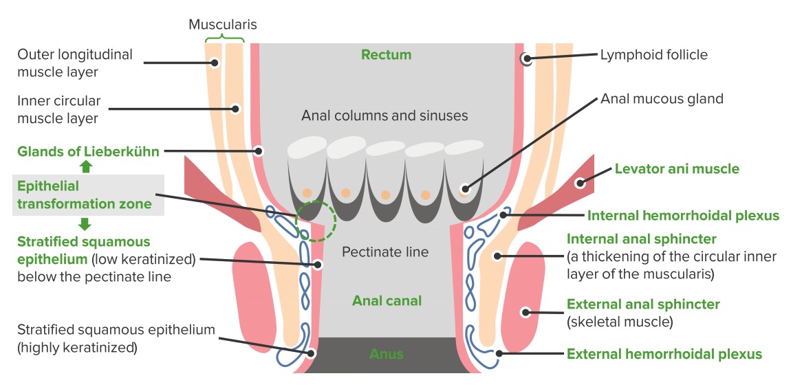
Anatomía del canal anal
Imagen por Lecturio.Factores de riesgo para cáncer anal:
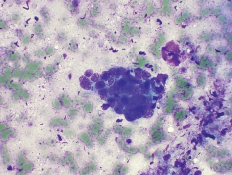
Carcinoma de células escamosas del canal anal:
La aspiración con aguja fina guiada por endoscopia revela un carcinoma de células escamosas poco diferenciado del canal anal. Se observan células nucleares periféricas en empalizada. (Tinción H&E, 40x)
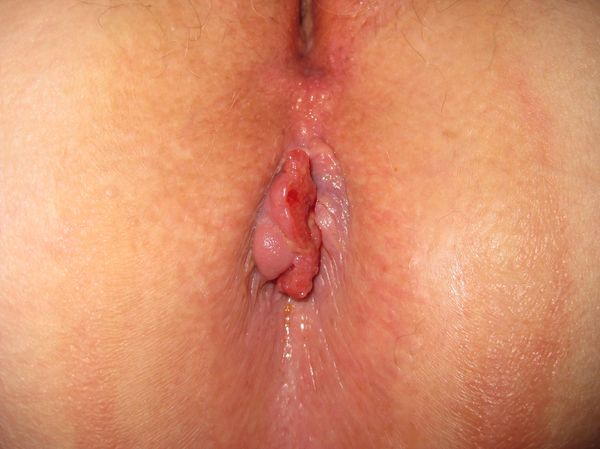
Carcinoma del canal anal
Imagen: “Anal canal carcinoma” por Dr. K.-H. Günther, Klinikum Main Spessart, Lohr am Main. Licencia: CC BY 3.0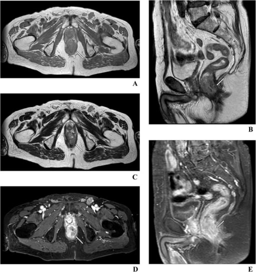
Imágenes de resonancia magnética de un cáncer anal con invasión del espacio interesfinteriano y del esfínter anal externo (flecha)
Imagen: “Anal cancer with invasion of the intersphincteric space” por St. John’s Cancer Centre, 7 Jaczewskiego Str., 20-090, Lublin, Poland. Licencia: CC BY 4.0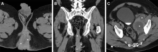
Imágenes de tomografía computarizada de un carcinoma de células escamosas del canal anal que se extiende desde la unión anorrectal hasta debajo del borde anal
Imagen: “Ulcero-fungating anal mass” por Massimo Tonolinicorresponding author and Roberto Bianco. Licencia: CC BY 4.0| Tumor Tumor Inflammation primario (T) | Nódulos (ganglios) linfáticos regionales (N) | Metástasis a distancia (M) |
|---|---|---|
| Tx: tumor Tumor Inflammation primario no evaluable | Nx: ganglios linfáticos regionales no evaluables | M0: sin metástasis a distancia |
| T0: sin evidencia de tumor Tumor Inflammation primario | N0: sin metástasis en EN Erythema nodosum is an immune-mediated panniculitis (inflammation of the subcutaneous fat) caused by a type IV (delayed-type) hypersensitivity reaction. It commonly manifests in young women as tender, erythematous nodules on the shins. Erythema Nodosum los LOS Neisseria ganglios linfáticos regionales | M1: metástasis a distancia |
| Tis: lesión escamosa intraepitelial de alto grado | N1: metástasis a
los
LOS
Neisseria ganglios inguinales, mesorrectales, ilíacos internos o ilíacos externos
|
|
| T1: tumor Tumor Inflammation ≤ 2 cm | ||
| T2: tumor Tumor Inflammation > 2 cm pero ≤ 5 cm | ||
| T3 T3 A T3 thyroid hormone normally synthesized and secreted by the thyroid gland in much smaller quantities than thyroxine (T4). Most T3 is derived from peripheral monodeiodination of T4 at the 5′ position of the outer ring of the iodothyronine nucleus. The hormone finally delivered and used by the tissues is mainly t3. Thyroid Hormones: tumor Tumor Inflammation > 5 cm | ||
| T4 T4 The major hormone derived from the thyroid gland. Thyroxine is synthesized via the iodination of tyrosines (monoiodotyrosine) and the coupling of iodotyrosines (diiodotyrosine) in the thyroglobulin. Thyroxine is released from thyroglobulin by proteolysis and secreted into the blood. Thyroxine is peripherally deiodinated to form triiodothyronine which exerts a broad spectrum of stimulatory effects on cell metabolism. Thyroid Hormones: tumor Tumor Inflammation de cualquier tamaño que invade órganos adyacentes, como la vagina Vagina The vagina is the female genital canal, extending from the vulva externally to the cervix uteri internally. The structures have sexual, reproductive, and urinary functions and a rich blood supply, mainly arising from the internal iliac artery. Vagina, Vulva, and Pelvic Floor: Anatomy, la uretra o la vejiga |
| Estadio | T | N | M |
|---|---|---|---|
| 0 | Tis | N0 | M0 |
| I | T1 | N0 | M0 |
| IIA | T2 | N0 | M0 |
| IIB | T3 T3 A T3 thyroid hormone normally synthesized and secreted by the thyroid gland in much smaller quantities than thyroxine (T4). Most T3 is derived from peripheral monodeiodination of T4 at the 5′ position of the outer ring of the iodothyronine nucleus. The hormone finally delivered and used by the tissues is mainly t3. Thyroid Hormones | N0 | M0 |
| IIIA | T1–2 | N1 | M0 |
| IIIB | T4 T4 The major hormone derived from the thyroid gland. Thyroxine is synthesized via the iodination of tyrosines (monoiodotyrosine) and the coupling of iodotyrosines (diiodotyrosine) in the thyroglobulin. Thyroxine is released from thyroglobulin by proteolysis and secreted into the blood. Thyroxine is peripherally deiodinated to form triiodothyronine which exerts a broad spectrum of stimulatory effects on cell metabolism. Thyroid Hormones | N0 | M0 |
| IIIC | T3 T3 A T3 thyroid hormone normally synthesized and secreted by the thyroid gland in much smaller quantities than thyroxine (T4). Most T3 is derived from peripheral monodeiodination of T4 at the 5′ position of the outer ring of the iodothyronine nucleus. The hormone finally delivered and used by the tissues is mainly t3. Thyroid Hormones–4 | N1 | M0 |
| IV | Cualquier T | Cualquier N | M1 |